• 文献检索
  • 文档翻译
  • 深度研究
  • 学术资讯
  • Suppr Zotero 插件Zotero 插件
  • 邀请有礼
  • 套餐&价格
  • 历史记录
应用&插件
Suppr Zotero 插件Zotero 插件浏览器插件Mac 客户端Windows 客户端微信小程序
定价
高级版会员购买积分包购买API积分包
服务
文献检索文档翻译深度研究API 文档MCP 服务
关于我们
关于 Suppr公司介绍联系我们用户协议隐私条款
关注我们

Suppr 超能文献

核心技术专利:CN118964589B侵权必究
粤ICP备2023148730 号-1Suppr @ 2026

文献检索

告别复杂PubMed语法,用中文像聊天一样搜索,搜遍4000万医学文献。AI智能推荐,让科研检索更轻松。

立即免费搜索

文件翻译

保留排版,准确专业,支持PDF/Word/PPT等文件格式,支持 12+语言互译。

免费翻译文档

深度研究

AI帮你快速写综述,25分钟生成高质量综述,智能提取关键信息,辅助科研写作。

立即免费体验

平板探测器灌注成像与常规多排探测器灌注成像在急性缺血性脑卒中患者中的对比研究。

Flat-panel Detector Perfusion Imaging and Conventional Multidetector Perfusion Imaging in Patients with Acute Ischemic Stroke : A Comparative Study.

机构信息

Department of Diagnostic and Interventional Neuroradiology, Inselspital, Bern University Hospital, University of Bern, Freiburgstrasse, 3010, Bern, Switzerland.

Department of Neuroradiology, Clinic for Radiology and Nuclear Medicine, University Hospital Basel, Petersgraben 4/Spitalstrasse 21, 4031, Basel, Switzerland.

出版信息

Clin Neuroradiol. 2024 Sep;34(3):625-635. doi: 10.1007/s00062-024-01401-7. Epub 2024 Mar 25.

DOI:10.1007/s00062-024-01401-7
PMID:38526586
原文链接:https://pmc.ncbi.nlm.nih.gov/articles/PMC11339100/
Abstract

PURPOSE

Flat-panel detector computed tomography (FDCT) is increasingly used in (neuro)interventional angiography suites. This study aimed to compare FDCT perfusion (FDCTP) with conventional multidetector computed tomography perfusion (MDCTP) in patients with acute ischemic stroke.

METHODS

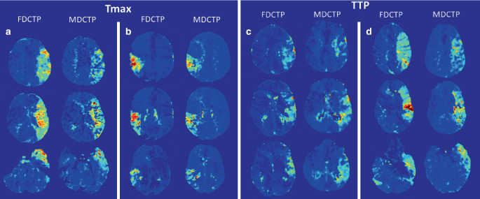
In this study, 19 patients with large vessel occlusion in the anterior circulation who had undergone mechanical thrombectomy, baseline MDCTP and pre-interventional FDCTP were included. Hypoperfused tissue volumes were manually segmented on time to maximum (Tmax) and time to peak (TTP) maps based on the maximum visible extent. Absolute and relative thresholds were applied to the maximum visible extent on Tmax and relative cerebral blood flow (rCBF) maps to delineate penumbra volumes and volumes with a high likelihood of irreversible infarcted tissue ("core"). Standard comparative metrics were used to evaluate the performance of FDCTP.

RESULTS

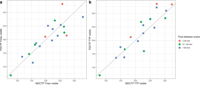
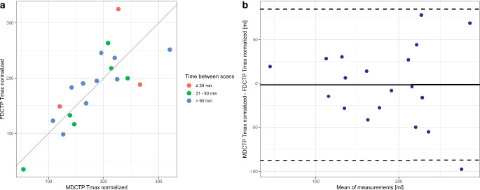
Strong correlations and robust agreement were found between manually segmented volumes on MDCTP and FDCTP Tmax maps (r = 0.85, 95% CI 0.65-0.94, p < 0.001; ICC = 0.85, 95% CI 0.69-0.94) and TTP maps (r = 0.91, 95% CI 0.78-0.97, p < 0.001; ICC = 0.90, 95% CI 0.78-0.96); however, direct quantitative comparisons using thresholding showed lower correlations and weaker agreement (MDCTP versus FDCTP Tmax 6 s: r = 0.35, 95% CI -0.13-0.69, p = 0.15; ICC = 0.32, 95% CI 0.07-0.75). Normalization techniques improved results for Tmax maps (r = 0.78, 95% CI 0.50-0.91, p < 0.001; ICC = 0.77, 95% CI 0.55-0.91). Bland-Altman analyses indicated a slight systematic underestimation of FDCTP Tmax maximum visible extent volumes and slight overestimation of FDCTP TTP maximum visible extent volumes compared to MDCTP.

CONCLUSION

FDCTP and MDCTP provide qualitatively comparable volumetric results on Tmax and TTP maps; however, direct quantitative measurements of infarct core and hypoperfused tissue volumes showed lower correlations and agreement.

摘要

目的

平板探测器 CT(FDCT)在(神经)介入血管造影室中越来越多地被使用。本研究旨在比较急性缺血性卒中患者的 FDCT 灌注(FDCTP)与常规多层 CT 灌注(MDCTP)。

方法

本研究纳入了 19 例接受机械血栓切除术的前循环大血管闭塞患者,进行了基线 MDCTP 和预介入 FDCTP。根据最大可见范围,在时间到最大值(Tmax)和时间到峰值(TTP)图上手动分割低灌注组织体积。在 Tmax 和相对脑血流量(rCBF)图上应用绝对和相对阈值来划定半暗带体积和高度可能不可逆梗死组织的体积(“核心”)。使用标准比较指标来评估 FDCTP 的性能。

结果

MDCTP 和 FDCTP Tmax 图(r=0.85,95%CI 0.65-0.94,p<0.001;ICC=0.85,95%CI 0.69-0.94)和 TTP 图(r=0.91,95%CI 0.78-0.97,p<0.001;ICC=0.90,95%CI 0.78-0.96)之间存在较强的相关性和稳健的一致性;然而,使用阈值的直接定量比较显示出较低的相关性和较弱的一致性(MDCTP 与 FDCTP Tmax 6s:r=0.35,95%CI-0.13-0.69,p=0.15;ICC=0.32,95%CI 0.07-0.75)。Tmax 图的归一化技术改善了结果(r=0.78,95%CI 0.50-0.91,p<0.001;ICC=0.77,95%CI 0.55-0.91)。Bland-Altman 分析表明,与 MDCTP 相比,FDCTP Tmax 最大可见范围体积存在轻微的系统低估,FDCTP TTP 最大可见范围体积存在轻微的高估。

结论

FDCTP 和 MDCTP 在 Tmax 和 TTP 图上提供了定性可比的容积结果;然而,对梗死核心和低灌注组织体积的直接定量测量显示出较低的相关性和一致性。

https://cdn.ncbi.nlm.nih.gov/pmc/blobs/2d46/11339100/d5a4f533e682/62_2024_1401_Fig3_HTML.jpg
https://cdn.ncbi.nlm.nih.gov/pmc/blobs/2d46/11339100/3b3f13fc1c1c/62_2024_1401_Fig1_HTML.jpg
https://cdn.ncbi.nlm.nih.gov/pmc/blobs/2d46/11339100/675ddb3dbc90/62_2024_1401_Fig2_HTML.jpg
https://cdn.ncbi.nlm.nih.gov/pmc/blobs/2d46/11339100/d5a4f533e682/62_2024_1401_Fig3_HTML.jpg
https://cdn.ncbi.nlm.nih.gov/pmc/blobs/2d46/11339100/3b3f13fc1c1c/62_2024_1401_Fig1_HTML.jpg
https://cdn.ncbi.nlm.nih.gov/pmc/blobs/2d46/11339100/675ddb3dbc90/62_2024_1401_Fig2_HTML.jpg
https://cdn.ncbi.nlm.nih.gov/pmc/blobs/2d46/11339100/d5a4f533e682/62_2024_1401_Fig3_HTML.jpg

相似文献

1
Flat-panel Detector Perfusion Imaging and Conventional Multidetector Perfusion Imaging in Patients with Acute Ischemic Stroke : A Comparative Study.平板探测器灌注成像与常规多排探测器灌注成像在急性缺血性脑卒中患者中的对比研究。
Clin Neuroradiol. 2024 Sep;34(3):625-635. doi: 10.1007/s00062-024-01401-7. Epub 2024 Mar 25.
2
A novel use of time separation technique to improve flat detector CT perfusion imaging in stroke patients.一种利用时间分离技术改善脑卒中患者平板探测器 CT 灌注成像的新方法。
Med Phys. 2022 Jun;49(6):3624-3637. doi: 10.1002/mp.15640. Epub 2022 Apr 20.
3
Pretreatment brain CT perfusion thresholds for predicting final infarct volume in distal medium vessel occlusions.预处理脑 CT 灌注阈值预测远端中等血管闭塞的最终梗死体积。
J Neuroimaging. 2023 Nov-Dec;33(6):968-975. doi: 10.1111/jon.13142. Epub 2023 Jun 25.
4
Angiography suite cone-beam CT perfusion for selection of thrombectomy patients: A pilot study.血管造影套房锥形束 CT 灌注用于选择血栓切除术患者:一项初步研究。
J Neuroimaging. 2022 May;32(3):493-501. doi: 10.1111/jon.12988. Epub 2022 Mar 21.
5
The Value of Whole-Brain Perfusion Parameters Combined with Multiphase Computed Tomography Angiography in Predicting Hemorrhagic Transformation in Ischemic Stroke.全脑灌注参数联合多相 CT 血管成像在预测缺血性脑卒中出血性转化中的价值。
J Stroke Cerebrovasc Dis. 2020 Apr;29(4):104690. doi: 10.1016/j.jstrokecerebrovasdis.2020.104690. Epub 2020 Feb 14.
6
In Acute Stroke, Can CT Perfusion-Derived Cerebral Blood Volume Maps Substitute for Diffusion-Weighted Imaging in Identifying the Ischemic Core?在急性卒中中,CT灌注衍生的脑血容量图能否在识别缺血核心方面替代弥散加权成像?
PLoS One. 2015 Jul 20;10(7):e0133566. doi: 10.1371/journal.pone.0133566. eCollection 2015.
7
Feasibility of deconvolution-based multiphase CT angiography perfusion maps in acute ischemic stroke: Simulation and concordance with CT perfusion.基于去卷积的多相 CT 血管造影灌注图在急性缺血性脑卒中的可行性:模拟与 CT 灌注的一致性。
J Stroke Cerebrovasc Dis. 2022 Dec;31(12):106844. doi: 10.1016/j.jstrokecerebrovasdis.2022.106844. Epub 2022 Oct 29.
8
Comparison of MRI IVIM and MR perfusion imaging in acute ischemic stroke due to large vessel occlusion.磁共振成像 IVIM 与 MR 灌注成像在大动脉闭塞性急性缺血性脑卒中的比较。
Int J Stroke. 2020 Apr;15(3):332-342. doi: 10.1177/1747493019873515. Epub 2019 Sep 3.
9
Noncontrast Computed Tomography e-Stroke Infarct Volume Is Similar to RAPID Computed Tomography Perfusion in Estimating Postreperfusion Infarct Volumes.非对比 CT 卒中梗死体积与 RAPID CT 灌注在估计再灌注后梗死体积方面相似。
Stroke. 2021 Jan;52(2):634-641. doi: 10.1161/STROKEAHA.120.031651. Epub 2021 Jan 12.
10
Insights into multilevel tissue-level collateral status using ColorViz maps from dual data sources in acute ischemic cerebrovascular diseases: A STARD-compliant retrospective study.利用急性缺血性脑血管病双数据源的 ColorViz 图谱深入了解多层次组织水平侧支状态:一项符合 STARD 标准的回顾性研究。
Medicine (Baltimore). 2024 Sep 20;103(38):e39787. doi: 10.1097/MD.0000000000039787.

本文引用的文献

1
Value of Immediate Flat Panel Perfusion Imaging after Endovascular Therapy (AFTERMATH): A Proof of Concept Study.血管内治疗后即刻平板灌注成像的价值(AFTERMATH):一项概念验证研究。
AJNR Am J Neuroradiol. 2024 Feb 7;45(2):163-170. doi: 10.3174/ajnr.A8103.
2
Clinical Applications of Conebeam CTP Imaging in Cerebral Disease: A Systematic Review.锥形束 CTP 成像在脑部疾病中的临床应用:系统评价。
AJNR Am J Neuroradiol. 2023 Aug;44(8):922-927. doi: 10.3174/ajnr.A7930. Epub 2023 Jul 6.
3
Extended Multimodal Flat Detector CT Imaging in Acute Ischemic Stroke: A Pilot Study.
扩展多模态平板探测器 CT 成像在急性缺血性脑卒中中的应用:一项初步研究。
J Digit Imaging. 2023 Jun;36(3):1198-1207. doi: 10.1007/s10278-022-00699-4. Epub 2023 Jan 17.
4
Importance of Delayed Reperfusions in Patients With Incomplete Thrombectomy.未能完全取栓患者实施延迟再灌注的重要性。
Stroke. 2022 Nov;53(11):3350-3358. doi: 10.1161/STROKEAHA.122.040063. Epub 2022 Oct 7.
5
Effective Dose Measurements of the Latest-Generation Angiographic System in Patients with Acute Stroke: A Comparison with the Newest Multidetector CT Generation.最新代血管造影系统在急性脑卒中患者中的有效剂量测量:与最新多排 CT 一代的比较。
AJNR Am J Neuroradiol. 2022 Nov;43(11):1621-1626. doi: 10.3174/ajnr.A7658. Epub 2022 Oct 6.
6
Correlation of Collateral Scores Derived from Whole-Brain Time-Resolved Flat Panel Detector Imaging in Acute Ischemic Stroke.基于全脑时间分辨平板探测器成像的侧支循环评分与急性缺血性脑卒中的相关性。
AJNR Am J Neuroradiol. 2022 Nov;43(11):1627-1632. doi: 10.3174/ajnr.A7657. Epub 2022 Oct 6.
7
Evaluation of time-resolved whole brain flat panel detector perfusion imaging using RAPID ANGIO in patients with acute stroke: comparison with CT perfusion imaging.使用 RAPID ANGIO 对急性脑卒中患者进行时间分辨全脑平板探测器灌注成像评估:与 CT 灌注成像的比较。
J Neurointerv Surg. 2023 Apr;15(4):387-392. doi: 10.1136/neurintsurg-2021-018464. Epub 2022 Apr 8.
8
Evaluation of Sine Spin flat detector CT imaging compared with multidetector CT.正弦单扇区探测器 CT 与多层螺旋 CT 的对比评估。
J Neurointerv Surg. 2023 Mar;15(3):292-297. doi: 10.1136/neurintsurg-2021-018312. Epub 2022 Mar 22.
9
Direct to angiography suite approaches for the triage of suspected acute stroke patients: a systematic review and meta-analysis.疑似急性卒中患者分诊的直接血管造影套件方法:系统评价与荟萃分析
Ther Adv Neurol Disord. 2022 Mar 2;15:17562864221078177. doi: 10.1177/17562864221078177. eCollection 2022.
10
Automated Brain Perfusion Imaging in Acute Ischemic Stroke: Interpretation Pearls and Pitfalls.急性缺血性脑卒中的自动脑灌注成像:解读要点及陷阱。
Stroke. 2021 Nov;52(11):3728-3738. doi: 10.1161/STROKEAHA.121.035049. Epub 2021 Sep 27.