• 文献检索
  • 文档翻译
  • 深度研究
  • 学术资讯
  • Suppr Zotero 插件Zotero 插件
  • 邀请有礼
  • 套餐&价格
  • 历史记录
应用&插件
Suppr Zotero 插件Zotero 插件浏览器插件Mac 客户端Windows 客户端微信小程序
定价
高级版会员购买积分包购买API积分包
服务
文献检索文档翻译深度研究API 文档MCP 服务
关于我们
关于 Suppr公司介绍联系我们用户协议隐私条款
关注我们

Suppr 超能文献

核心技术专利:CN118964589B侵权必究
粤ICP备2023148730 号-1Suppr @ 2026

文献检索

告别复杂PubMed语法,用中文像聊天一样搜索,搜遍4000万医学文献。AI智能推荐,让科研检索更轻松。

立即免费搜索

文件翻译

保留排版,准确专业,支持PDF/Word/PPT等文件格式,支持 12+语言互译。

免费翻译文档

深度研究

AI帮你快速写综述,25分钟生成高质量综述,智能提取关键信息,辅助科研写作。

立即免费体验

提高糖尿病性黄斑水肿的治疗效果:探索ESASO分类和光学相干断层扫描结构生物标志物

Enhancing Diabetic Macular Edema Treatment Outcomes: Exploring the ESASO Classification and Structural OCT Biomarkers.

作者信息

Panozzo Giacomo, Cicinelli Maria V, Dalla Mura Giulia, Giannarelli Diana, Vadalà Maria, Bonfiglio Vincenza, Bellisario Giovanni, Bandello Francesco

机构信息

Ophthalmology Unit, Clinica San Francesco, Verona, Italy.

ESASO, European School of Advanced Studies in Ophthalmology, Lugano, Switzerland.

出版信息

Ophthalmol Ther. 2024 May;13(5):1383-1398. doi: 10.1007/s40123-024-00925-y. Epub 2024 Mar 26.

DOI:10.1007/s40123-024-00925-y
PMID:38530567
原文链接:https://pmc.ncbi.nlm.nih.gov/articles/PMC11039609/
Abstract

INTRODUCTION

This study assessed the European School of Advanced Studies in Ophthalmology (ESASO) classification's prognostic value for diabetic macular edema (DME) in predicting intravitreal therapy outcomes.

METHODS

In this retrospective, multicenter study, patients aged > 50 years with type 1 or 2 diabetes and DME received intravitreal antivascular endothelial growth factor (anti-VEGF) agents (ranibizumab, bevacizumab, and aflibercept) or steroids (dexamethasone). The primary outcome was visual acuity (VA) change post-treatment, termed as functional response, measured 4-6 weeks post-third anti-VEGF or 12-16 weeks post-steroid injection, stratified by initial DME stage.

RESULTS

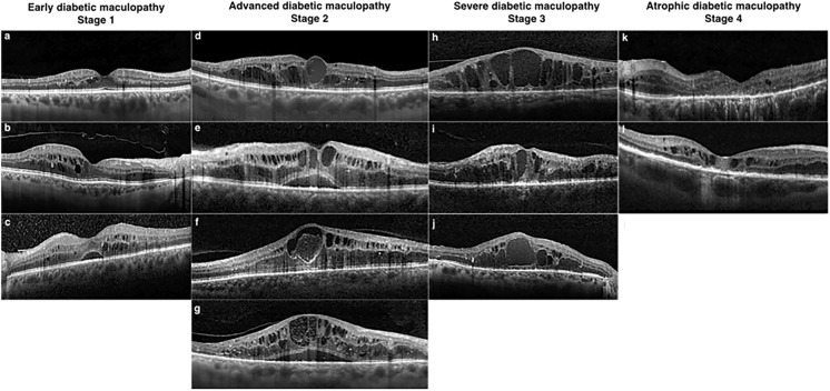
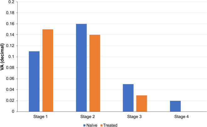
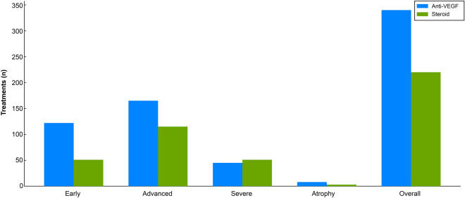
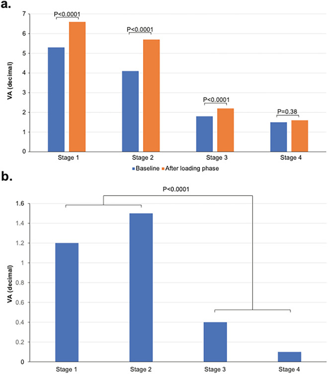
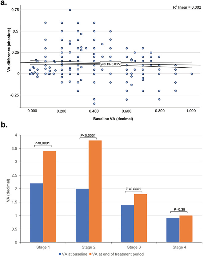
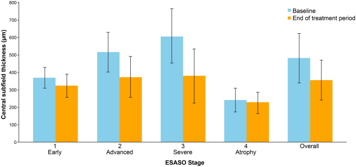
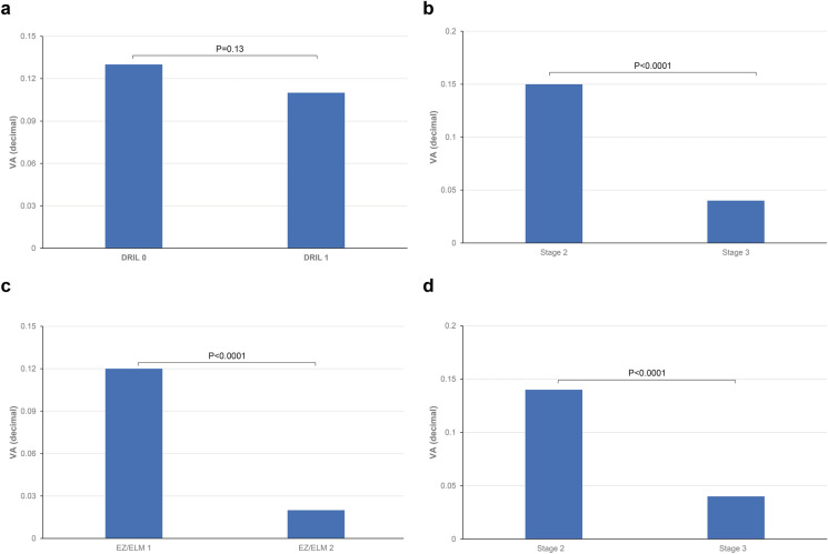
Of the 560 eyes studied (62% male, mean age 66.7 years), 31% were classified as stage 1 (early), 50% stage 2 (advanced), 17% stage 3 (severe), and 2% stage 4 (atrophic). Visual acuity (VA; decimal) improved by 0.12-0.15 decimals in stages 1-2 but only 0.03 decimal in stage 3 (all p < 0.0001) and 0.01 in stage 4 (p = 0.38). Even in eyes with low baseline VA ≤ 0.3, improvements were significant only in stages 1 and 2 (0.12 and 0.17 decimals, respectively). Central subfield thickness (CST) improvement was greatest in stage 3 (-229 µm, 37.6%, p < 0.0001), but uncorrelated with VA gains, unlike stages 1 and 2 (respectively: -142 µm, 27.4%; - 5 µm, 12%; both p < 0.0001). Stage 4 showed no significant CST change. Baseline disorganization of retinal inner layers and focal damage of the ellipsoid zone/external limiting membrane did not influence VA improvement in stages 1 and 2. Treatment patterns varied, with 61% receiving anti-VEGF and 39% dexamethasone, influenced by DME stage, with no significant differences between therapeutic agents.

CONCLUSION

The ESASO classification, which views the retina as a neurovascular unit and integrates multiple biomarkers, surpasses single biomarkers in predicting visual outcomes. Significant functional improvement occurred only in stages 1 and 2, suggesting reversible damage, whereas stages 3 and 4 likely reflect irreversible damage.

摘要

引言

本研究评估了欧洲高级眼科学术院(ESASO)分类法对糖尿病性黄斑水肿(DME)预测玻璃体内治疗效果的预后价值。

方法

在这项回顾性多中心研究中,年龄大于50岁的1型或2型糖尿病合并DME患者接受玻璃体内抗血管内皮生长因子(抗VEGF)药物(雷珠单抗、贝伐单抗和阿柏西普)或类固醇(地塞米松)治疗。主要结局是治疗后视力(VA)变化,即功能反应,在第三次抗VEGF治疗后4 - 6周或类固醇注射后12 - 16周测量,并按初始DME阶段分层。

结果

在研究的560只眼中(62%为男性,平均年龄66.7岁),31%被分类为1期(早期),50%为2期(进展期),17%为3期(重度),2%为4期(萎缩期)。1 - 2期视力(VA;小数)提高了0.12 - 0.15,但3期仅提高了0.03(所有p < 0.0001),4期提高了0.01(p = 0.38)。即使在基线视力≤0.3的眼中,仅1期和2期有显著改善(分别为0.12和0.17)。中心子野厚度(CST)改善在3期最大(-229 µm,37.6%,p < 0.0001),但与视力提高无关,与1期和2期不同(分别为:-142 µm,27.4%;-5 µm,12%;均p < 0.0001)。4期CST无显著变化变化。视网膜内层的基线紊乱和椭圆体带/外界膜的局灶性损伤在1期和2期不影响视力改善。治疗模式各不相同,61%接受抗VEGF治疗,39%接受地塞米松治疗,受DME阶段影响,治疗药物之间无显著差异。

结论

ESASO分类法将视网膜视为神经血管单元并整合多种生物标志物,在预测视力结果方面优于单一生物标志物。仅1期和2期有显著的功能改善,提示损伤可逆,而3期和4期可能反映不可逆损伤。

https://cdn.ncbi.nlm.nih.gov/pmc/blobs/26c3/11039609/b6ce4f8481f3/40123_2024_925_Fig7_HTML.jpg
https://cdn.ncbi.nlm.nih.gov/pmc/blobs/26c3/11039609/e2d7cfe5b46a/40123_2024_925_Fig1_HTML.jpg
https://cdn.ncbi.nlm.nih.gov/pmc/blobs/26c3/11039609/3129d9d41c47/40123_2024_925_Fig2_HTML.jpg
https://cdn.ncbi.nlm.nih.gov/pmc/blobs/26c3/11039609/47e5e9e4eab1/40123_2024_925_Fig3_HTML.jpg
https://cdn.ncbi.nlm.nih.gov/pmc/blobs/26c3/11039609/4b909512dd4d/40123_2024_925_Fig4_HTML.jpg
https://cdn.ncbi.nlm.nih.gov/pmc/blobs/26c3/11039609/8f05c8488a91/40123_2024_925_Fig5_HTML.jpg
https://cdn.ncbi.nlm.nih.gov/pmc/blobs/26c3/11039609/0da7bd76cae5/40123_2024_925_Fig6_HTML.jpg
https://cdn.ncbi.nlm.nih.gov/pmc/blobs/26c3/11039609/b6ce4f8481f3/40123_2024_925_Fig7_HTML.jpg

相似文献

1
Enhancing Diabetic Macular Edema Treatment Outcomes: Exploring the ESASO Classification and Structural OCT Biomarkers.提高糖尿病性黄斑水肿的治疗效果:探索ESASO分类和光学相干断层扫描结构生物标志物
Ophthalmol Ther. 2024 May;13(5):1383-1398. doi: 10.1007/s40123-024-00925-y. Epub 2024 Mar 26.
2
Optical Coherence Tomography Biomarkers in Bilateral Diabetic Macular Edema Patients with Asymmetric anti-VEGF Response.双侧糖尿病性黄斑水肿患者抗VEGF反应不对称时的光学相干断层扫描生物标志物
Semin Ophthalmol. 2021 Aug 18;36(5-6):444-451. doi: 10.1080/08820538.2021.1907423. Epub 2021 Mar 29.
3
Dril Influences Short-term Visual Outcome after Intravitreal Corticosteroid Injection for Refractory Diabetic Macular Edema.玻璃体内注射皮质类固醇治疗难治性糖尿病性黄斑水肿后,DRIL影响短期视觉预后。
Curr Eye Res. 2021 Sep;46(9):1378-1386. doi: 10.1080/02713683.2021.1878540. Epub 2021 Jan 31.
4
Factors Associated With Visual Acuity and Central Subfield Thickness Changes When Treating Diabetic Macular Edema With Anti-Vascular Endothelial Growth Factor Therapy: An Exploratory Analysis of the Protocol T Randomized Clinical Trial.抗血管内皮生长因子治疗糖尿病黄斑水肿时与视力和中央视网膜神经纤维层厚度变化相关的因素:Protocol T 随机临床试验的探索性分析。
JAMA Ophthalmol. 2019 Apr 1;137(4):382-389. doi: 10.1001/jamaophthalmol.2018.6786.
5
Disorganization of the retinal inner layers as a predictor of visual acuity in eyes with center-involved diabetic macular edema.视网膜内层结构紊乱作为累及黄斑中心的糖尿病性黄斑水肿患眼视力的预测指标。
JAMA Ophthalmol. 2014 Nov;132(11):1309-16. doi: 10.1001/jamaophthalmol.2014.2350.
6
OCT biomarkers as predictors of visual improvement in diabetic macular edema eyes receiving dexamethasone implants.光学相干断层扫描(OCT)生物标志物作为接受地塞米松植入物治疗的糖尿病性黄斑水肿眼视力改善的预测指标。
Int J Retina Vitreous. 2023 Jun 14;9(1):35. doi: 10.1186/s40942-023-00473-w.
7
Does HbA1c Level or Glomerular Filtration Rate Affect the Clinical Response to Endothelial Growth Factor Therapy (Ranibizumab or Aflibercept) in Diabetic Macular Edema? A Real-Life Experience.糖化血红蛋白水平或肾小球滤过率是否会影响糖尿病性黄斑水肿患者对内皮生长因子治疗(雷珠单抗或阿柏西普)的临床反应?一项真实病例经验。
Ophthalmol Ther. 2023 Oct;12(5):2657-2670. doi: 10.1007/s40123-023-00758-1. Epub 2023 Jul 28.
8
Optical Coherence Tomography Biomarkers in Predicting Treatment Outcomes of Diabetic Macular Edema after Ranibizumab Injections.光学相干断层扫描生物标志物预测雷珠单抗治疗糖尿病黄斑水肿的疗效。
Medicina (Kaunas). 2023 Mar 21;59(3):629. doi: 10.3390/medicina59030629.
9
Baseline Factors Associated With 6-Month Visual Acuity and Retinal Thickness Outcomes in Patients With Macular Edema Secondary to Central Retinal Vein Occlusion or Hemiretinal Vein Occlusion: SCORE2 Study Report 4.视网膜中央静脉阻塞或半侧视网膜静脉阻塞继发黄斑水肿患者6个月视力和视网膜厚度结果的基线因素:SCORE2研究报告4
JAMA Ophthalmol. 2017 Jun 1;135(6):639-649. doi: 10.1001/jamaophthalmol.2017.1141.
10
Retinal changes after fluocinolone acetonide implant (ILUVIEN®) for DME: SD-OCT imaging assessment using ESASO classification.DME 患者应用氟轻松玻璃体内植入剂(ILUVIEN®)后的视网膜变化:采用 ESASO 分类的 SD-OCT 成像评估。
Eur J Ophthalmol. 2024 Jan;34(1):233-244. doi: 10.1177/11206721231183471. Epub 2023 Jul 20.

引用本文的文献

1
Predictive Role of Complete Blood Count-Derived Inflammation Indices and Optical Coherence Tomography Biomarkers for Early Response to Intravitreal Anti-VEGF in Diabetic Macular Edema.全血细胞计数衍生的炎症指标和光学相干断层扫描生物标志物对糖尿病性黄斑水肿玻璃体内抗VEGF治疗早期反应的预测作用
Biomedicines. 2025 May 27;13(6):1308. doi: 10.3390/biomedicines13061308.
2
Ultrastructural imaging biomarkers in diabetic macular edema: A major review.糖尿病性黄斑水肿中的超微结构成像生物标志物:一项主要综述。
Indian J Ophthalmol. 2025 Jan 1;73(Suppl 1):S7-S23. doi: 10.4103/IJO.IJO_878_24. Epub 2024 Dec 24.

本文引用的文献

1
From diagnosis to prognosis: A paradigm shift for multimodal imaging in assessing diabetic macular edema.从诊断到预后:评估糖尿病性黄斑水肿的多模态成像的范式转变。
Eur J Ophthalmol. 2024 Jan;34(1):7-10. doi: 10.1177/11206721231199149. Epub 2023 Aug 30.
2
Retinal changes after fluocinolone acetonide implant (ILUVIEN®) for DME: SD-OCT imaging assessment using ESASO classification.DME 患者应用氟轻松玻璃体内植入剂(ILUVIEN®)后的视网膜变化:采用 ESASO 分类的 SD-OCT 成像评估。
Eur J Ophthalmol. 2024 Jan;34(1):233-244. doi: 10.1177/11206721231183471. Epub 2023 Jul 20.
3
Validation of Esaso Classification of Diabetic Maculopathy.
Esaso 分类法对糖尿病性黄斑病变的验证。
Eur J Ophthalmol. 2024 Mar;34(2):497-501. doi: 10.1177/11206721231186649. Epub 2023 Jul 16.
4
Persistent diabetic macular edema: Definition, incidence, biomarkers, and treatment methods.持续性糖尿病性黄斑水肿:定义、发生率、生物标志物和治疗方法。
Surv Ophthalmol. 2023 Mar-Apr;68(2):147-174. doi: 10.1016/j.survophthal.2022.11.008. Epub 2022 Nov 24.
5
Early DMO: a predictor of poor outcomes following cataract surgery in diabetic patients. The DICAT-II study.早期 DMO:糖尿病患者白内障手术后不良结局的预测指标。DICAT-II 研究。
Eye (Lond). 2022 Aug;36(8):1687-1693. doi: 10.1038/s41433-021-01718-4. Epub 2021 Aug 3.
6
Systemic Factors Associated with Treatment Response in Diabetic Macular Edema.与糖尿病性黄斑水肿治疗反应相关的全身因素
J Ophthalmol. 2020 Mar 19;2020:1875860. doi: 10.1155/2020/1875860. eCollection 2020.
7
An optical coherence tomography-based grading of diabetic maculopathy proposed by an international expert panel: The European School for Advanced Studies in Ophthalmology classification.一个国际专家小组提出的基于光学相干断层扫描的糖尿病性黄斑病变分级:欧洲高级眼科学术研究学会分类法
Eur J Ophthalmol. 2020 Jan;30(1):8-18. doi: 10.1177/1120672119880394. Epub 2019 Nov 12.
8
Evaluation of markers of outcome in real-world treatment of diabetic macular edema.糖尿病性黄斑水肿实际治疗中预后指标的评估
Eye Vis (Lond). 2018 Oct 11;5:27. doi: 10.1186/s40662-018-0119-9. eCollection 2018.
9
Choroidal thickness changes stratified by outcome in real-world treatment of diabetic macular edema.在糖尿病性黄斑水肿的实际治疗中,按结局分层的脉络膜厚度变化
Graefes Arch Clin Exp Ophthalmol. 2018 Oct;256(10):1857-1865. doi: 10.1007/s00417-018-4072-z. Epub 2018 Jul 23.
10
Optical Coherence Tomography Features in Diabetic Macular Edema and the Impact on Anti-VEGF Response.糖尿病性黄斑水肿的光学相干断层扫描特征及其对抗血管内皮生长因子治疗反应的影响
Ophthalmic Surg Lasers Imaging Retina. 2016 Oct 1;47(10):908-913. doi: 10.3928/23258160-20161004-03.