Center for Advanced Retinal and Ocular Therapeutics (CAROT), University of Pennsylvania, Philadelphia, PA 19104, USA.
Department of Ophthalmology, University of Pennsylvania, Philadelphia, PA 19104, USA.
Brain. 2024 Sep 3;147(9):3234-3246. doi: 10.1093/brain/awae096.
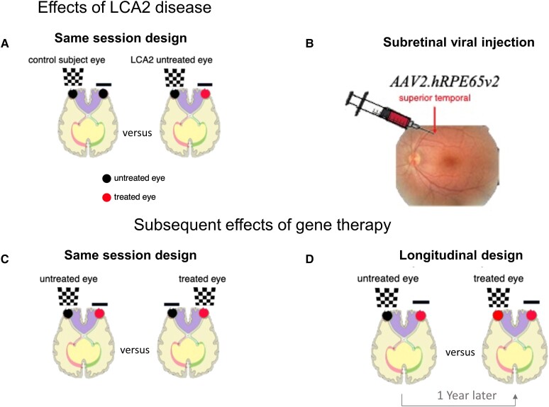
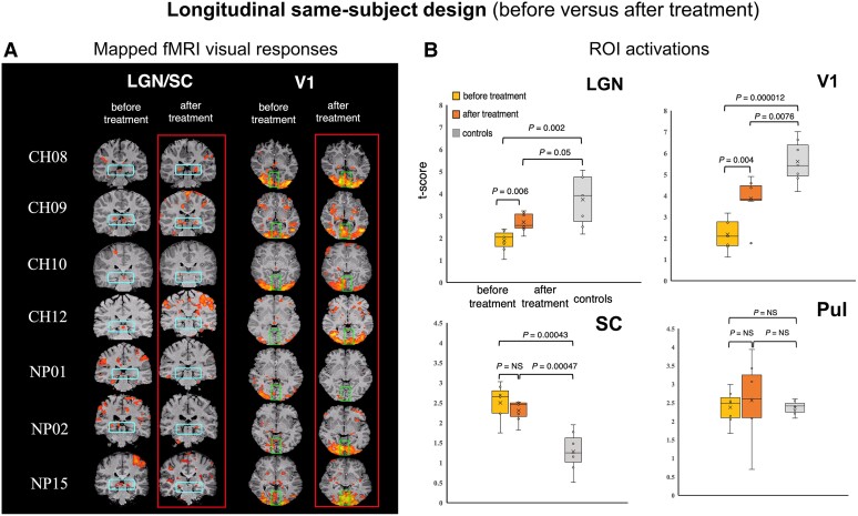
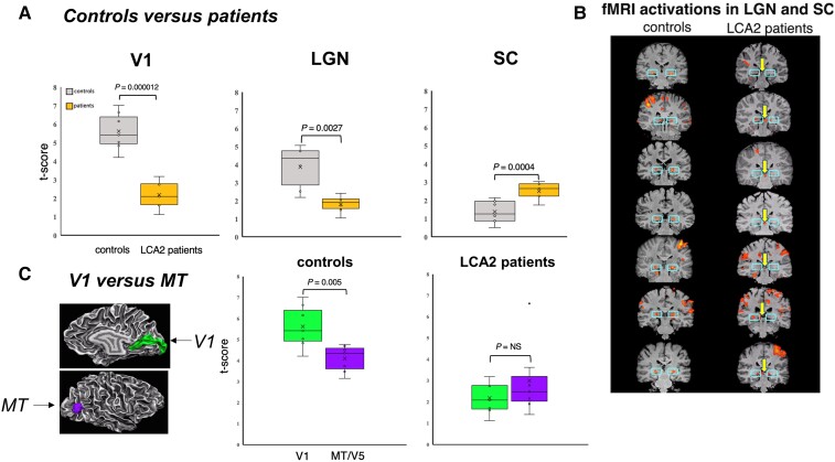
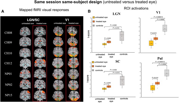
Genetic diseases affecting the retina can result in partial or complete loss of visual function. Leber's congenital amaurosis (LCA) is a rare blinding disease, usually inherited in an autosomally recessive manner, with no cure. Retinal gene therapy has been shown to improve vision in LCA patients caused by mutations in the RPE65 gene (LCA2). However, little is known about how activity in central visual pathways is affected by the disease or by subsequent gene therapy. Functional MRI (fMRI) was used to assess retinal signal transmission in cortical and subcortical visual structures before and 1 year after retinal intervention. The fMRI paradigm consisted of 15-s blocks of flickering (8 Hz) black and white checkerboards interleaved with 15 s of blank (black) screen. Visual activation in the brain was assessed using the general linear model, with multiple comparisons corrected using the false discovery rate method. Response to visual stimulation through untreated eyes of LCA2 patients showed heightened fMRI responses in the superior colliculus and diminished activities in the lateral geniculate nucleus (LGN) compared to controls, indicating a shift in the patients' visual processing towards the retinotectal pathway. Following gene therapy, stimuli presented to the treated eye elicited significantly stronger fMRI responses in the LGN and primary visual cortex, indicating some re-engagement of the geniculostriate pathway (GS) pathway. Across patients, the post-treatment LGN fMRI responses correlated significantly with performance on a clinical test measuring light sensitivity. Our results demonstrate that the low vision observed in LCA2 patients involves a shift in visual processing toward the retinotectal pathway, and that gene therapy partially reinstates visual transmission through the GS pathway. This selective boosting of retinal output through the GS pathway and its correlation to improved visual performance, following several years of degenerative retinal disease, is striking. However, while retinal gene therapy and other ocular interventions have given hope to RPE65 patients, it may take years before development of therapies tailored to treat the diseases in other low vision patients are available. Our demonstration of a shift toward the retinotectal pathway in these patients may spur the development of new tools and rehabilitation strategies to help maximize the use of residual visual abilities and augment experience-dependent plasticity.
遗传性视网膜疾病可导致部分或完全丧失视觉功能。莱伯先天性黑蒙症(LCA)是一种罕见的致盲性疾病,通常以常染色体隐性遗传方式遗传,尚无治愈方法。视网膜基因治疗已被证明可改善 RPE65 基因突变引起的 LCA 患者的视力(LCA2)。然而,对于疾病或随后的基因治疗如何影响中央视觉通路的活动,我们知之甚少。功能磁共振成像(fMRI)用于评估视网膜干预前后皮质和皮质下视觉结构中的视网膜信号传递。fMRI 范式由闪烁(8 Hz)黑白棋盘的 15 秒块组成,每隔 15 秒插入 15 秒空白(黑色)屏幕。使用广义线性模型评估大脑中的视觉激活,使用错误发现率方法校正多重比较。与对照组相比,LCA2 患者未经治疗的眼睛对视觉刺激的反应显示出上丘的 fMRI 反应增强,外侧膝状体核(LGN)的活动减少,表明患者的视觉处理向视网膜-顶盖通路转移。基因治疗后,给予治疗的眼睛的刺激会引起 LGN 和初级视觉皮层的 fMRI 反应明显增强,表明视束-纹状通路(GS)通路的重新参与。在患者中,治疗后 LGN fMRI 反应与测量光敏感性的临床测试的表现显著相关。我们的研究结果表明,LCA2 患者的低视力涉及视觉处理向视网膜-顶盖通路的转移,基因治疗部分恢复了 GS 通路的视觉传递。在退行性视网膜疾病发生数年之后,GS 通路的这种选择性增强视网膜输出及其与视觉表现改善的相关性令人瞩目。然而,虽然视网膜基因治疗和其他眼部干预措施为 RPE65 患者带来了希望,但可能需要数年时间才能开发出针对其他低视力患者疾病的治疗方法。我们在这些患者中观察到向视网膜-顶盖通路的转变,这可能会促使开发新的工具和康复策略,以帮助最大限度地利用残留的视觉能力并增强经验依赖性可塑性。