• 文献检索
  • 文档翻译
  • 深度研究
  • 学术资讯
  • Suppr Zotero 插件Zotero 插件
  • 邀请有礼
  • 套餐&价格
  • 历史记录
应用&插件
Suppr Zotero 插件Zotero 插件浏览器插件Mac 客户端Windows 客户端微信小程序
定价
高级版会员购买积分包购买API积分包
服务
文献检索文档翻译深度研究API 文档MCP 服务
关于我们
关于 Suppr公司介绍联系我们用户协议隐私条款
关注我们

Suppr 超能文献

核心技术专利:CN118964589B侵权必究
粤ICP备2023148730 号-1Suppr @ 2026

文献检索

告别复杂PubMed语法,用中文像聊天一样搜索,搜遍4000万医学文献。AI智能推荐,让科研检索更轻松。

立即免费搜索

文件翻译

保留排版,准确专业,支持PDF/Word/PPT等文件格式,支持 12+语言互译。

免费翻译文档

深度研究

AI帮你快速写综述,25分钟生成高质量综述,智能提取关键信息,辅助科研写作。

立即免费体验

使用低剂量锥形束计算机断层扫描采集协议评估3D打印腭裂阻塞器:一项概念验证研究。

Evaluation of a 3D-Printed Cleft Palate Obturator Using a Low-Dose Cone Beam Computed Tomography Acquisition Protocol: A Proof-of-Concept Study.

作者信息

Nelson Thomas, Cobos Santiago F, Gandhi Vaibhav, Katechia Bina, Yadav Sumit, Tadinada Aditya

机构信息

School of Dentistry, University of Connecticut Health, Farmington, USA.

Oral and Maxillofacial Radiology, University of Connecticut Health, Farmington, USA.

出版信息

Cureus. 2024 Apr 4;16(4):e57602. doi: 10.7759/cureus.57602. eCollection 2024 Apr.

DOI:10.7759/cureus.57602
PMID:38577166
原文链接:https://pmc.ncbi.nlm.nih.gov/articles/PMC10994164/
Abstract

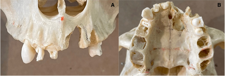
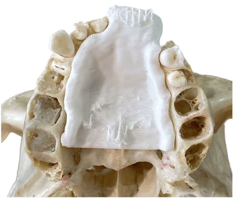
Cone beam computed tomography (CBCT) technology is increasingly utilized in the head and neck region and is valuable in treatment planning for cleft palate patients, potentially enabling the creation of 3D-printed obturators to assist with feeding and speech. This technical report investigates the feasibility of using data from a 360-degree CBCT scan to accurately produce a cleft palate obturator and assesses whether a lower-dose 180-degree CBCT scan can achieve a comparable result. A simulated cleft palate was crafted on a dehydrated human skull, which was then scanned using both 360-degree and 180-degree CBCT scanning protocols. Two obturators were digitally designed based on the segmented images from each scan and subsequently 3D printed. Evaluation of the segmented images and 3D-printed obturators from both protocols demonstrated clear visualization of anatomical landmarks and identical scores across all parameters, suggesting that the 180-degree CBCT scan can produce an obturator of comparable quality to that of the 360-degree scan, with the added benefit of reduced radiation exposure.

摘要

锥形束计算机断层扫描(CBCT)技术在头颈部区域的应用越来越广泛,对腭裂患者的治疗规划具有重要价值,有可能制作出3D打印的阻塞器以辅助进食和言语功能。本技术报告研究了使用360度CBCT扫描数据精确制作腭裂阻塞器的可行性,并评估低剂量180度CBCT扫描是否能取得可比结果。在一个脱水的人类颅骨上制作了模拟腭裂,然后分别使用360度和180度CBCT扫描协议进行扫描。根据每次扫描的分割图像进行了两个阻塞器的数字化设计,随后进行3D打印。对两种扫描协议的分割图像和3D打印阻塞器的评估表明,解剖标志清晰可见,所有参数的得分相同,这表明180度CBCT扫描能够制作出与360度扫描质量相当的阻塞器,且具有减少辐射暴露的额外优势。

https://cdn.ncbi.nlm.nih.gov/pmc/blobs/eecd/10994164/8541775d4332/cureus-0016-00000057602-i04.jpg
https://cdn.ncbi.nlm.nih.gov/pmc/blobs/eecd/10994164/6c5ff0531bc9/cureus-0016-00000057602-i01.jpg
https://cdn.ncbi.nlm.nih.gov/pmc/blobs/eecd/10994164/6306f83eb9f0/cureus-0016-00000057602-i02.jpg
https://cdn.ncbi.nlm.nih.gov/pmc/blobs/eecd/10994164/0876ff9e05f6/cureus-0016-00000057602-i03.jpg
https://cdn.ncbi.nlm.nih.gov/pmc/blobs/eecd/10994164/8541775d4332/cureus-0016-00000057602-i04.jpg

相似文献

1
Evaluation of a 3D-Printed Cleft Palate Obturator Using a Low-Dose Cone Beam Computed Tomography Acquisition Protocol: A Proof-of-Concept Study.使用低剂量锥形束计算机断层扫描采集协议评估3D打印腭裂阻塞器:一项概念验证研究。
Cureus. 2024 Apr 4;16(4):e57602. doi: 10.7759/cureus.57602. eCollection 2024 Apr.
2
The usefulness of cone beam computed tomography according to age in cleft lip and palate.根据年龄评估腭裂患者锥形束 CT 的有用性。
J Med Life. 2022 Sep;15(9):1136-1142. doi: 10.25122/jml-2022-0209.
3
Optimization of orofacial cleft imaging protocols using device-specific low-dose cone-beam computed tomography.使用特定设备的低剂量锥形束计算机断层扫描技术优化口腔颌面裂影像学检查方案。
J Oral Rehabil. 2024 Sep;51(9):1712-1720. doi: 10.1111/joor.13745. Epub 2024 Jun 14.
4
Evaluation of the Differences Between Conventional and Digitally Developed Models Used for Prosthetic Rehabilitation in a Case of Untreated Palatal Cleft.未治疗的腭裂病例中用于修复性康复的传统模型与数字开发模型之间差异的评估
Cleft Palate Craniofac J. 2021 Mar;58(3):386-390. doi: 10.1177/1055665620950074. Epub 2020 Aug 18.
5
An Investigation of the Feasibility and Utility of a Low-dose Cone-beam Computed Tomography Scan Protocol for Head and Neck Cancer Patients.头颈部癌症患者低剂量锥形束计算机断层扫描方案的可行性和实用性研究
J Med Imaging Radiat Sci. 2015 Jun;46(2):141-147. doi: 10.1016/j.jmir.2014.10.005. Epub 2015 Jan 31.
6
3D evaluation of the maxillary sinus volumes in patients with bilateral cleft lip and palate.双侧唇腭裂患者上颌窦容积的 3D 评估。
J Clin Pediatr Dent. 2024 Mar;48(2):173-180. doi: 10.22514/jocpd.2024.045. Epub 2024 Mar 3.
7
Evaluation of Maxillary Sinus Characteristics in Patients With Cleft Lip and Palate Using Cone Beam Computed Tomography.使用锥形束计算机断层扫描评估唇腭裂患者的上颌窦特征
Cleft Palate Craniofac J. 2022 May;59(5):589-594. doi: 10.1177/10556656211023239. Epub 2021 Jun 21.
8
Evaluation of the Nasopalatine Canal of Patients With and Without Cleft Lip and Palate in CBCT Exams.CBCT 检查中唇腭裂患者与非唇腭裂患者的鼻腭管评估。
Cleft Palate Craniofac J. 2024 Apr;61(4):610-619. doi: 10.1177/10556656221134146. Epub 2022 Oct 22.
9
Three-dimensional evaluation of maxillary sinus and maxilla for adolescent patients with unilateral cleft lip and palate using cone-beam computed tomography.使用锥形束计算机断层扫描对单侧唇腭裂青少年患者的上颌窦和上颌骨进行三维评估。
Int J Pediatr Otorhinolaryngol. 2020 Aug;135:110085. doi: 10.1016/j.ijporl.2020.110085. Epub 2020 May 5.
10
Creation of Dimicleft radiological cleft phantom skulls using reversed virtual planning technique.采用逆向虚拟规划技术制作 DIMICLEFT 放射性裂缝颅骨体模。
Dentomaxillofac Radiol. 2023 Oct;52(7):20230121. doi: 10.1259/dmfr.20230121. Epub 2023 Jun 22.

引用本文的文献

1
Redefining Digital Dentistry: Multidisciplinary Applications of 3D Printing for Personalized Dental Care.重新定义数字牙科:3D打印在个性化牙科护理中的多学科应用。
Cureus. 2025 Jun 26;17(6):e86791. doi: 10.7759/cureus.86791. eCollection 2025 Jun.

本文引用的文献

1
Newly Developed Low Dose 180-degree CBCT Protocol Reduces Radiation Dose Without Compromising Diagnostic Value.新开发的低剂量180度锥形束CT方案在不影响诊断价值的情况下降低了辐射剂量。
Int J Oral Maxillofac Implants. 2023 Dec 12;38(6):1161-1167. doi: 10.11607/jomi.10364.
2
The usefulness of cone beam computed tomography according to age in cleft lip and palate.根据年龄评估腭裂患者锥形束 CT 的有用性。
J Med Life. 2022 Sep;15(9):1136-1142. doi: 10.25122/jml-2022-0209.
3
Cone beam computed tomography: basics and applications in dentistry.
锥形束计算机断层扫描:牙科领域的基础与应用
J Istanb Univ Fac Dent. 2017 Dec 2;51(3 Suppl 1):S102-S121. doi: 10.17096/jiufd.00289. eCollection 2017.
4
Epidemiology, Etiology, and Treatment of Isolated Cleft Palate.孤立性腭裂的流行病学、病因学及治疗
Front Physiol. 2016 Mar 1;7:67. doi: 10.3389/fphys.2016.00067. eCollection 2016.
5
Effective radiation dose and eye lens dose in dental cone beam CT: effect of field of view and angle of rotation.牙科锥形束CT中的有效辐射剂量和晶状体剂量:视野和旋转角度的影响
Br J Radiol. 2014 Oct;87(1042):20130654. doi: 10.1259/bjr.20130654.
6
Clinical utility of dental cone-beam computed tomography: current perspectives.牙科锥形束计算机断层扫描的临床应用:当前观点。
Clin Cosmet Investig Dent. 2014 Apr 2;6:29-43. doi: 10.2147/CCIDE.S41621. eCollection 2014.
7
Genome-wide analysis of parent-of-origin effects in non-syndromic orofacial clefts.非综合征性口腔颌面部裂隙中亲本来源效应的全基因组分析。
Eur J Hum Genet. 2014 Jun;22(6):822-30. doi: 10.1038/ejhg.2013.235. Epub 2013 Oct 30.
8
Updated National Birth Prevalence estimates for selected birth defects in the United States, 2004-2006.2004 - 2006年美国部分出生缺陷的最新全国出生患病率估计
Birth Defects Res A Clin Mol Teratol. 2010 Dec;88(12):1008-16. doi: 10.1002/bdra.20735. Epub 2010 Sep 28.
9
Review of the role of potential teratogens in the origin of human nonsyndromic oral clefts.潜在致畸因素在人类非综合征性口腔裂隙起源中的作用综述。
Teratology. 1996 May;53(5):309-17. doi: 10.1002/(SICI)1096-9926(199605)53:5<309::AID-TERA5>3.0.CO;2-W.
10
A report on the hazards encountered when taking neonatal cleft palate impressions (1983-1992).关于获取新生儿腭裂印模时遇到的风险的报告(1983 - 1992年)
Br J Orthod. 1995 Nov;22(4):299-307. doi: 10.1179/bjo.22.4.299.