• 文献检索
  • 文档翻译
  • 深度研究
  • 学术资讯
  • Suppr Zotero 插件Zotero 插件
  • 邀请有礼
  • 套餐&价格
  • 历史记录
应用&插件
Suppr Zotero 插件Zotero 插件浏览器插件Mac 客户端Windows 客户端微信小程序
定价
高级版会员购买积分包购买API积分包
服务
文献检索文档翻译深度研究API 文档MCP 服务
关于我们
关于 Suppr公司介绍联系我们用户协议隐私条款
关注我们

Suppr 超能文献

核心技术专利:CN118964589B侵权必究
粤ICP备2023148730 号-1Suppr @ 2026

文献检索

告别复杂PubMed语法,用中文像聊天一样搜索,搜遍4000万医学文献。AI智能推荐,让科研检索更轻松。

立即免费搜索

文件翻译

保留排版,准确专业,支持PDF/Word/PPT等文件格式,支持 12+语言互译。

免费翻译文档

深度研究

AI帮你快速写综述,25分钟生成高质量综述,智能提取关键信息,辅助科研写作。

立即免费体验

新型 Fuchs 内皮角膜营养不良小鼠模型的特性研究。

Characterization of a Novel Mouse Model for Fuchs Endothelial Corneal Dystrophy.

机构信息

Vision Science Program, School of Optometry, Indiana University Bloomington, Indiana, United States.

Department of Biology, Indiana University Bloomington, Indiana, United States.

出版信息

Invest Ophthalmol Vis Sci. 2024 Apr 1;65(4):18. doi: 10.1167/iovs.65.4.18.

DOI:10.1167/iovs.65.4.18
PMID:38587441
原文链接:https://pmc.ncbi.nlm.nih.gov/articles/PMC11005065/
Abstract

PURPOSE

Fuchs endothelial corneal dystrophy (FECD) is a progressive blinding disorder, characterized by increased corneal endothelial excrescences (guttae), corneal endothelial cell loss, and edema. These symptoms are hypothesized to be caused by changes in the extracellular matrix (ECM) and mitochondrial dysfunction in the corneal endothelium. Despite this clinical and biological relevance, a comprehensive animal model that recapitulates all the major disease characteristics is currently unavailable. In this study, we develop such a model to improve our understanding of the signaling pathways involved in the FECD progression and develop strategies for early intervention.

METHOD

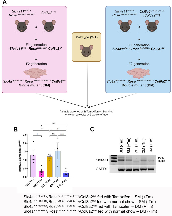
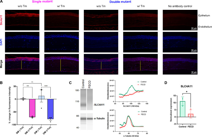
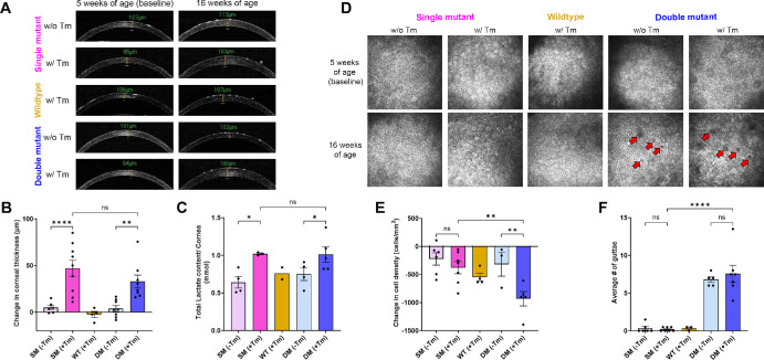
To generate a comprehensive FECD model, we generated a double mutant mouse bearing tamoxifen-inducible knockdown of Slc4a11 and the Col8a2 (Q455K) mutation. We performed optical coherence tomography (OCT) and in vivo confocal microscopy using the Heidelberg Retinal Tomography 3 - Rostock Cornea module (HRT3-RCM) on the mice at 5 weeks of age before tamoxifen feeding to establish baseline values for corneal thickness, endothelial cell density, and test for the presence of guttae. We measured these parameters again post-tamoxifen treatment at 16 weeks of age. We collected corneas at 16 weeks to perform histopathology, immunofluorescence staining for tight junctions, adherens junctions, and oxidative stress. We evaluated endothelial pump function using a lactate assay.

RESULTS

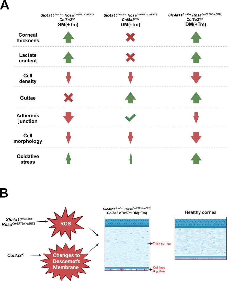
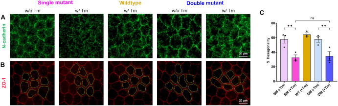
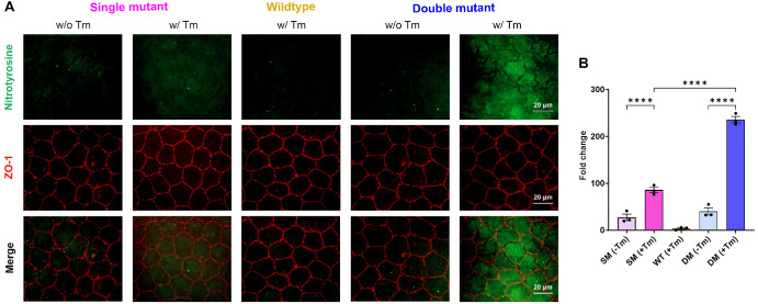
The double mutant tamoxifen-fed animals showed the presence of guttae, and displayed increased corneal thickness and decreased endothelial cell density. Endothelial cells showed altered morphology with disrupted adherens junctions and elevated reactive oxygen species (ROS). Finally, we found that stromal lactate concentrations were elevated in the double mutant mice, indicative of compromised endothelial pump function.

CONCLUSIONS

Overall, this mouse model recapitulates all the important phenotypic features associated with FECD.

摘要

目的

Fuchs 内皮角膜营养不良(FECD)是一种进行性致盲疾病,其特征为角膜内皮赘生物(guttae)增加、角膜内皮细胞丧失和水肿。这些症状被认为是由角膜内皮细胞外基质(ECM)变化和线粒体功能障碍引起的。尽管具有这种临床和生物学相关性,但目前尚无全面再现所有主要疾病特征的动物模型。在这项研究中,我们开发了这样一种模型,以提高我们对参与 FECD 进展的信号通路的理解,并制定早期干预策略。

方法

为了生成一种全面的 FECD 模型,我们生成了一种双突变小鼠,其携带 tamoxifen 诱导的 Slc4a11 敲低和 Col8a2(Q455K)突变。我们在喂食 tamoxifen 前,使用 Heidelberg Retinal Tomography 3 - Rostock Cornea 模块(HRT3-RCM)对 5 周龄的小鼠进行光学相干断层扫描(OCT)和体内共聚焦显微镜检查,以建立角膜厚度、内皮细胞密度和 guttae 存在的基线值。我们在 16 周龄时再次测量这些参数,然后在喂食 tamoxifen 后测量。我们在 16 周时收集角膜进行组织病理学检查、紧密连接、黏着连接和氧化应激的免疫荧光染色。我们使用乳酸测定法评估内皮泵功能。

结果

双突变型 tamoxifen 喂养的动物表现出 guttae 的存在,并显示出角膜厚度增加和内皮细胞密度降低。内皮细胞形态发生改变,黏着连接中断,活性氧(ROS)水平升高。最后,我们发现双突变小鼠的基质中乳酸浓度升高,表明内皮泵功能受损。

结论

总的来说,这种小鼠模型再现了与 FECD 相关的所有重要表型特征。

https://cdn.ncbi.nlm.nih.gov/pmc/blobs/5af5/11005065/3c4ebf3f0f21/iovs-65-4-18-f006.jpg
https://cdn.ncbi.nlm.nih.gov/pmc/blobs/5af5/11005065/81be1bdc6765/iovs-65-4-18-f001.jpg
https://cdn.ncbi.nlm.nih.gov/pmc/blobs/5af5/11005065/3a9c9922b2f4/iovs-65-4-18-f002.jpg
https://cdn.ncbi.nlm.nih.gov/pmc/blobs/5af5/11005065/11ce101d2f97/iovs-65-4-18-f003.jpg
https://cdn.ncbi.nlm.nih.gov/pmc/blobs/5af5/11005065/6e8307121018/iovs-65-4-18-f004.jpg
https://cdn.ncbi.nlm.nih.gov/pmc/blobs/5af5/11005065/d17a7e055205/iovs-65-4-18-f005.jpg
https://cdn.ncbi.nlm.nih.gov/pmc/blobs/5af5/11005065/3c4ebf3f0f21/iovs-65-4-18-f006.jpg

相似文献

1
Characterization of a Novel Mouse Model for Fuchs Endothelial Corneal Dystrophy.新型 Fuchs 内皮角膜营养不良小鼠模型的特性研究。
Invest Ophthalmol Vis Sci. 2024 Apr 1;65(4):18. doi: 10.1167/iovs.65.4.18.
2
An alpha 2 collagen VIII transgenic knock-in mouse model of Fuchs endothelial corneal dystrophy shows early endothelial cell unfolded protein response and apoptosis.Fuchs 内皮角膜营养不良的 alpha 2 胶原 VIII 转基因敲入小鼠模型显示早期内皮细胞未折叠蛋白反应和细胞凋亡。
Hum Mol Genet. 2012 Jan 15;21(2):384-93. doi: 10.1093/hmg/ddr473. Epub 2011 Oct 14.
3
Biomechanical changes to Descemet's membrane precede endothelial cell loss in an early-onset murine model of Fuchs endothelial corneal dystrophy.生物力学变化对 Descemet 膜的影响早于内皮细胞丢失在一个早期发病的鼠模型的 Fuchs 内皮角膜营养不良。
Exp Eye Res. 2019 Mar;180:18-22. doi: 10.1016/j.exer.2018.11.021. Epub 2018 Nov 22.
4
Endothelial Cdkn1a (p21) overexpression and accelerated senescence in a mouse model of Fuchs endothelial corneal dystrophy.内皮细胞 Cdkn1a(p21)过表达和加速衰老在 Fuchs 内皮角膜营养不良的小鼠模型中。
Invest Ophthalmol Vis Sci. 2012 Sep 28;53(10):6718-27. doi: 10.1167/iovs.12-9669.
5
L450W and Q455K Col8a2 knock-in mouse models of Fuchs endothelial corneal dystrophy show distinct phenotypes and evidence for altered autophagy.L450W 和 Q455K Col8a2 knock-in 鼠模型的 Fuchs 内皮角膜营养不良显示出不同的表型,并证明存在自噬改变。
Invest Ophthalmol Vis Sci. 2013 Mar 28;54(3):1887-97. doi: 10.1167/iovs.12-11021.
6
Lithium treatment increases endothelial cell survival and autophagy in a mouse model of Fuchs endothelial corneal dystrophy.锂治疗可增加 Fuchs 内皮角膜营养不良小鼠模型中内皮细胞的存活和自噬。
Br J Ophthalmol. 2013 Aug;97(8):1068-73. doi: 10.1136/bjophthalmol-2012-302881. Epub 2013 Jun 12.
7
Endothelial cell whole genome expression analysis in a mouse model of early-onset Fuchs' endothelial corneal dystrophy.在早发性 Fuchs 内皮角膜营养不良的小鼠模型中进行内皮细胞全基因组表达分析。
Invest Ophthalmol Vis Sci. 2013 Mar 15;54(3):1931-40. doi: 10.1167/iovs.12-10898.
8
Loss of ion transporters and increased unfolded protein response in Fuchs' dystrophy.富克斯角膜内皮营养不良中离子转运蛋白的丧失及未折叠蛋白反应增强
Mol Vis. 2014 Dec 12;20:1668-79. eCollection 2014.
9
Fuchs endothelial corneal dystrophy: The vicious cycle of Fuchs pathogenesis.Fuchs 内皮角膜营养不良:Fuchs 发病机制的恶性循环。
Prog Retin Eye Res. 2021 Jan;80:100863. doi: 10.1016/j.preteyeres.2020.100863. Epub 2020 May 8.
10
NQO1 downregulation potentiates menadione-induced endothelial-mesenchymal transition during rosette formation in Fuchs endothelial corneal dystrophy.NQO1 下调增强了在 Fuchs 内皮角膜营养不良的玫瑰花结形成过程中,亚甲二氢叶酸还原酶诱导的内皮-间充质转化。
Free Radic Biol Med. 2018 Feb 20;116:19-30. doi: 10.1016/j.freeradbiomed.2017.12.036. Epub 2017 Dec 30.

引用本文的文献

1
Generation of FECD Phenotypes in the Mouse Cornea by UVA Exposure and Surgical Removal of its Corneal Endothelial Layer.通过紫外线A照射和手术切除角膜内皮层在小鼠角膜中产生Fuchs内皮性角膜营养不良(FECD)表型
Bio Protoc. 2025 Mar 20;15(6):e5249. doi: 10.21769/BioProtoc.5249.
2
Preclinical Models for Studying Fuchs Endothelial Corneal Dystrophy.用于研究富克斯角膜内皮营养不良的临床前模型
Cells. 2025 Mar 28;14(7):505. doi: 10.3390/cells14070505.
3
METTL3-mA-mediated TGF-β signaling promotes Fuchs endothelial corneal dystrophy via regulating corneal endothelial-to-mesenchymal transition.

本文引用的文献

1
Mice Deficient in TAZ (Wwtr1) Demonstrate Clinical Features of Late-Onset Fuchs' Endothelial Corneal Dystrophy.TAZ(WWTR1)缺陷小鼠表现出迟发性 Fuchs 内皮角膜营养不良的临床特征。
Invest Ophthalmol Vis Sci. 2023 Apr 3;64(4):22. doi: 10.1167/iovs.64.4.22.
2
Update on the genetics of corneal endothelial dystrophies.角膜内皮营养不良的遗传学研究进展。
Indian J Ophthalmol. 2022 Jul;70(7):2239-2248. doi: 10.4103/ijo.IJO_992_22.
3
The H Transporter SLC4A11: Roles in Metabolism, Oxidative Stress and Mitochondrial Uncoupling.H 转运蛋白 SLC4A11:在代谢、氧化应激和线粒体解偶联中的作用。
METTL3介导的N6-甲基腺苷(m6A)通过调节角膜内皮-间充质转化促进Fuchs内皮性角膜营养不良。
Cell Death Discov. 2025 Mar 15;11(1):104. doi: 10.1038/s41420-025-02384-1.
4
Looking to the Future of Viral Vectors in Ocular Gene Therapy: Clinical Review.展望眼部基因治疗中病毒载体的未来:临床综述
Biomedicines. 2025 Feb 5;13(2):365. doi: 10.3390/biomedicines13020365.
Cells. 2022 Jan 7;11(2):197. doi: 10.3390/cells11020197.
4
Mitochondrial Targeting of the Ammonia-Sensitive Uncoupler SLC4A11 by the Chaperone-Mediated Carrier Pathway in Corneal Endothelium.角膜内皮细胞中伴侣介导的载体途径对氨敏感解偶联剂 SLC4A11 的线粒体靶向作用。
Invest Ophthalmol Vis Sci. 2021 Sep 2;62(12):4. doi: 10.1167/iovs.62.12.4.
5
Inducible Slc4a11 Knockout Triggers Corneal Edema Through Perturbation of Corneal Endothelial Pump.诱导型 Slc4a11 基因敲除通过干扰角膜内皮泵导致角膜水肿。
Invest Ophthalmol Vis Sci. 2021 Jun 1;62(7):28. doi: 10.1167/iovs.62.7.28.
6
Corneal endothelial dysfunction: Evolving understanding and treatment options.角膜内皮功能障碍:不断发展的认识和治疗选择。
Prog Retin Eye Res. 2021 May;82:100904. doi: 10.1016/j.preteyeres.2020.100904. Epub 2020 Sep 22.
7
Five-Year Follow-up of First 11 Patients Undergoing Injection of Cultured Corneal Endothelial Cells for Corneal Endothelial Failure.首批11例接受培养角膜内皮细胞注射治疗角膜内皮失代偿患者的五年随访
Ophthalmology. 2021 Apr;128(4):504-514. doi: 10.1016/j.ophtha.2020.09.002. Epub 2020 Sep 6.
8
Fuchs endothelial corneal dystrophy: The vicious cycle of Fuchs pathogenesis.Fuchs 内皮角膜营养不良:Fuchs 发病机制的恶性循环。
Prog Retin Eye Res. 2021 Jan;80:100863. doi: 10.1016/j.preteyeres.2020.100863. Epub 2020 May 8.
9
Corneal Endothelial Pump Coupling to Lactic Acid Efflux in the Rabbit and Mouse.兔和鼠眼角膜内皮泵与乳酸外排的偶联关系。
Invest Ophthalmol Vis Sci. 2020 Feb 7;61(2):7. doi: 10.1167/iovs.61.2.7.
10
Aberrant DNA methylation of miRNAs in Fuchs endothelial corneal dystrophy.Fuchs 内皮角膜营养不良中 miRNA 的异常 DNA 甲基化。
Sci Rep. 2019 Nov 8;9(1):16385. doi: 10.1038/s41598-019-52727-z.