Sharma Susmita, Sahoo Rudranarayan, Chawla Saurabh, Das Sujata, Alone Debasmita Pankaj
School of Biological Sciences, National Institute of Science Education and Research (NISER) Bhubaneswar, P.O. Bhimpur- Padanpur, Jatni, Khurda, Odisha, India.
Homi Bhabha National Institute (HBNI), Training School Complex, Anushaktinagar, Mumbai, India.
Bio Protoc. 2025 Mar 20;15(6):e5249. doi: 10.21769/BioProtoc.5249.
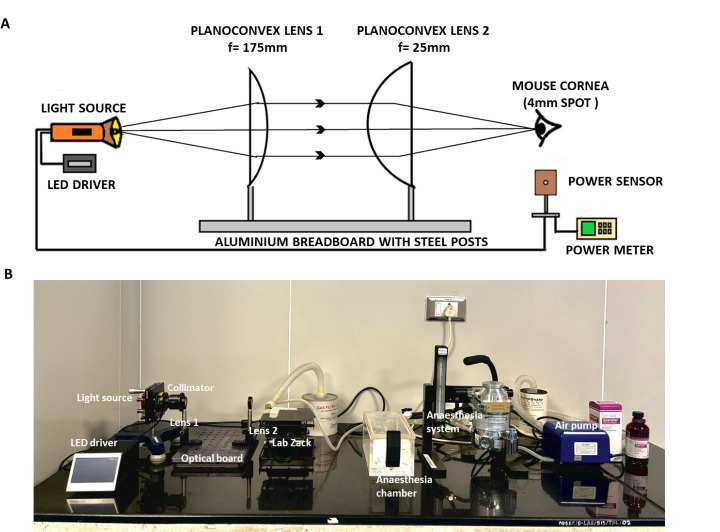
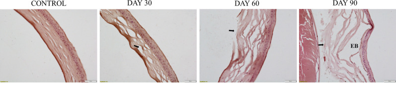
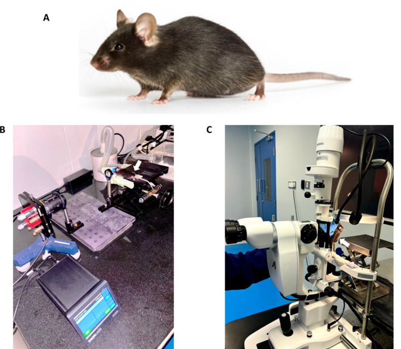
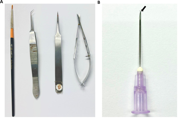
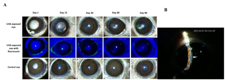
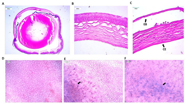
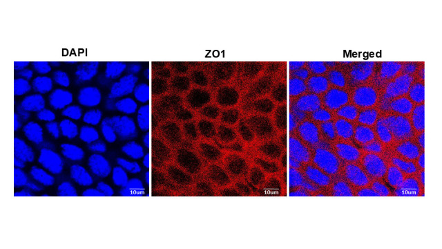
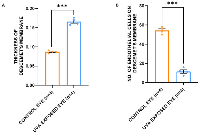
Fuchs endothelial corneal dystrophy (FECD) is a rare and multifactorial disorder leading to cell death in the innermost layer of the cornea, i.e., the endothelium; UV radiation is reported as the major environmental risk for the disease. Establishing an animal model for this disease has remained challenging in FECD research. We have developed a detailed protocol for the establishment of a UVA-induced FECD mouse model and removal of corneal endothelium from the eye for further molecular and histological studies by taking references from previous studies. UVA light of 500 J/cm was focused on the C57BL/6J female mouse cornea and kept for an observation period of 90 days. The animal developed corneal scarring by the end of three months. Slit-lamp microscopy and alizarin red-trypan blue staining confirmed endothelial cell death and formation of corneal guttae in the endothelium. Surgical removal of the endothelial layer was successfully done in the diseased mouse, and the result was confirmed by immunofluorescence. This study is relevant for in-depth research using a FECD mouse model, which will surpass the limitation of human tissue scarcity and can be used for in vivo drug targeting to develop therapeutics to cure FECD. Key features • UVA radiation induces FECD only in the exposed eye of female mice. • Females are more affected and develop the FECD phenotype. • This protocol will help dissect the endothelium layer with Descemet's membrane (DM) from the mouse cornea, which is equivalent to human surgical tissue.
富克斯内皮性角膜营养不良(FECD)是一种罕见的多因素疾病,可导致角膜最内层即内皮细胞死亡;据报道,紫外线辐射是该疾病的主要环境风险因素。在FECD研究中,建立这种疾病的动物模型一直具有挑战性。我们参考以往研究,制定了一份详细的方案,用于建立紫外线A(UVA)诱导的FECD小鼠模型,并从眼中去除角膜内皮,以进行进一步的分子和组织学研究。将500 J/cm的UVA光聚焦在C57BL/6J雌性小鼠的角膜上,并持续观察90天。三个月结束时,该动物出现了角膜瘢痕形成。裂隙灯显微镜检查和茜素红-台盼蓝染色证实了内皮细胞死亡以及内皮中角膜小滴的形成。在患病小鼠中成功完成了内皮层的手术切除,免疫荧光证实了结果。这项研究对于使用FECD小鼠模型进行深入研究具有重要意义,该模型将突破人体组织稀缺的限制,可用于体内药物靶向研究,以开发治疗FECD的疗法。关键特征 • UVA辐射仅在雌性小鼠的暴露眼中诱导FECD。 • 雌性受影响更大并出现FECD表型。 • 该方案将有助于从小鼠角膜中分离出带有后弹力层(DM)的内皮层,这相当于人类手术组织。