Department of Pediatrics, School for Oncology and Reproduction (GROW), Maastricht University, 6229 ER Maastricht, The Netherlands.
Department of Pediatrics, School of Nutrition and Translational Research in Metabolism (NUTRIM), Maastricht University, 6229 ER Maastricht, The Netherlands.
Int J Mol Sci. 2024 Apr 3;25(7):4000. doi: 10.3390/ijms25074000.
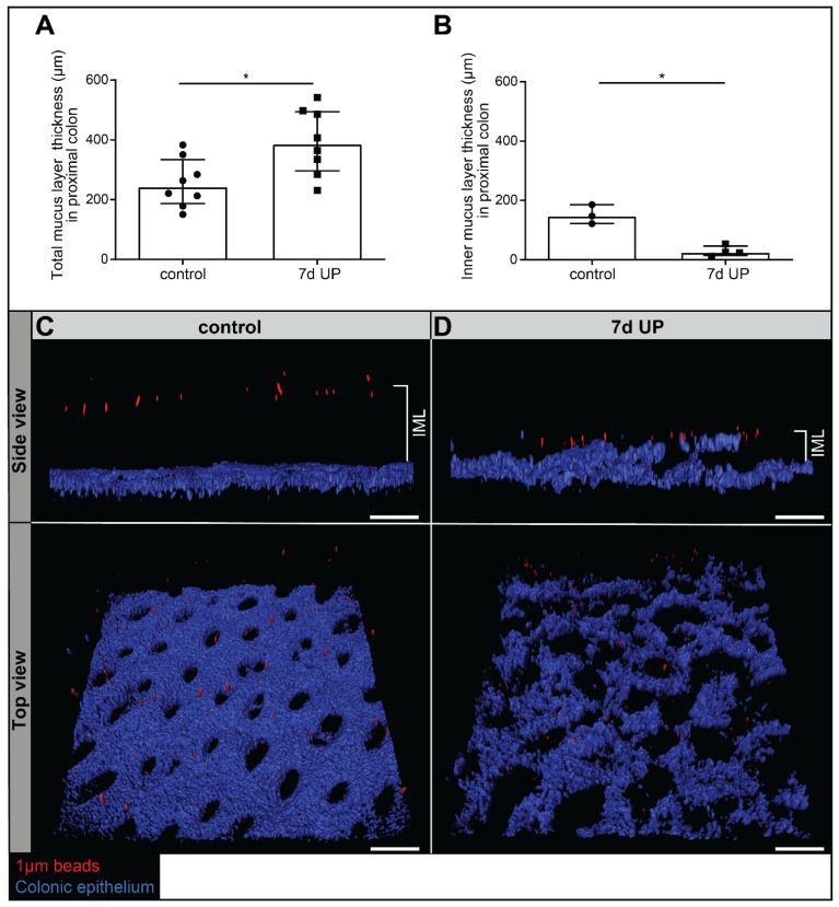
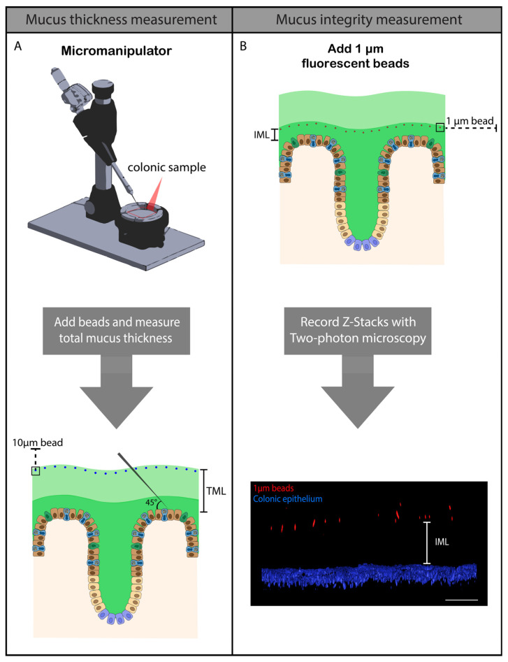
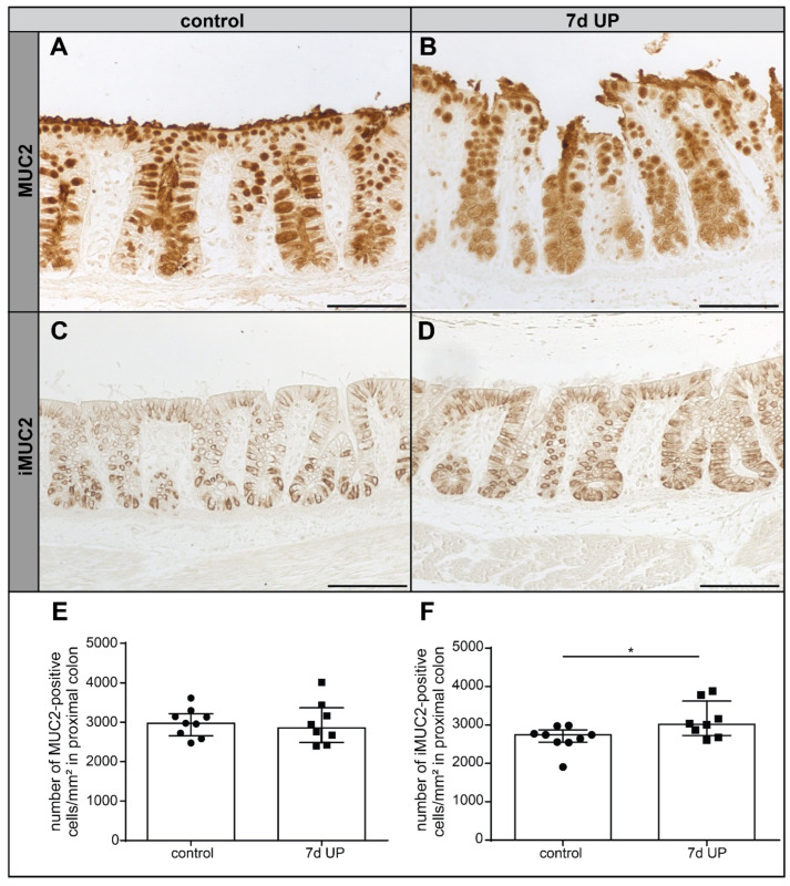
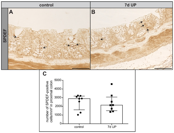
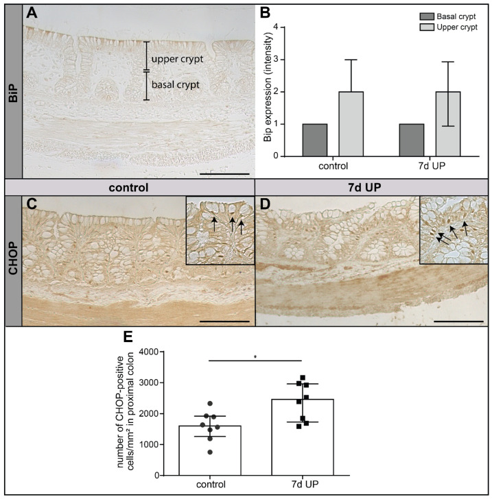
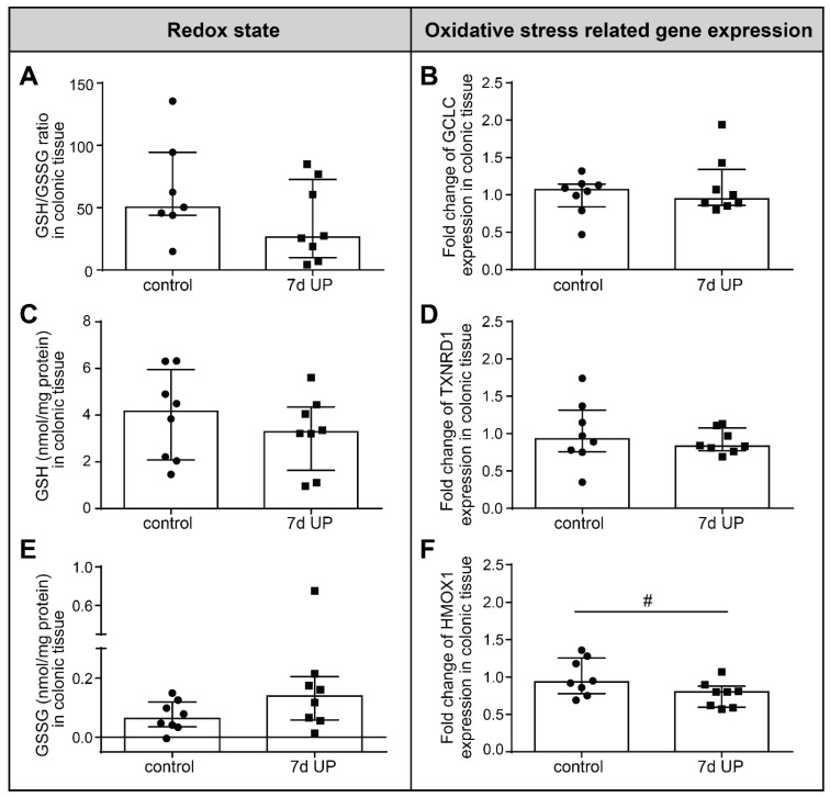
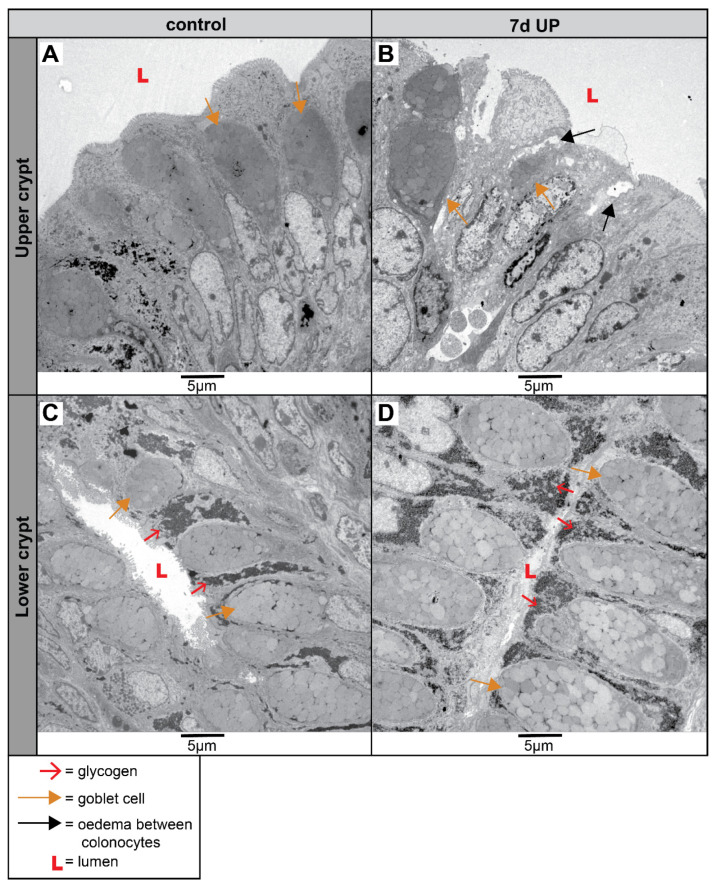
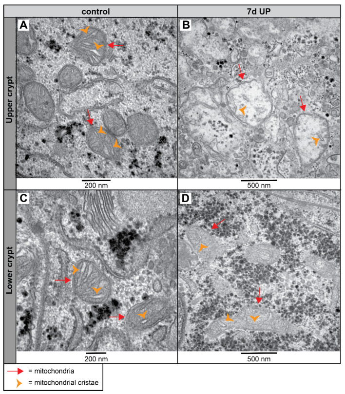
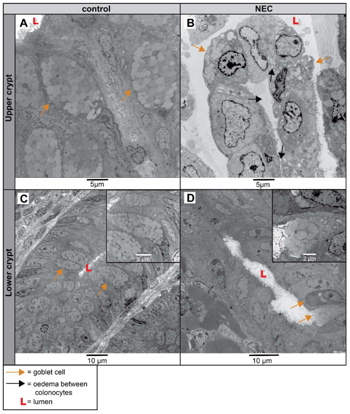
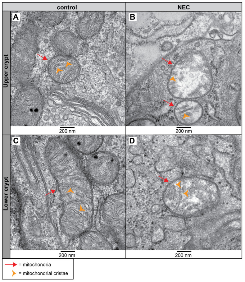
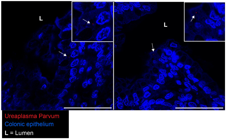
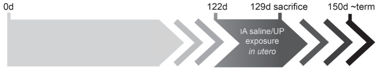
Chorioamnionitis is a risk factor for necrotizing enterocolitis (NEC). (UP) is clinically the most isolated microorganism in chorioamnionitis, but its pathogenicity remains debated. Chorioamnionitis is associated with ileal barrier changes, but colonic barrier alterations, including those of the mucus barrier, remain under-investigated, despite their importance in NEC pathophysiology. Therefore, in this study, the hypothesis that antenatal UP exposure disturbs colonic mucus barrier integrity, thereby potentially contributing to NEC pathogenesis, was investigated. In an established ovine chorioamnionitis model, lambs were intra-amniotically exposed to UP or saline for 7 d from 122 to 129 d gestational age. Thereafter, colonic mucus layer thickness and functional integrity, underlying mechanisms, including endoplasmic reticulum (ER) stress and redox status, and cellular morphology by transmission electron microscopy were studied. The clinical significance of the experimental findings was verified by examining colon samples from NEC patients and controls. UP-exposed lambs have a thicker but dysfunctional colonic mucus layer in which bacteria-sized beads reach the intestinal epithelium, indicating undesired bacterial contact with the epithelium. This is paralleled by disturbed goblet cell MUC2 folding, pro-apoptotic ER stress and signs of mitochondrial dysfunction in the colonic epithelium. Importantly, the colonic epithelium from human NEC patients showed comparable mitochondrial aberrations, indicating that NEC-associated intestinal barrier injury already occurs during chorioamnionitis. This study underlines the pathogenic potential of UP during pregnancy; it demonstrates that antenatal UP infection leads to severe colonic mucus barrier deficits, providing a mechanistic link between antenatal infections and postnatal NEC development.
绒毛膜羊膜炎是坏死性小肠结肠炎(NEC)的一个危险因素。(UP)是临床上在绒毛膜羊膜炎中最常被分离到的微生物,但它的致病性仍存在争议。绒毛膜羊膜炎与回肠屏障变化有关,但结肠屏障的改变,包括黏液屏障的改变,尽管它们在 NEC 病理生理学中很重要,但仍未得到充分研究。因此,在这项研究中,假设产前 UP 暴露会破坏结肠黏液屏障的完整性,从而可能导致 NEC 的发病机制,这一假说得到了研究。在已建立的羊绒毛膜羊膜炎模型中,从妊娠 122 至 129 天,羊通过羊膜内途径将 UP 或盐水暴露于胎儿体内 7 天。此后,研究了结肠黏液层厚度和功能完整性、潜在机制,包括内质网(ER)应激和氧化还原状态,以及透射电子显微镜下的细胞形态。通过检查 NEC 患者和对照者的结肠样本,验证了实验结果的临床意义。暴露于 UP 的羔羊的结肠黏液层较厚,但功能失调,其中细菌大小的珠子可到达肠上皮,表明细菌与上皮细胞之间发生了不希望发生的接触。这与杯状细胞 MUC2 折叠紊乱、促凋亡 ER 应激和结肠上皮中线粒体功能障碍的迹象相平行。重要的是,来自人类 NEC 患者的结肠上皮表现出类似的线粒体异常,表明 NEC 相关的肠屏障损伤在绒毛膜羊膜炎期间已经发生。这项研究强调了 UP 在妊娠期间的致病潜力;它表明产前 UP 感染会导致严重的结肠黏液屏障缺陷,为产前感染与产后 NEC 发展之间提供了一个机制联系。