Ma Yujuan, Zhao Xuebo, Chen Xianxia
Tianshui Maternity and Child Healthcare Hospital, Tianshui, China.
Graduate School of Qinghai University, Xining, China.
Front Oncol. 2024 Apr 3;14:1301900. doi: 10.3389/fonc.2024.1301900. eCollection 2024.
Contrast-enhanced ultrasound (CEUS) and elastography are of great value in the diagnosis of cervical cancer (CC). However, there is limited research on the role of contrast-enhanced ultrasound combined with elastography in predicting concurrent chemoradiotherapy and disease progression for cervical cancer. The purpose of this study was to evaluate the feasibility of contrast-enhanced ultrasound combined with elastography and tumor prognosis.
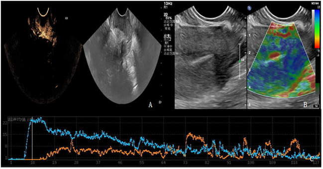
MRI was performed on 98 patients with cervical cancer before and after treatment. Before, during, and 1 week after the treatment, contrast-enhanced ultrasound and elastography were conducted, and the alterations of ultrasound-related parameters at each time point of the treatment were compared. The correlation between contrast-enhanced ultrasound combined with elastic imaging and oncological outcome was assessed.
There was no notable difference in overall clinical data between the complete remission (CR) group and the partial remission (PR) group (P>0.05). Before treatment, there were no statistically significant differences in elasticity score, time to peak (TTP), and peak intensity (PI) between the CR group and the PR group. However, there were no statistical differences in elastic strain ratio (SR) and area under the curve (AUC) before and after treatment between the CR group and the PR group, and there were also no statistical differences in the elastic strain ratio (SR) and area under the curve (AUC) of contrast-enhanced ultrasound parameters between the CR group and the PR group before and during treatment. There was a statistically significant difference after treatment (P<0.05).At present, the follow-up of patients is about 1 year, 7 patients were excluded due to loss to follow-up, and 91 patients were included in the follow-up study. Through the review of the cases and combined with MRI (version RECIST1.1) and serology and other related examinations, if the patient has a new lesion or the lesion is larger than before, the tumor marker Squamous cell carcinoma antigen (SCC-Ag) is significantly increased twice in a row, and the patient is divided into progressive disease (PD). Those who did not see significant changes were divided into stable disease (SD) group. The relationship between clinical characteristics, ultrasound parameters and disease progression in 91 patients was compared. There was no significant difference in age and clinical stage between the two groups (P>0.05), but there was a significant difference in the elevation of tumor marker squamous cell carcinoma antigen (SCC-Ag) between the two groups (P<0.05).With the growth of tumors, TTP decreased, elasticity score and PI increased, and the difference was statistically significant (P<0.05). The AUC of SCC-Ag was 0.655, the sensitivity was 85.3%, and the specificity was 45.6%.The AUC, sensitivity and specificity of ultrasound parameters combined with SCC-Ag predicted disease progression was 0.959, 91.2% and 94.8%.
Using contrast-enhanced ultrasound and elastography to predict the efficacy and disease progression of concurrent chemoradiotherapy is feasible. In addition, the combination of SCC-Ag with contrast-enhanced ultrasound and elastography can further enhance the efficiency of predicting disease progression.

超声造影(CEUS)和弹性成像在宫颈癌(CC)诊断中具有重要价值。然而,关于超声造影联合弹性成像在预测宫颈癌同步放化疗及疾病进展方面的作用研究有限。本研究旨在评估超声造影联合弹性成像的可行性及肿瘤预后情况。
对98例宫颈癌患者在治疗前后进行磁共振成像(MRI)检查。在治疗前、治疗期间及治疗后1周进行超声造影和弹性成像检查,并比较治疗各时间点超声相关参数的变化。评估超声造影联合弹性成像与肿瘤学结局之间的相关性。
完全缓解(CR)组和部分缓解(PR)组的总体临床数据无显著差异(P>0.05)。治疗前,CR组和PR组在弹性评分、达峰时间(TTP)和峰值强度(PI)方面无统计学显著差异。然而,CR组和PR组治疗前后的弹性应变率(SR)和曲线下面积(AUC)无统计学差异,且CR组和PR组治疗前及治疗期间超声造影参数的弹性应变率(SR)和曲线下面积(AUC)也无统计学差异。治疗后有统计学显著差异(P<0.05)。目前,患者随访约1年,7例因失访被排除,91例患者纳入随访研究。通过病例回顾并结合MRI(版本RECIST1.1)及血清学等相关检查,若患者出现新病灶或病灶较前增大,肿瘤标志物鳞状细胞癌抗原(SCC-Ag)连续两次显著升高,则将患者分为疾病进展(PD)组。未见明显变化者分为疾病稳定(SD)组。比较91例患者的临床特征、超声参数与疾病进展之间的关系。两组患者年龄和临床分期无显著差异(P>0.05),但两组肿瘤标志物鳞状细胞癌抗原(SCC-Ag)升高情况有显著差异(P<0.05)。随着肿瘤生长,TTP降低,弹性评分和PI升高,差异有统计学意义(P<0.05)。SCC-Ag的AUC为0.655,灵敏度为85.3%,特异度为45.6%。超声参数联合SCC-Ag预测疾病进展的AUC、灵敏度和特异度分别为0.959、91.2%和94.8%。
利用超声造影和弹性成像预测同步放化疗的疗效及疾病进展是可行的。此外,SCC-Ag与超声造影和弹性成像联合可进一步提高预测疾病进展的效率。