Maller Vijetha V, Johnson Jason N, Boston Umar, Knott-Craig Christopher
Department of Radiology, LeBonheur Children's Hospital, University of Tennessee Health Science Center, 848 Adams Avenue, Radiology G216, Memphis, TN, USA.
Division of Pediatric Cardiology, Pediatrics, Heart institute, LeBonheur Children's Hospital, University of Tennessee Health Science Center, Memphis, TN, USA.
Pediatr Radiol. 2024 Jul;54(8):1261-1269. doi: 10.1007/s00247-024-05911-x. Epub 2024 Apr 19.
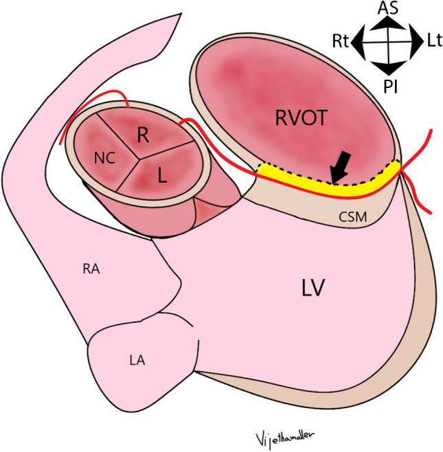
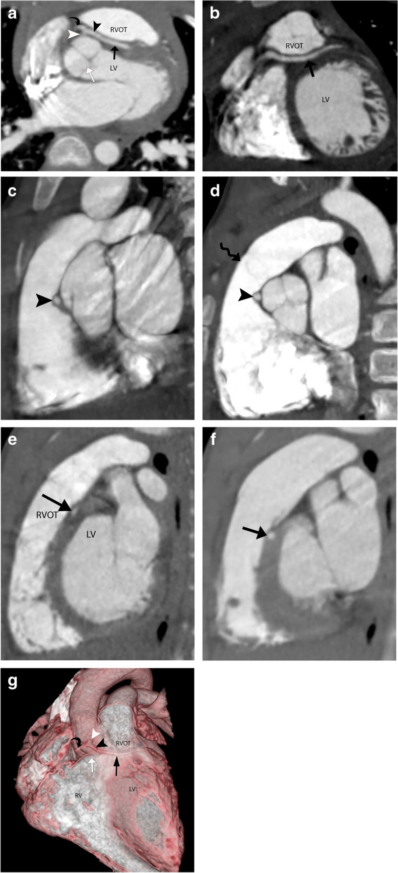
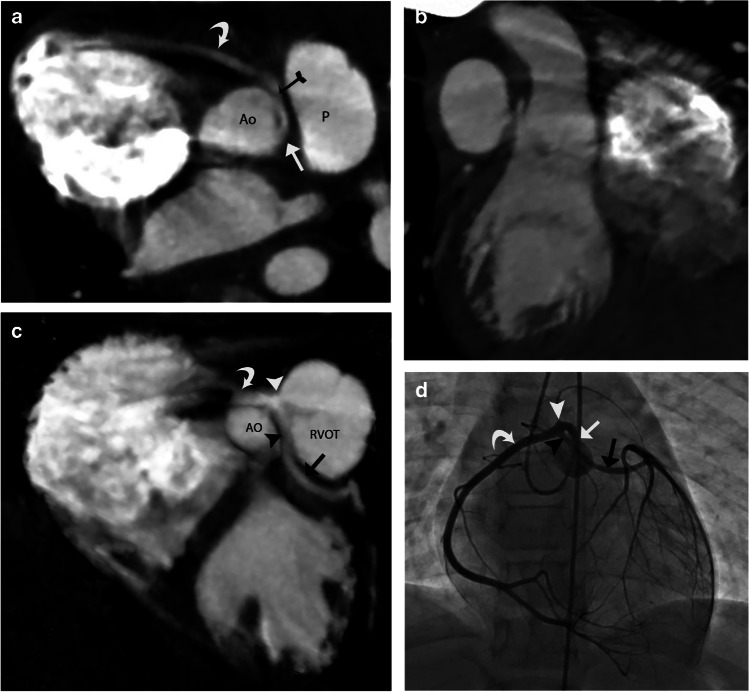
A transseptal coronary artery course, also known as a transconal course, is an anomalous course of the left main coronary artery (LMCA) or the left anterior descending artery (LAD) through the conal septal myocardium. The conal septal myocardium is the posterior wall of the right ventricular outflow tract (RVOT), acting as a dividing myocardial wall between the subaortic and subpulmonary outflow tracts. The initial segment of a transseptal coronary artery has an extraconal course between the aorta and the RVOT cranial to the true intramyocardial segment. The transseptal coronary artery then emerges out of the conal septal myocardium at the epicardial surface on the lateral aspect of the RVOT. Many consider the transseptal coronary artery to be a benign entity. However, there are few case reports of severe cardiac symptoms such as myocardial ischemia, arrhythmia, and even sudden cardiac deaths due to potential coronary artery compression in the systolic phase. In this article, we seek to describe the imaging findings of transseptal coronary artery course on coronary computed tomography angiography (CTA), discuss their clinical analysis, and briefly discuss the management of these lesions.
经间隔冠状动脉走行,也称为经圆锥走行,是左主干冠状动脉(LMCA)或左前降支动脉(LAD)通过圆锥间隔心肌的异常走行。圆锥间隔心肌是右心室流出道(RVOT)的后壁,作为主动脉下和肺动脉下流出道之间的分隔心肌壁。经间隔冠状动脉的起始段在主动脉和RVOT之间有一段位于真正心肌内段上方的心外圆锥走行。然后,经间隔冠状动脉在心外膜表面从RVOT外侧的圆锥间隔心肌中穿出。许多人认为经间隔冠状动脉是一种良性病变。然而,有少数病例报告显示,由于收缩期潜在的冠状动脉受压,出现了严重的心脏症状,如心肌缺血、心律失常,甚至心源性猝死。在本文中,我们旨在描述经间隔冠状动脉走行在冠状动脉计算机断层扫描血管造影(CTA)上的影像学表现,讨论其临床分析,并简要讨论这些病变的处理。