• 文献检索
  • 文档翻译
  • 深度研究
  • 学术资讯
  • Suppr Zotero 插件Zotero 插件
  • 邀请有礼
  • 套餐&价格
  • 历史记录
应用&插件
Suppr Zotero 插件Zotero 插件浏览器插件Mac 客户端Windows 客户端微信小程序
定价
高级版会员购买积分包购买API积分包
服务
文献检索文档翻译深度研究API 文档MCP 服务
关于我们
关于 Suppr公司介绍联系我们用户协议隐私条款
关注我们

Suppr 超能文献

核心技术专利:CN118964589B侵权必究
粤ICP备2023148730 号-1Suppr @ 2026

文献检索

告别复杂PubMed语法,用中文像聊天一样搜索,搜遍4000万医学文献。AI智能推荐,让科研检索更轻松。

立即免费搜索

文件翻译

保留排版,准确专业,支持PDF/Word/PPT等文件格式,支持 12+语言互译。

免费翻译文档

深度研究

AI帮你快速写综述,25分钟生成高质量综述,智能提取关键信息,辅助科研写作。

立即免费体验

细菌 NADPH 氧化酶的 X 射线结构和酶学研究突出了真核细胞 NOX 的激活机制。

X-ray structure and enzymatic study of a bacterial NADPH oxidase highlight the activation mechanism of eukaryotic NOX.

机构信息

Univ. Grenoble Alpes, CNRS, CEA, Institut de Biologie Structurale, Grenoble, France.

European Molecular Biology Laboratory, Grenoble, France.

出版信息

Elife. 2024 Apr 19;13:RP93759. doi: 10.7554/eLife.93759.

DOI:10.7554/eLife.93759
PMID:38640072
原文链接:https://pmc.ncbi.nlm.nih.gov/articles/PMC11031084/
Abstract

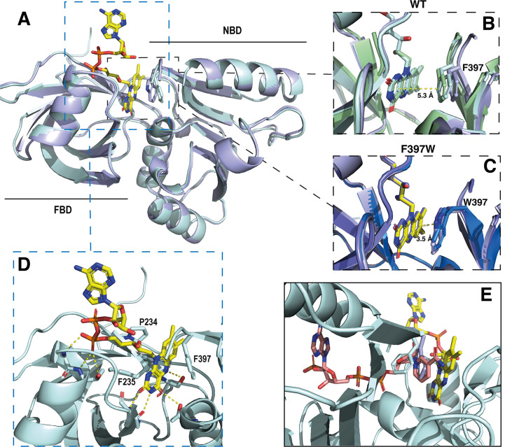
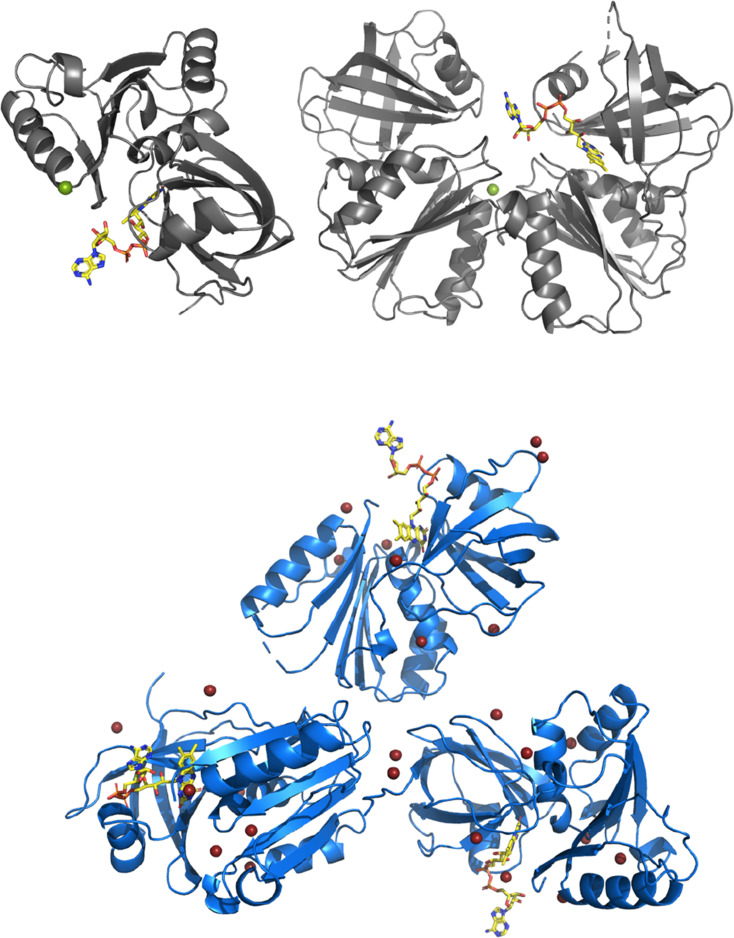
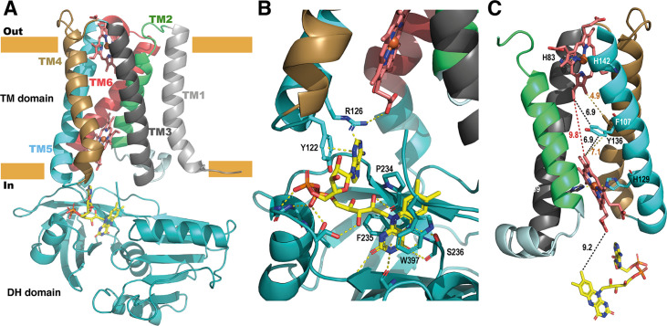
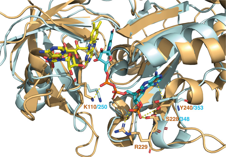
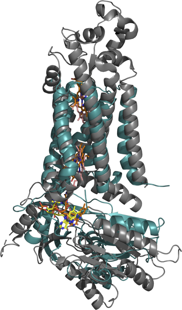
NADPH oxidases (NOX) are transmembrane proteins, widely spread in eukaryotes and prokaryotes, that produce reactive oxygen species (ROS). Eukaryotes use the ROS products for innate immune defense and signaling in critical (patho)physiological processes. Despite the recent structures of human NOX isoforms, the activation of electron transfer remains incompletely understood. SpNOX, a homolog from , can serves as a robust model for exploring electron transfers in the NOX family thanks to its constitutive activity. Crystal structures of SpNOX full-length and dehydrogenase (DH) domain constructs are revealed here. The isolated DH domain acts as a flavin reductase, and both constructs use either NADPH or NADH as substrate. Our findings suggest that hydride transfer from NAD(P)H to FAD is the rate-limiting step in electron transfer. We identify significance of F397 in nicotinamide access to flavin isoalloxazine and confirm flavin binding contributions from both DH and Transmembrane (TM) domains. Comparison with related enzymes suggests that distal access to heme may influence the final electron acceptor, while the relative position of DH and TM does not necessarily correlate with activity, contrary to previous suggestions. It rather suggests requirement of an internal rearrangement, within the DH domain, to switch from a resting to an active state. Thus, SpNOX appears to be a good model of active NOX2, which allows us to propose an explanation for NOX2's requirement for activation.

摘要

NADPH 氧化酶(NOX)是一种跨膜蛋白,广泛存在于真核生物和原核生物中,可产生活性氧(ROS)。真核生物利用 ROS 产物进行先天免疫防御和关键(病理)生理过程中的信号转导。尽管最近已经解析了人类 NOX 同工型的结构,但电子传递的激活仍未完全了解。来自 的 SpNOX 同源物可以作为探索 NOX 家族中电子传递的强大模型,因为其具有组成型活性。本文揭示了全长 SpNOX 和脱氢酶(DH)结构域构建体的晶体结构。分离的 DH 结构域充当黄素还原酶,并且这两种构建体都可以使用 NADPH 或 NADH 作为底物。我们的研究结果表明,从 NAD(P)H 到 FAD 的氢化物转移是电子传递的限速步骤。我们确定了 F397 在烟酰胺进入黄素异咯嗪中的重要性,并证实了 DH 和跨膜(TM)结构域都对黄素结合有贡献。与相关酶的比较表明,远端进入血红素可能影响最终电子受体,而 DH 和 TM 的相对位置不一定与活性相关,这与之前的建议相反。它表明需要在 DH 结构域内进行内部重排,以从静止状态切换到激活状态。因此,SpNOX 似乎是活性 NOX2 的良好模型,这使我们能够对 NOX2 的激活要求提出解释。

https://cdn.ncbi.nlm.nih.gov/pmc/blobs/7769/11031084/9a26cf170d4e/elife-93759-sa3-fig1.jpg
https://cdn.ncbi.nlm.nih.gov/pmc/blobs/7769/11031084/a7d2d471151d/elife-93759-fig1.jpg
https://cdn.ncbi.nlm.nih.gov/pmc/blobs/7769/11031084/2888814740e7/elife-93759-fig1-figsupp1.jpg
https://cdn.ncbi.nlm.nih.gov/pmc/blobs/7769/11031084/a3d435657869/elife-93759-fig2.jpg
https://cdn.ncbi.nlm.nih.gov/pmc/blobs/7769/11031084/c6fbaa4767ec/elife-93759-fig2-figsupp1.jpg
https://cdn.ncbi.nlm.nih.gov/pmc/blobs/7769/11031084/5bab802cbe24/elife-93759-fig3.jpg
https://cdn.ncbi.nlm.nih.gov/pmc/blobs/7769/11031084/11cc5b6ef8db/elife-93759-fig4.jpg
https://cdn.ncbi.nlm.nih.gov/pmc/blobs/7769/11031084/437ccc189f00/elife-93759-fig4-figsupp1.jpg
https://cdn.ncbi.nlm.nih.gov/pmc/blobs/7769/11031084/2effd972d71b/elife-93759-fig5.jpg
https://cdn.ncbi.nlm.nih.gov/pmc/blobs/7769/11031084/dbd018c0da94/elife-93759-fig5-figsupp1.jpg
https://cdn.ncbi.nlm.nih.gov/pmc/blobs/7769/11031084/ee7a5f7bfa87/elife-93759-fig5-figsupp2.jpg
https://cdn.ncbi.nlm.nih.gov/pmc/blobs/7769/11031084/23a814c080b0/elife-93759-fig6.jpg
https://cdn.ncbi.nlm.nih.gov/pmc/blobs/7769/11031084/572377083fd9/elife-93759-fig6-figsupp1.jpg
https://cdn.ncbi.nlm.nih.gov/pmc/blobs/7769/11031084/7039845d1fe5/elife-93759-fig7.jpg
https://cdn.ncbi.nlm.nih.gov/pmc/blobs/7769/11031084/9bd2a135bda3/elife-93759-fig8.jpg
https://cdn.ncbi.nlm.nih.gov/pmc/blobs/7769/11031084/e2a6c9e8e6c0/elife-93759-fig9.jpg
https://cdn.ncbi.nlm.nih.gov/pmc/blobs/7769/11031084/bf92d4c7ce52/elife-93759-fig9-figsupp1.jpg
https://cdn.ncbi.nlm.nih.gov/pmc/blobs/7769/11031084/2da14c50622d/elife-93759-fig9-figsupp2.jpg
https://cdn.ncbi.nlm.nih.gov/pmc/blobs/7769/11031084/9a26cf170d4e/elife-93759-sa3-fig1.jpg

相似文献

1
X-ray structure and enzymatic study of a bacterial NADPH oxidase highlight the activation mechanism of eukaryotic NOX.细菌 NADPH 氧化酶的 X 射线结构和酶学研究突出了真核细胞 NOX 的激活机制。
Elife. 2024 Apr 19;13:RP93759. doi: 10.7554/eLife.93759.
2
Constitutive NADPH-dependent electron transferase activity of the Nox4 dehydrogenase domain.Nox4 脱氢酶结构域的组成型 NADPH 依赖的电子转移酶活性。
Biochemistry. 2010 Mar 23;49(11):2433-42. doi: 10.1021/bi9022285.
3
The NOX Family of Proteins Is Also Present in Bacteria.NOX 蛋白家族也存在于细菌中。
mBio. 2017 Nov 7;8(6):e01487-17. doi: 10.1128/mBio.01487-17.
4
An Elegant Four-Helical Fold in NOX and STEAP Enzymes Facilitates Electron Transport across Biomembranes-Similar Vehicle, Different Destination.NOX 和 STEAP 酶中的优雅四螺旋折叠促进电子跨生物膜运输——类似的载体,不同的目的地。
Acc Chem Res. 2020 Sep 15;53(9):1969-1980. doi: 10.1021/acs.accounts.0c00400. Epub 2020 Aug 20.
5
Structural and mechanistic insights into Streptococcus pneumoniae NADPH oxidase.肺炎链球菌 NADPH 氧化酶的结构与机制研究进展
Nat Struct Mol Biol. 2024 Nov;31(11):1769-1777. doi: 10.1038/s41594-024-01348-w. Epub 2024 Jul 22.
6
High-resolution studies of hydride transfer in the ferredoxin:NADP reductase superfamily.铁氧化还原蛋白:NADP还原酶超家族中氢化物转移的高分辨率研究。
FEBS J. 2017 Oct;284(19):3302-3319. doi: 10.1111/febs.14190. Epub 2017 Aug 29.
7
Structure of human phagocyte NADPH oxidase in the resting state.人吞噬细胞 NADPH 氧化酶在静息状态下的结构。
Elife. 2022 Nov 22;11:e83743. doi: 10.7554/eLife.83743.
8
Stopped-flow kinetic studies of flavin reduction in human cytochrome P450 reductase and its component domains.人细胞色素P450还原酶及其组成结构域中黄素还原的停流动力学研究。
Biochemistry. 2001 Feb 20;40(7):1964-75. doi: 10.1021/bi001719m.
9
Crystal structures and atomic model of NADPH oxidase.NADPH 氧化酶的晶体结构和原子模型。
Proc Natl Acad Sci U S A. 2017 Jun 27;114(26):6764-6769. doi: 10.1073/pnas.1702293114. Epub 2017 Jun 12.
10
Architecture of the NADPH oxidase family of enzymes.NADPH 氧化酶家族酶的结构。
Redox Biol. 2022 Jun;52:102298. doi: 10.1016/j.redox.2022.102298. Epub 2022 Mar 18.

引用本文的文献

1
X-ray diffraction images for two membrane protein crystals presenting high anisotropy; the ABC transporter BmrA and the NADPH oxidase.两种呈现高各向异性的膜蛋白晶体的X射线衍射图像;ABC转运蛋白BmrA和NADPH氧化酶。
IUCrdata. 2025 Jul 4;10(Pt 7):x250591. doi: 10.1107/S2414314625005917. eCollection 2025 Jul.
2
NADPH oxidases: redox regulation of cell homeostasis and disease.烟酰胺腺嘌呤二核苷酸磷酸氧化酶:细胞稳态与疾病的氧化还原调节
Physiol Rev. 2025 Jul 1;105(3):1291-1428. doi: 10.1152/physrev.00034.2023. Epub 2025 Jan 15.
3
To activate NAD(P)H oxidase with a brief pulse of photodynamic action.

本文引用的文献

1
Structural and mechanistic insights into Streptococcus pneumoniae NADPH oxidase.肺炎链球菌 NADPH 氧化酶的结构与机制研究进展
Nat Struct Mol Biol. 2024 Nov;31(11):1769-1777. doi: 10.1038/s41594-024-01348-w. Epub 2024 Jul 22.
2
Structure of human phagocyte NADPH oxidase in the activated state.人吞噬细胞 NADPH 氧化酶在激活状态下的结构。
Nature. 2024 Mar;627(8002):189-195. doi: 10.1038/s41586-024-07056-1. Epub 2024 Feb 14.
3
VEO-IBD NOX1 variant highlights a structural region essential for NOX/DUOX catalytic activity.
通过短暂的光动力作用脉冲激活NAD(P)H氧化酶。
FASEB J. 2024 Dec 15;38(23):e70246. doi: 10.1096/fj.202402292R.
4
Structural profiles of the full phagocyte NADPH oxidase unveiled by combining computational biology and experimental knowledge.结合计算生物学与实验知识揭示的全吞噬细胞NADPH氧化酶的结构概况
J Biol Chem. 2024 Dec;300(12):107943. doi: 10.1016/j.jbc.2024.107943. Epub 2024 Oct 29.
5
Pharmacological mechanisms by which baicalin ameliorates cardiovascular disease.黄芩苷改善心血管疾病的药理机制。
Front Pharmacol. 2024 Aug 9;15:1415971. doi: 10.3389/fphar.2024.1415971. eCollection 2024.
6
Structural and mechanistic insights into Streptococcus pneumoniae NADPH oxidase.肺炎链球菌 NADPH 氧化酶的结构与机制研究进展
Nat Struct Mol Biol. 2024 Nov;31(11):1769-1777. doi: 10.1038/s41594-024-01348-w. Epub 2024 Jul 22.
VEO-IBD 中 NOX1 变体突出了一个对 NOX/ DUOX 催化活性至关重要的结构区域。
Redox Biol. 2023 Nov;67:102905. doi: 10.1016/j.redox.2023.102905. Epub 2023 Sep 27.
4
Structure of human phagocyte NADPH oxidase in the resting state.人吞噬细胞 NADPH 氧化酶在静息状态下的结构。
Elife. 2022 Nov 22;11:e83743. doi: 10.7554/eLife.83743.
5
Structure of the core human NADPH oxidase NOX2.核心人类 NADPH 氧化酶 NOX2 的结构。
Nat Commun. 2022 Oct 14;13(1):6079. doi: 10.1038/s41467-022-33711-0.
6
Specific Xray diffraction patterns of membrane proteins caused by secondary structure collinearity.膜蛋白二级结构共线性引起的特殊 X 射线衍射图谱。
Biochim Biophys Acta Biomembr. 2023 Jan 1;1865(1):184065. doi: 10.1016/j.bbamem.2022.184065. Epub 2022 Oct 4.
7
Evolutionary and structural analyses of the NADPH oxidase family in eukaryotes reveal an initial calcium dependency.真核生物 NADPH 氧化酶家族的进化和结构分析显示出初始的钙离子依赖性。
Redox Biol. 2022 Oct;56:102436. doi: 10.1016/j.redox.2022.102436. Epub 2022 Aug 12.
8
Membrane-Bound Flavocytochrome MsrQ Is a Substrate of the Flavin Reductase Fre in .膜结合黄素细胞色素 MsrQ 是黄素还原酶 Fre 的底物。
ACS Chem Biol. 2021 Nov 19;16(11):2547-2559. doi: 10.1021/acschembio.1c00613. Epub 2021 Sep 22.
9
A guide to the Michaelis-Menten equation: steady state and beyond.米氏方程指南:稳态及其他。
FEBS J. 2022 Oct;289(20):6086-6098. doi: 10.1111/febs.16124. Epub 2021 Jul 31.
10
Highly accurate protein structure prediction with AlphaFold.利用 AlphaFold 进行高精度蛋白质结构预测。
Nature. 2021 Aug;596(7873):583-589. doi: 10.1038/s41586-021-03819-2. Epub 2021 Jul 15.