Chang Yi-Shin, Huang Kai, Lee Jessica M, Vagts Christen L, Ascoli Christian, Amin Md-Ruhul, Ghassemi Mahmood, Lora Claudia M, Edafetanure-Ibeh Russell, Huang Yue, Cherian Ruth A, Sarup Nandini, Warpecha Samantha R, Hwang Sunghyun, Goel Rhea, Turturice Benjamin A, Schott Cody, Hernandez Montserrat, Chen Yang, Jorgensen Julianne, Wang Wangfei, Rasic Mladen, Novak Richard M, Finn Patricia W, Perkins David L
Department of Medicine, University of Illinois at Chicago, Chicago, United States.
Department of Bioengineering, University of Illinois at Chicago, Chicago, United States.
Elife. 2024 Apr 24;13:e83641. doi: 10.7554/eLife.83641.
End-stage renal disease (ESRD) patients experience immune compromise characterized by complex alterations of both innate and adaptive immunity, and results in higher susceptibility to infection and lower response to vaccination. This immune compromise, coupled with greater risk of exposure to infectious disease at hemodialysis (HD) centers, underscores the need for examination of the immune response to the COVID-19 mRNA-based vaccines.
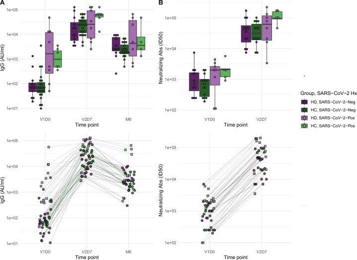
The immune response to the COVID-19 BNT162b2 mRNA vaccine was assessed in 20 HD patients and cohort-matched controls. RNA sequencing of peripheral blood mononuclear cells was performed longitudinally before and after each vaccination dose for a total of six time points per subject. Anti-spike antibody levels were quantified prior to the first vaccination dose (V1D0) and 7 d after the second dose (V2D7) using anti-spike IgG titers and antibody neutralization assays. Anti-spike IgG titers were additionally quantified 6 mo after initial vaccination. Clinical history and lab values in HD patients were obtained to identify predictors of vaccination response.
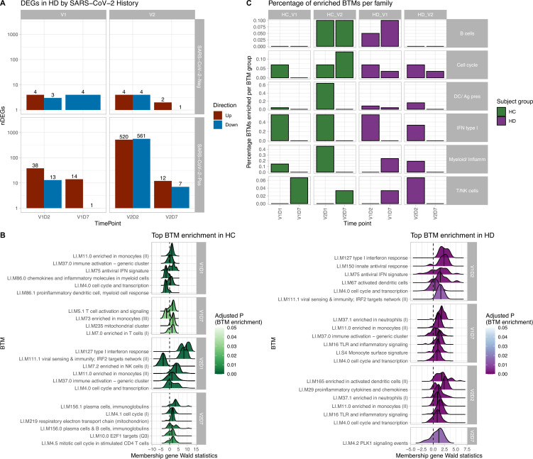
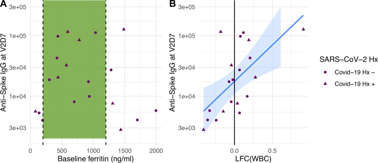
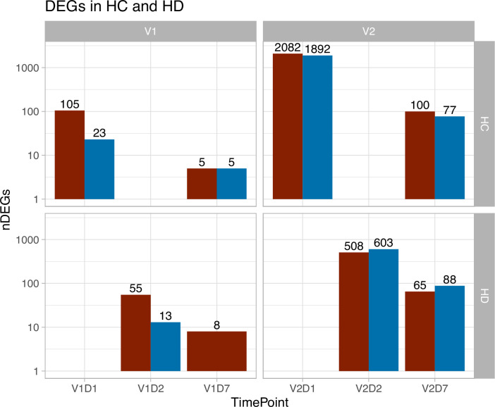
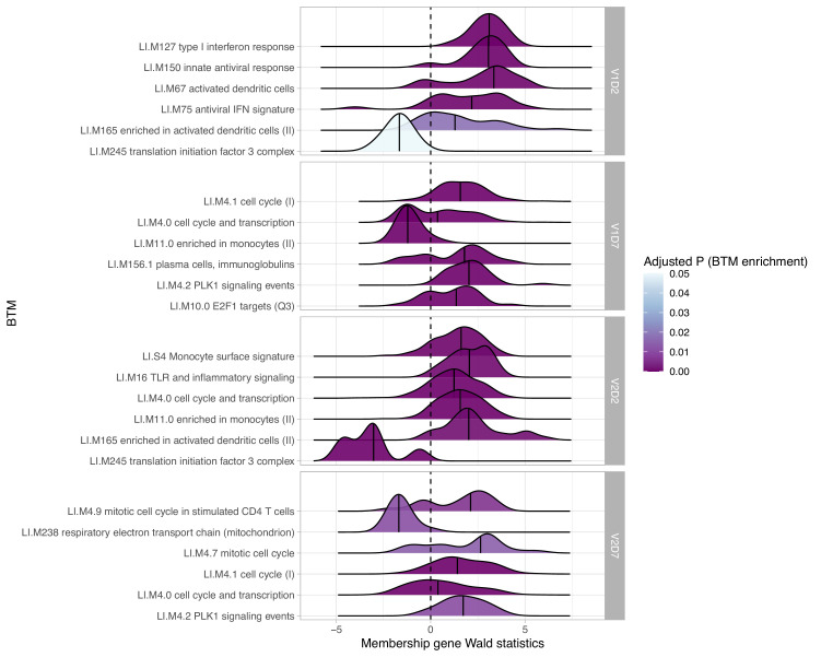
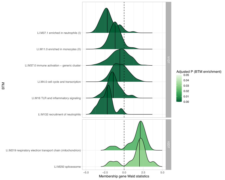
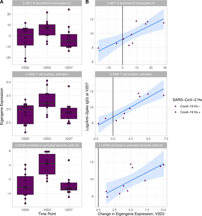
Transcriptomic analyses demonstrated differing time courses of immune responses, with prolonged myeloid cell activity in HD at 1 wk after the first vaccination dose. HD also demonstrated decreased metabolic activity and decreased antigen presentation compared to controls after the second vaccination dose. Anti-spike IgG titers and neutralizing function were substantially elevated in both controls and HD at V2D7, with a small but significant reduction in titers in HD groups (p<0.05). Anti-spike IgG remained elevated above baseline at 6 mo in both subject groups. Anti-spike IgG titers at V2D7 were highly predictive of 6-month titer levels. Transcriptomic biomarkers after the second vaccination dose and clinical biomarkers including ferritin levels were found to be predictive of antibody development.
Overall, we demonstrate differing time courses of immune responses to the BTN162b2 mRNA COVID-19 vaccination in maintenance HD subjects comparable to healthy controls and identify transcriptomic and clinical predictors of anti-spike IgG titers in HD. Analyzing vaccination as an in vivo perturbation, our results warrant further characterization of the immune dysregulation of ESRD.
F30HD102093, F30HL151182, T32HL144909, R01HL138628. This research has been funded by the University of Illinois at Chicago Center for Clinical and Translational Science (CCTS) award UL1TR002003.
终末期肾病(ESRD)患者存在免疫功能受损,其特征为固有免疫和适应性免疫均发生复杂改变,导致患者更易感染且对疫苗接种的反应较低。这种免疫功能受损,再加上在血液透析(HD)中心接触传染病的风险更高,凸显了检查对基于mRNA的新冠疫苗免疫反应的必要性。
评估了20名HD患者和队列匹配对照对新冠BNT162b2 mRNA疫苗的免疫反应。在每次接种疫苗剂量前后纵向对外周血单个核细胞进行RNA测序,每位受试者共六个时间点。在首次接种疫苗剂量前(V1D0)和第二次接种剂量后7天(V2D7),使用抗刺突IgG滴度和抗体中和试验对抗刺突抗体水平进行定量。在初次接种疫苗6个月后,还额外对抗刺突IgG滴度进行了定量。获取HD患者的临床病史和实验室值,以确定疫苗接种反应的预测因素。
转录组分析显示免疫反应的时间进程不同,首次接种疫苗剂量后1周,HD患者的髓系细胞活性延长。与对照组相比,第二次接种疫苗剂量后,HD患者还表现出代谢活性降低和抗原呈递减少。在V2D7时,对照组和HD患者的抗刺突IgG滴度和中和功能均大幅升高,HD组的滴度有小幅但显著的降低(p<0.05)。在两个受试者组中,抗刺突IgG在6个月时仍高于基线水平。V2D7时的抗刺突IgG滴度高度预测6个月时的滴度水平。发现第二次接种疫苗剂量后的转录组生物标志物和包括铁蛋白水平在内的临床生物标志物可预测抗体产生。
总体而言,我们证明了维持性HD受试者对BTN162b2 mRNA新冠疫苗的免疫反应时间进程与健康对照不同,并确定了HD患者抗刺突IgG滴度的转录组和临床预测因素。将疫苗接种作为一种体内扰动进行分析,我们的结果值得进一步描述ESRD患者的免疫失调情况。
F30HD102093、F30HL151182、T32HL144909、R01HL138628。本研究由伊利诺伊大学芝加哥分校临床与转化科学中心(CCTS)授予的UL1TR002003资助。