• 文献检索
  • 文档翻译
  • 深度研究
  • 学术资讯
  • Suppr Zotero 插件Zotero 插件
  • 邀请有礼
  • 套餐&价格
  • 历史记录
应用&插件
Suppr Zotero 插件Zotero 插件浏览器插件Mac 客户端Windows 客户端微信小程序
定价
高级版会员购买积分包购买API积分包
服务
文献检索文档翻译深度研究API 文档MCP 服务
关于我们
关于 Suppr公司介绍联系我们用户协议隐私条款
关注我们

Suppr 超能文献

核心技术专利:CN118964589B侵权必究
粤ICP备2023148730 号-1Suppr @ 2026

文献检索

告别复杂PubMed语法,用中文像聊天一样搜索,搜遍4000万医学文献。AI智能推荐,让科研检索更轻松。

立即免费搜索

文件翻译

保留排版,准确专业,支持PDF/Word/PPT等文件格式,支持 12+语言互译。

免费翻译文档

深度研究

AI帮你快速写综述,25分钟生成高质量综述,智能提取关键信息,辅助科研写作。

立即免费体验

揭示源自子孢子的细胞外囊泡:分析、起源和蛋白质组成。

Unveiling sporozoite-derived extracellular vesicles: profiling, origin, and protein composition.

作者信息

Bertuccini Lucia, Boussadia Zaira, Salzano Anna Maria, Vanni Ilaria, Passerò Ilaria, Nocita Emanuela, Scaloni Andrea, Sanchez Massimo, Sargiacomo Massimo, Fiani Maria Luisa, Tosini Fabio

机构信息

Core Facilities, Istituto Superiore di Sanità, Rome, Italy.

National Center for Drug Research and Evaluation, Istituto Superiore di Sanità, Rome, Italy.

出版信息

Front Cell Infect Microbiol. 2024 Apr 10;14:1367359. doi: 10.3389/fcimb.2024.1367359. eCollection 2024.

DOI:10.3389/fcimb.2024.1367359
PMID:38660488
原文链接:https://pmc.ncbi.nlm.nih.gov/articles/PMC11039866/
Abstract

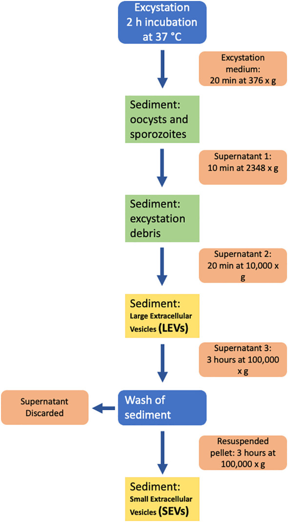
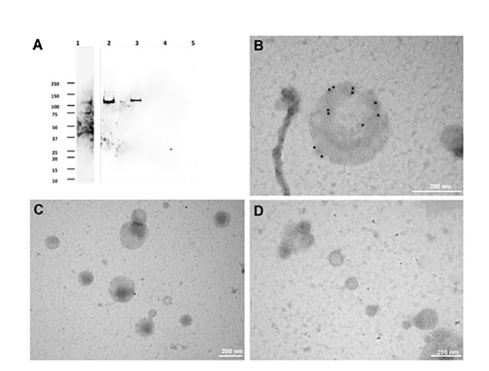
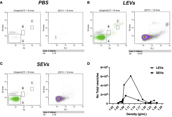
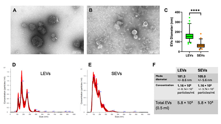
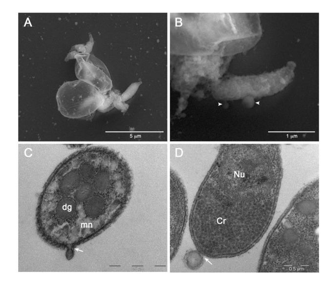
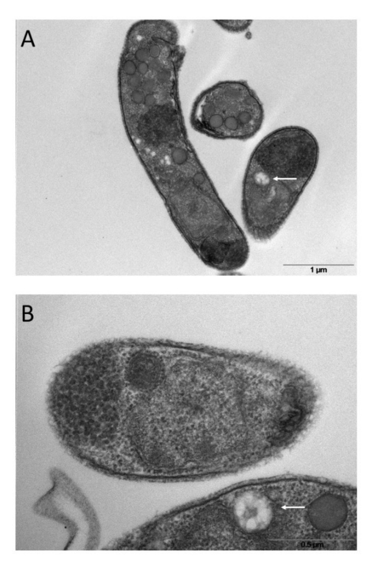
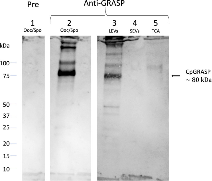
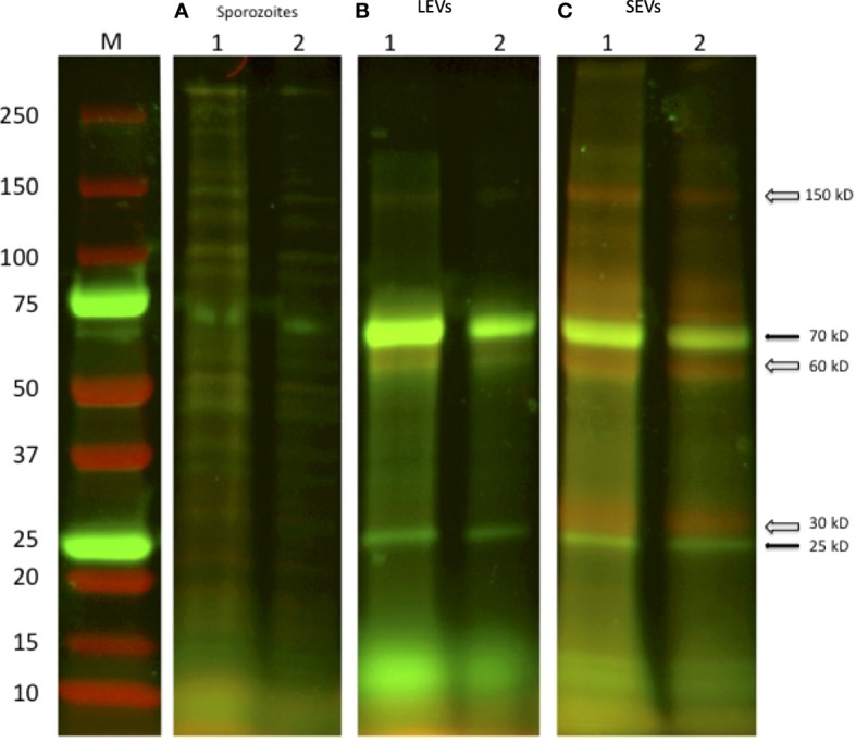
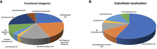
is a common cause of a zoonotic disease and a main cause of diarrhea in newborns. Effective drugs or vaccines are still lacking. Oocyst is the infective form of the parasite; after its ingestion, the oocyst excysts and releases four sporozoites into the host intestine that rapidly attack the enterocytes. The membrane protein CpRom1 is a large rhomboid protease that is expressed by sporozoites and recognized as antigen by the host immune system. In this study, we observed the release of CpRom1 with extracellular vesicles (EVs) that was not previously described. To investigate this phenomenon, we isolated and resolved EVs from the excystation medium by differential ultracentrifugation. Fluorescence flow cytometry and transmission electron microscopy (TEM) experiments identified two types of sporozoite-derived vesicles: large extracellular vesicles (LEVs) and small extracellular vesicles (SEVs). Nanoparticle tracking analysis (NTA) revealed mode diameter of 181 nm for LEVs and 105 nm for SEVs, respectively. Immunodetection experiments proved the presence of CpRom1 and the Golgi protein CpGRASP in LEVs, while immune-electron microscopy trials demonstrated the localization of CpRom1 on the LEVs surface. TEM and scanning electron microscopy (SEM) showed that LEVs were generated by means of the budding of the outer membrane of sporozoites; conversely, the origin of SEVs remained uncertain. Distinct protein compositions were observed between LEVs and SEVs as evidenced by their corresponding electrophoretic profiles. Indeed, a dedicated proteomic analysis identified 5 and 16 proteins unique for LEVs and SEVs, respectively. Overall, 60 proteins were identified in the proteome of both types of vesicles and most of these proteins (48 in number) were already identified in the molecular cargo of extracellular vesicles from other organisms. Noteworthy, we identified 12 proteins unique to spp. and this last group included the immunodominant parasite antigen glycoprotein GP60, which is one of the most abundant proteins in both LEVs and SEVs.

摘要

是一种人畜共患病的常见病因,也是新生儿腹泻的主要原因。目前仍缺乏有效的药物或疫苗。卵囊是该寄生虫的感染形式;摄入后,卵囊脱囊并向宿主肠道释放四个子孢子,这些子孢子会迅速攻击肠上皮细胞。膜蛋白CpRom1是一种大型类菱形蛋白酶,由子孢子表达并被宿主免疫系统识别为抗原。在本研究中,我们观察到CpRom1与细胞外囊泡(EVs)一起释放,这一现象此前未见报道。为了研究这一现象,我们通过差速超速离心从脱囊培养基中分离并解析了EVs。荧光流式细胞术和透射电子显微镜(TEM)实验鉴定出两种源自子孢子的囊泡:大型细胞外囊泡(LEVs)和小型细胞外囊泡(SEVs)。纳米颗粒跟踪分析(NTA)显示,LEVs的平均直径为181nm,SEVs的平均直径为105nm。免疫检测实验证明LEVs中存在CpRom1和高尔基体蛋白CpGRASP,而免疫电子显微镜试验表明CpRom1定位于LEVs表面。TEM和扫描电子显微镜(SEM)显示,LEVs是通过子孢子外膜出芽产生的;相反,SEVs的来源仍不确定。LEVs和SEVs之间观察到不同的蛋白质组成,这由它们相应的电泳图谱证明。事实上,一项专门的蛋白质组学分析分别鉴定出LEVs和SEVs特有的5种和16种蛋白质。总体而言,在两种囊泡的蛋白质组中鉴定出60种蛋白质,其中大多数蛋白质(48种)已在其他生物体的细胞外囊泡的分子货物中鉴定出来。值得注意的是,我们鉴定出12种 属特有的蛋白质,最后一组包括免疫显性寄生虫抗原糖蛋白GP60,它是LEVs和SEVs中最丰富的蛋白质之一。

https://cdn.ncbi.nlm.nih.gov/pmc/blobs/60cf/11039866/c18d4d1a6f6f/fcimb-14-1367359-g012.jpg
https://cdn.ncbi.nlm.nih.gov/pmc/blobs/60cf/11039866/8246ac4f26fb/fcimb-14-1367359-g001.jpg
https://cdn.ncbi.nlm.nih.gov/pmc/blobs/60cf/11039866/09cf25d1ed20/fcimb-14-1367359-g002.jpg
https://cdn.ncbi.nlm.nih.gov/pmc/blobs/60cf/11039866/251e78f8056a/fcimb-14-1367359-g003.jpg
https://cdn.ncbi.nlm.nih.gov/pmc/blobs/60cf/11039866/c12135befd98/fcimb-14-1367359-g004.jpg
https://cdn.ncbi.nlm.nih.gov/pmc/blobs/60cf/11039866/2dc8271ace63/fcimb-14-1367359-g005.jpg
https://cdn.ncbi.nlm.nih.gov/pmc/blobs/60cf/11039866/8548d8cab1e4/fcimb-14-1367359-g006.jpg
https://cdn.ncbi.nlm.nih.gov/pmc/blobs/60cf/11039866/d55396c29894/fcimb-14-1367359-g007.jpg
https://cdn.ncbi.nlm.nih.gov/pmc/blobs/60cf/11039866/55399415d6cc/fcimb-14-1367359-g008.jpg
https://cdn.ncbi.nlm.nih.gov/pmc/blobs/60cf/11039866/4dd73870d0f0/fcimb-14-1367359-g009.jpg
https://cdn.ncbi.nlm.nih.gov/pmc/blobs/60cf/11039866/6fb4c6b177ed/fcimb-14-1367359-g010.jpg
https://cdn.ncbi.nlm.nih.gov/pmc/blobs/60cf/11039866/ffabae9abf9a/fcimb-14-1367359-g011.jpg
https://cdn.ncbi.nlm.nih.gov/pmc/blobs/60cf/11039866/c18d4d1a6f6f/fcimb-14-1367359-g012.jpg

相似文献

1
Unveiling sporozoite-derived extracellular vesicles: profiling, origin, and protein composition.揭示源自子孢子的细胞外囊泡:分析、起源和蛋白质组成。
Front Cell Infect Microbiol. 2024 Apr 10;14:1367359. doi: 10.3389/fcimb.2024.1367359. eCollection 2024.
2
Proteomics analysis and protein expression during sporozoite excystation of Cryptosporidium parvum (Coccidia, Apicomplexa).微小隐孢子虫(球虫纲,顶复门)子孢子脱囊过程中的蛋白质组学分析及蛋白质表达
Mol Cell Proteomics. 2007 Feb;6(2):346-55. doi: 10.1074/mcp.M600372-MCP200. Epub 2006 Nov 23.
3
Comparative proteomic analysis of three major extracellular vesicle classes secreted from human primary and metastatic colorectal cancer cells: Exosomes, microparticles, and shed midbody remnants.三种主要的细胞外囊泡(外泌体、微粒体和脱落中体残片)在人原发性和转移性结直肠癌细胞中的分泌的比较蛋白质组学分析。
Proteomics. 2024 Jun;24(11):e2300057. doi: 10.1002/pmic.202300057. Epub 2023 Jul 28.
4
Extracellular Vesicle Subtypes Released From Activated or Apoptotic T-Lymphocytes Carry a Specific and Stimulus-Dependent Protein Cargo.从活化或凋亡的 T 淋巴细胞释放的细胞外囊泡亚型携带特定的、刺激依赖性的蛋白质货物。
Front Immunol. 2018 Mar 15;9:534. doi: 10.3389/fimmu.2018.00534. eCollection 2018.
5
Evidence for mucin-like glycoproteins that tether sporozoites of Cryptosporidium parvum to the inner surface of the oocyst wall.关于将微小隐孢子虫子孢子拴系于卵囊壁内表面的黏蛋白样糖蛋白的证据。
Eukaryot Cell. 2010 Jan;9(1):84-96. doi: 10.1128/EC.00288-09. Epub 2009 Nov 30.
6
Elongation factor-1α is a novel protein associated with host cell invasion and a potential protective antigen of Cryptosporidium parvum.延长因子-1α 是一种与宿主细胞入侵相关的新型蛋白,也是微小隐孢子虫的一种潜在保护性抗原。
J Biol Chem. 2013 Nov 22;288(47):34111-34120. doi: 10.1074/jbc.M113.515544. Epub 2013 Sep 30.
7
The mucin-like, secretory type-I transmembrane glycoprotein GP900 in the apicomplexan Cryptosporidium parvum is cleaved in the secretory pathway and likely plays a lubrication role.黏蛋白样、分泌型 I 型跨膜糖蛋白 GP900 在顶复门微小隐孢子虫中在分泌途径中被切割,可能发挥润滑作用。
Parasit Vectors. 2022 May 17;15(1):170. doi: 10.1186/s13071-022-05286-8.
8
Charting the proteome of Cryptosporidium parvum sporozoites using sequence similarity-based BLAST searching.利用基于序列相似性的BLAST搜索绘制微小隐孢子虫子孢子的蛋白质组图谱。
J Vet Sci. 2009 Sep;10(3):203-10. doi: 10.4142/jvs.2009.10.3.203.
9
Requirement of microtubules for secretion of a micronemal protein CpTSP4 in the invasive stage of the apicomplexan .微管对于顶复门生物的侵袭阶段中微线蛋白 CpTSP4 的分泌的需求。
mBio. 2024 Feb 14;15(2):e0315823. doi: 10.1128/mbio.03158-23. Epub 2024 Jan 24.
10
Discovery of New Microneme Proteins in and Implication of the Roles of a Rhomboid Membrane Protein (CpROM1) in Host-Parasite Interaction.疟原虫中新微线蛋白的发现及菱形膜蛋白(CpROM1)在宿主-寄生虫相互作用中的作用探讨
Front Vet Sci. 2021 Dec 13;8:778560. doi: 10.3389/fvets.2021.778560. eCollection 2021.

引用本文的文献

1
Extracellular vesicles in intestinal protozoa: hidden mediators of host-parasite communication.肠道原生动物中的细胞外囊泡:宿主-寄生虫通讯的隐藏介质
Gut Pathog. 2025 Aug 28;17(1):69. doi: 10.1186/s13099-025-00747-8.

本文引用的文献

1
Metabolic labelling of a subpopulation of small extracellular vesicles using a fluorescent palmitic acid analogue.使用荧光棕榈酸类似物对一小部分细胞外囊泡进行代谢标记。
J Extracell Vesicles. 2023 Dec;12(12):e12392. doi: 10.1002/jev2.12392.
2
Cryptosporidium uses multiple distinct secretory organelles to interact with and modify its host cell.隐孢子虫利用多种不同的分泌细胞器与宿主细胞相互作用并进行修饰。
Cell Host Microbe. 2023 Apr 12;31(4):650-664.e6. doi: 10.1016/j.chom.2023.03.001. Epub 2023 Mar 22.
3
The state of the art of extracellular vesicle research in protozoan infection.
原生动物感染中细胞外囊泡研究的现状
Front Genet. 2022 Aug 12;13:941561. doi: 10.3389/fgene.2022.941561. eCollection 2022.
4
The exosome journey: from biogenesis to uptake and intracellular signalling.外泌体的旅程:从生物发生到摄取和细胞内信号转导。
Cell Commun Signal. 2021 Apr 23;19(1):47. doi: 10.1186/s12964-021-00730-1.
5
Fluorescent labeling of extracellular vesicles.细胞外囊泡的荧光标记。
Methods Enzymol. 2020;645:15-42. doi: 10.1016/bs.mie.2020.09.002. Epub 2020 Sep 29.
6
Perils and Promises of Pathogenic Protozoan Extracellular Vesicles.致病性原生动物细胞外囊泡的风险与前景
Front Cell Infect Microbiol. 2020 Aug 14;10:371. doi: 10.3389/fcimb.2020.00371. eCollection 2020.
7
The role of rhomboid superfamily members in protein homeostasis: Mechanistic insight and physiological implications.菱形超家族成员在蛋白质平衡中的作用:机制见解和生理意义。
Biochim Biophys Acta Mol Cell Res. 2020 Oct;1867(10):118793. doi: 10.1016/j.bbamcr.2020.118793. Epub 2020 Jul 6.
8
Molecular epidemiologic tools for waterborne pathogens spp. and .用于水生病原体物种的分子流行病学工具
Food Waterborne Parasitol. 2017 Sep 29;8-9:14-32. doi: 10.1016/j.fawpar.2017.09.002. eCollection 2017 Sep-Dec.
9
Intramembrane proteolysis at a glance: from signalling to protein degradation.跨膜蛋白水解:从信号转导到蛋白降解。
J Cell Sci. 2019 Aug 15;132(16):jcs217745. doi: 10.1242/jcs.217745.
10
New Insights Into the Golgi Stacking Proteins.对高尔基体堆叠蛋白的新见解
Front Cell Dev Biol. 2019 Jul 16;7:131. doi: 10.3389/fcell.2019.00131. eCollection 2019.