• 文献检索
  • 文档翻译
  • 深度研究
  • 学术资讯
  • Suppr Zotero 插件Zotero 插件
  • 邀请有礼
  • 套餐&价格
  • 历史记录
应用&插件
Suppr Zotero 插件Zotero 插件浏览器插件Mac 客户端Windows 客户端微信小程序
定价
高级版会员购买积分包购买API积分包
服务
文献检索文档翻译深度研究API 文档MCP 服务
关于我们
关于 Suppr公司介绍联系我们用户协议隐私条款
关注我们

Suppr 超能文献

核心技术专利:CN118964589B侵权必究
粤ICP备2023148730 号-1Suppr @ 2026

文献检索

告别复杂PubMed语法,用中文像聊天一样搜索,搜遍4000万医学文献。AI智能推荐,让科研检索更轻松。

立即免费搜索

文件翻译

保留排版,准确专业,支持PDF/Word/PPT等文件格式,支持 12+语言互译。

免费翻译文档

深度研究

AI帮你快速写综述,25分钟生成高质量综述,智能提取关键信息,辅助科研写作。

立即免费体验

与锥形束计算机断层扫描相比,评估头颅侧位片在量化三维咽气道形态方面的准确性。

Assessing the Accuracy of Lateral Cephalogram in Quantifying Three-Dimensional Pharyngeal Airway Morphology Compared to Cone-Beam Computed Tomography.

作者信息

Ashique Abdulhameed Syed, Riyaz Ss Mohamed Abdulcader, Almutairy Mohammed, Khan N Salman, Jayakumar Saikarthik, Gaonkar Prachi

机构信息

Department of Orthodontics and Dentofacial Orthopaedics, Meenakshi Ammal Dental Vollege and Hospital, Chennai, IND.

Department of Maxillofacial Surgery and Diagnostic Science, College of Dentistry, Qassim University, Ar Rass, SAU.

出版信息

Cureus. 2024 Mar 31;16(3):e57301. doi: 10.7759/cureus.57301. eCollection 2024 Mar.

DOI:10.7759/cureus.57301
PMID:38690459
原文链接:https://pmc.ncbi.nlm.nih.gov/articles/PMC11059114/
Abstract

BACKGROUND

When it comes to orthodontic diagnosis and treatment planning, the structures of the upper and lower airway space are crucial because of the role they play in craniofacial development.

AIM

The major objective of this study was to evaluate the accuracy of lateral cephalogram in the evaluation of upper and lower pharyngeal space by comparing it to clinical usage of cone-beam computed tomography (CBCT) in quantifying the 3D morphology of the pharyngeal airway.

METHODS AND MATERIALS

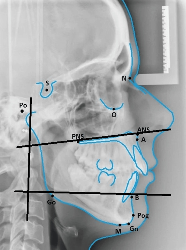
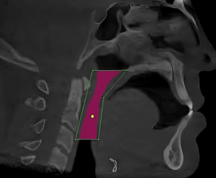
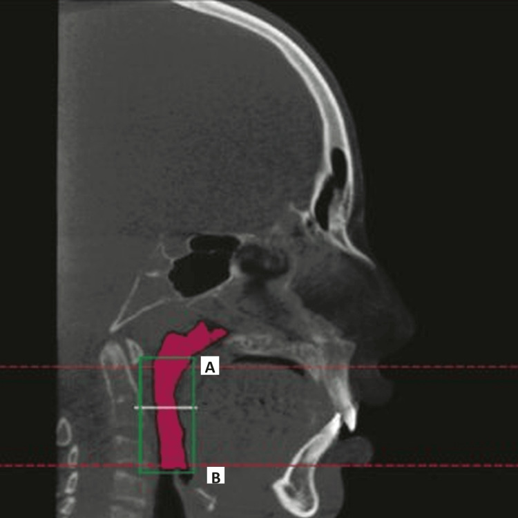
In total, 70 patients were included in the study. They had both a CBCT scan and a lateral cephalogram performed within a week of each other. Different cephalometric landmarks have been utilized to estimate linear and area dimensions for use in lateral cephalogram airway investigations. By superimposing the lateral cephalogram measurement of the vertical height of the pharyngeal airway over axial CBCT slices of 0.8 to 1 mm in thickness, airway volumes were calculated. For this study, we measured the pharyngeal airway space in each patient in two dimensions (2D) using the airway area from the lateral cephalogram and in three dimensions (3D) using the airway volume from the CBCT scan over the same region of interest, using a uniform scale and magnification throughout all split 3D volumes.

RESULTS

The mean value of the area of pharyngeal space calculated by lateral cephalograph analysis (LCA) was 336.35 ± 86.49 mm. The maximum value was 551.234 mm. The minimum value was 206.32 mm. The mean value of the volume of the same area calculated using CBCT was 3409.11 ± 1237.96 mm. The maximum value was 5887.23 mm. When the area calculated using LCA was compared with the volume calculated using CBCT, the correlation between them was significant statistically (r=0.831, p-value =0.000). The mean values of volume evaluated in 3D CBCT in males were 4198±1008 mm while for females it was 2980±1134.5 mm. During the statistical analysis, these observations were found to have a positive correlation with increased volume of pharyngeal space in males as compared to that of females (p=0.006). The values of the area of pharyngeal space calculated using LCA in males was 370.1±60.9 mm while it was 301.9±88 mm  in females.

CONCLUSION

The area estimated for the pharyngeal airway on LCA correlates strongly with the volume determined by a CBCT scan. Since we have considered pharyngeal space analysis using CBCT to be a reliable and standard methodology, therefore a positive correlation of area calculated using LCA with volume calculated using CBCT shows that the analysis made by LCA can be reliable.

摘要

背景

在正畸诊断和治疗计划中,上下气道空间的结构至关重要,因为它们在颅面发育中发挥着作用。

目的

本研究的主要目的是通过将头颅侧位片与锥形束计算机断层扫描(CBCT)在量化咽气道三维形态的临床应用进行比较,评估头颅侧位片在评估上下咽腔空间方面的准确性。

方法和材料

本研究共纳入70例患者。他们在一周内分别进行了CBCT扫描和头颅侧位片检查。使用了不同的头影测量标志点来估计线性和面积尺寸,用于头颅侧位片气道研究。通过将头颅侧位片测量的咽气道垂直高度叠加在厚度为0.8至1毫米的轴向CBCT切片上,计算气道体积。在本研究中,我们使用统一的比例和放大倍数,在所有分割的三维体积的相同感兴趣区域,使用头颅侧位片的气道面积在二维(2D)上测量每位患者的咽气道空间,并使用CBCT扫描的气道体积在三维(3D)上测量。

结果

头颅侧位片分析(LCA)计算的咽腔空间面积平均值为336.35±86.49平方毫米。最大值为551.234平方毫米。最小值为206.32平方毫米。使用CBCT计算的同一区域体积平均值为3409.11±1237.96立方毫米。最大值为5887.23立方毫米。当将LCA计算的面积与CBCT计算的体积进行比较时,它们之间的相关性具有统计学意义(r = 0.831,p值 = 0.000)。男性在三维CBCT中评估的体积平均值为4198±1008立方毫米,而女性为2980±1134.5立方毫米。在统计分析中,发现这些观察结果与男性咽腔空间体积增加呈正相关,与女性相比(p = 0.006)。男性使用LCA计算的咽腔空间面积值为370.1±60.9平方毫米,而女性为301.9±88平方毫米。

结论

LCA估计的咽气道面积与CBCT扫描确定的体积密切相关。由于我们认为使用CBCT进行咽腔空间分析是一种可靠的标准方法,因此LCA计算的面积与CBCT计算的体积呈正相关表明LCA进行的分析是可靠的。

https://cdn.ncbi.nlm.nih.gov/pmc/blobs/c0cb/11059114/d95bfe72b13e/cureus-0016-00000057301-i03.jpg
https://cdn.ncbi.nlm.nih.gov/pmc/blobs/c0cb/11059114/bbaf917d912a/cureus-0016-00000057301-i01.jpg
https://cdn.ncbi.nlm.nih.gov/pmc/blobs/c0cb/11059114/d0703d13dc48/cureus-0016-00000057301-i02.jpg
https://cdn.ncbi.nlm.nih.gov/pmc/blobs/c0cb/11059114/d95bfe72b13e/cureus-0016-00000057301-i03.jpg
https://cdn.ncbi.nlm.nih.gov/pmc/blobs/c0cb/11059114/bbaf917d912a/cureus-0016-00000057301-i01.jpg
https://cdn.ncbi.nlm.nih.gov/pmc/blobs/c0cb/11059114/d0703d13dc48/cureus-0016-00000057301-i02.jpg
https://cdn.ncbi.nlm.nih.gov/pmc/blobs/c0cb/11059114/d95bfe72b13e/cureus-0016-00000057301-i03.jpg

相似文献

1
Assessing the Accuracy of Lateral Cephalogram in Quantifying Three-Dimensional Pharyngeal Airway Morphology Compared to Cone-Beam Computed Tomography.与锥形束计算机断层扫描相比,评估头颅侧位片在量化三维咽气道形态方面的准确性。
Cureus. 2024 Mar 31;16(3):e57301. doi: 10.7759/cureus.57301. eCollection 2024 Mar.
2
Analysis of Pharyngeal Airway Using Lateral Cephalogram vs CBCT Images: A Cross-sectional Retrospective Study.使用头颅侧位片与锥形束计算机断层扫描(CBCT)图像分析咽气道:一项横断面回顾性研究。
Open Dent J. 2015 Jul 31;9:263-6. doi: 10.2174/1874210601509010263. eCollection 2015.
3
Effect of Craniofacial Morphology on Pharyngeal Airway Volume Measured Using Cone-Beam Computed Tomography (CBCT)-A Retrospective Pilot Study.颅面形态对锥形束 CT(CBCT)测量的咽气道容积的影响:一项回顾性初步研究。
Int J Environ Res Public Health. 2021 May 10;18(9):5040. doi: 10.3390/ijerph18095040.
4
No correlation between two-dimensional measurements and three-dimensional configuration of the pharyngeal upper airway space in cone-beam computed tomography.锥形束计算机断层扫描中咽上气道空间的二维测量与三维结构之间无相关性。
J Craniomaxillofac Surg. 2017 Mar;45(3):371-376. doi: 10.1016/j.jcms.2017.01.004. Epub 2017 Jan 10.
5
Study of pharyngeal airway morphology with CBCT: Benefits of four premolar extraction orthodontic treatments.锥形束 CT 研究咽气道形态:四前磨牙拔除正畸治疗的优势。
Niger J Clin Pract. 2022 Dec;25(12):1955-1962. doi: 10.4103/njcp.njcp_1815_21.
6
From 2D to 3D: an algorithm to derive normal values for 3-dimensional computerized assessment.从 2D 到 3D:一种用于三维计算机评估的正常数值推导算法。
Angle Orthod. 2011 Jan;81(1):3-10. doi: 10.2319/032910-173.1.
7
Evaluation of pharyngeal airway space amongst different skeletal patterns.评价不同骨骼类型的咽腔气道空间。
Int J Oral Maxillofac Surg. 2012 Jul;41(7):814-9. doi: 10.1016/j.ijom.2012.01.015. Epub 2012 Feb 24.
8
Comparative study of cephalometric measurements using 3 imaging modalities.三种影像学方法测量头影测量指标的比较研究。
J Am Dent Assoc. 2017 Dec;148(12):913-921. doi: 10.1016/j.adaj.2017.07.030. Epub 2017 Oct 16.
9
Comparison of pharyngeal airway changes on plain radiography and cone-beam computed tomography after orthognathic surgery.正颌外科手术后X线平片与锥形束计算机断层扫描对咽部气道变化的比较。
J Oral Maxillofac Surg. 2011 Nov;69(11):e385-94. doi: 10.1016/j.joms.2011.03.015. Epub 2011 Jul 20.
10
Comparison of pharyngeal airway volume in different skeletal facial patterns using cone beam computed tomography.使用锥形束计算机断层扫描比较不同骨骼面部模式下的咽气道容积。
J Clin Exp Dent. 2018 Oct 1;10(10):e1017-e1028. doi: 10.4317/jced.55033. eCollection 2018 Oct.

引用本文的文献

1
Predictors of disease alleviation with mandibular advancement devices in obstructive sleep apnea: a retrospective cohort study.下颌前移装置治疗阻塞性睡眠呼吸暂停疾病缓解的预测因素:一项回顾性队列研究。
Head Face Med. 2025 Apr 14;21(1):26. doi: 10.1186/s13005-025-00504-x.
2
Retrospective analysis of the upper airway anatomy and Sella turcica morphology across different skeletal malocclusions: a computerized technique.回顾性分析不同骨骼畸形患者的上气道解剖结构和蝶鞍形态:一种计算机技术。
BMC Oral Health. 2024 Sep 19;24(1):1110. doi: 10.1186/s12903-024-04867-6.
3
Application of Artificial Intelligence in Cone-Beam Computed Tomography for Airway Analysis: A Narrative Review.

本文引用的文献

1
Basic training requirements for the use of dental CBCT by dentists: a position paper prepared by the European Academy of DentoMaxilloFacial Radiology.牙科 CBCT 使用的基本培训要求:欧洲口腔颌面放射学会制定的立场文件。
Dentomaxillofac Radiol. 2014;43(1):20130291. doi: 10.1259/dmfr.20130291. Epub 2013 Oct 16.
2
Teenage sleep-disordered breathing: recurrence of syndrome.青少年睡眠呼吸障碍:综合征复发。
Sleep Med. 2013 Jan;14(1):37-44. doi: 10.1016/j.sleep.2012.08.010. Epub 2012 Sep 29.
3
Cone beam computed tomography: craniofacial and airway analysis.
人工智能在锥形束计算机断层扫描气道分析中的应用:一篇叙述性综述。
Diagnostics (Basel). 2024 Aug 30;14(17):1917. doi: 10.3390/diagnostics14171917.
锥形束计算机断层扫描:颅面及气道分析
Dent Clin North Am. 2012 Apr;56(2):343-57. doi: 10.1016/j.cden.2012.02.002. Epub 2012 Feb 20.
4
Evaluation of pharyngeal airway space amongst different skeletal patterns.评价不同骨骼类型的咽腔气道空间。
Int J Oral Maxillofac Surg. 2012 Jul;41(7):814-9. doi: 10.1016/j.ijom.2012.01.015. Epub 2012 Feb 24.
5
Evaluation of upper airway obstruction in Class II children with fluid-mechanical simulation.应用流固力学模拟评价 II 类错颌儿童的上气道阻塞。
Am J Orthod Dentofacial Orthop. 2011 Feb;139(2):e135-45. doi: 10.1016/j.ajodo.2010.08.014.
6
An analysis of different approaches to the assessment of upper airway morphology: a CBCT study.一种评估上呼吸道形态方法的分析:一项 CBCT 研究。
Orthod Craniofac Res. 2010 May;13(2):96-105. doi: 10.1111/j.1601-6343.2010.01482.x.
7
Three-dimensional analysis of pharyngeal airway in preadolescent children with different anteroposterior skeletal patterns.不同前后向骨骼模式的青春期前儿童咽气道的三维分析。
Am J Orthod Dentofacial Orthop. 2010 Mar;137(3):306.e1-11; discussion 306-7. doi: 10.1016/j.ajodo.2009.10.025.
8
Comparison of cone-beam CT parameters and sleep questionnaires in sleep apnea patients and control subjects.睡眠呼吸暂停患者与对照受试者的锥束CT参数与睡眠问卷比较。
Oral Surg Oral Med Oral Pathol Oral Radiol Endod. 2010 Feb;109(2):285-93. doi: 10.1016/j.tripleo.2009.09.033.
9
Computed tomographic evaluation of mouth breathers among paediatric patients.儿童张口呼吸患者的计算机断层扫描评估。
Dentomaxillofac Radiol. 2010 Jan;39(1):1-10. doi: 10.1259/dmfr/80778956.
10
Soft palate length and upper airway relationship in OSA and non-OSA subjects.软腭长度与 OSA 和非 OSA 患者上呼吸道的关系。
Sleep Breath. 2010 Dec;14(4):353-8. doi: 10.1007/s11325-009-0318-7. Epub 2009 Dec 8.