• 文献检索
  • 文档翻译
  • 深度研究
  • 学术资讯
  • Suppr Zotero 插件Zotero 插件
  • 邀请有礼
  • 套餐&价格
  • 历史记录
应用&插件
Suppr Zotero 插件Zotero 插件浏览器插件Mac 客户端Windows 客户端微信小程序
定价
高级版会员购买积分包购买API积分包
服务
文献检索文档翻译深度研究API 文档MCP 服务
关于我们
关于 Suppr公司介绍联系我们用户协议隐私条款
关注我们

Suppr 超能文献

核心技术专利:CN118964589B侵权必究
粤ICP备2023148730 号-1Suppr @ 2026

文献检索

告别复杂PubMed语法,用中文像聊天一样搜索,搜遍4000万医学文献。AI智能推荐,让科研检索更轻松。

立即免费搜索

文件翻译

保留排版,准确专业,支持PDF/Word/PPT等文件格式,支持 12+语言互译。

免费翻译文档

深度研究

AI帮你快速写综述,25分钟生成高质量综述,智能提取关键信息,辅助科研写作。

立即免费体验

心肌梗死后心脏内皮细胞转录组分析揭示了时间变化和长期缺陷。

Transcriptome analysis of cardiac endothelial cells after myocardial infarction reveals temporal changes and long-term deficits.

机构信息

Division of Cardiovascular Medicine, Department of Medicine, Vanderbilt University Medical Center, Nashville, TN, USA.

Vascular Medicine Institute, University of Pittsburgh, Pittsburgh, PA, USA.

出版信息

Sci Rep. 2024 May 1;14(1):9991. doi: 10.1038/s41598-024-59155-8.

DOI:10.1038/s41598-024-59155-8
PMID:38693202
原文链接:https://pmc.ncbi.nlm.nih.gov/articles/PMC11063162/
Abstract

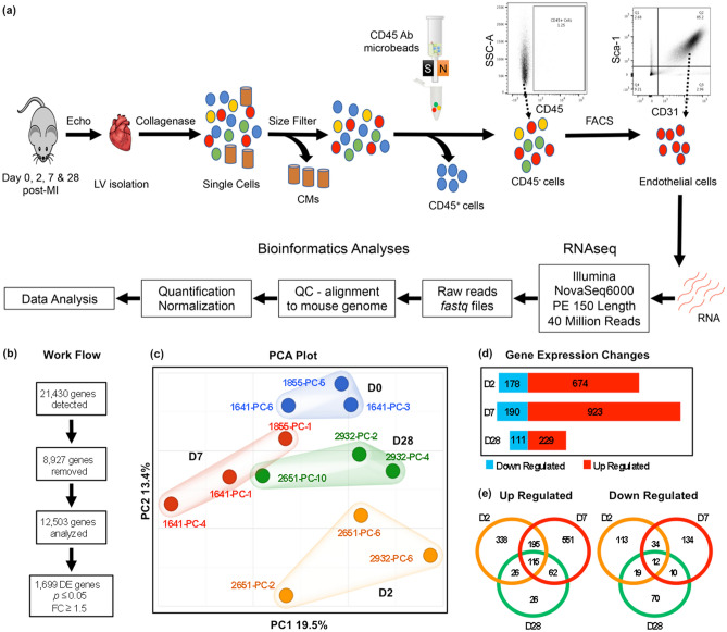
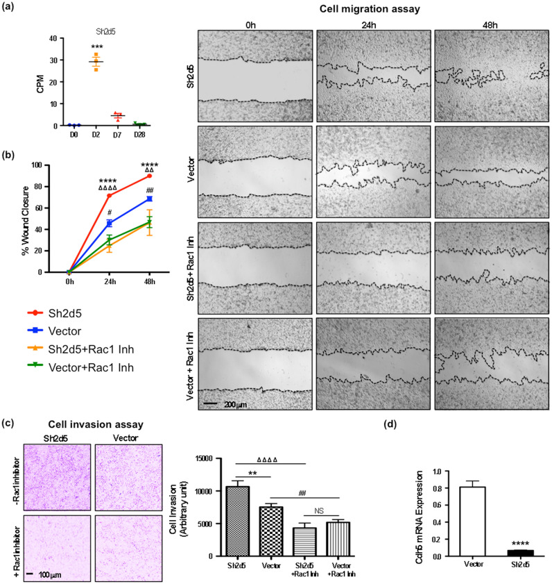
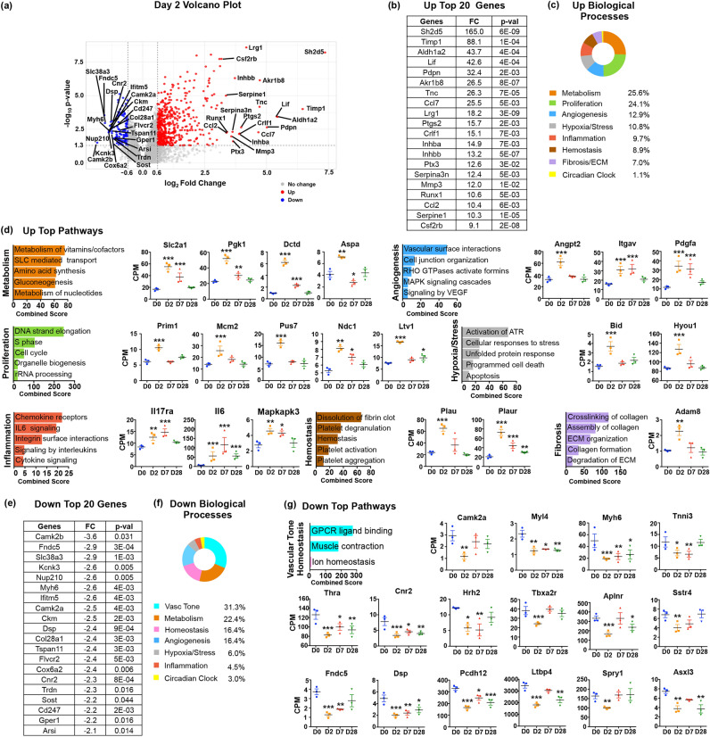
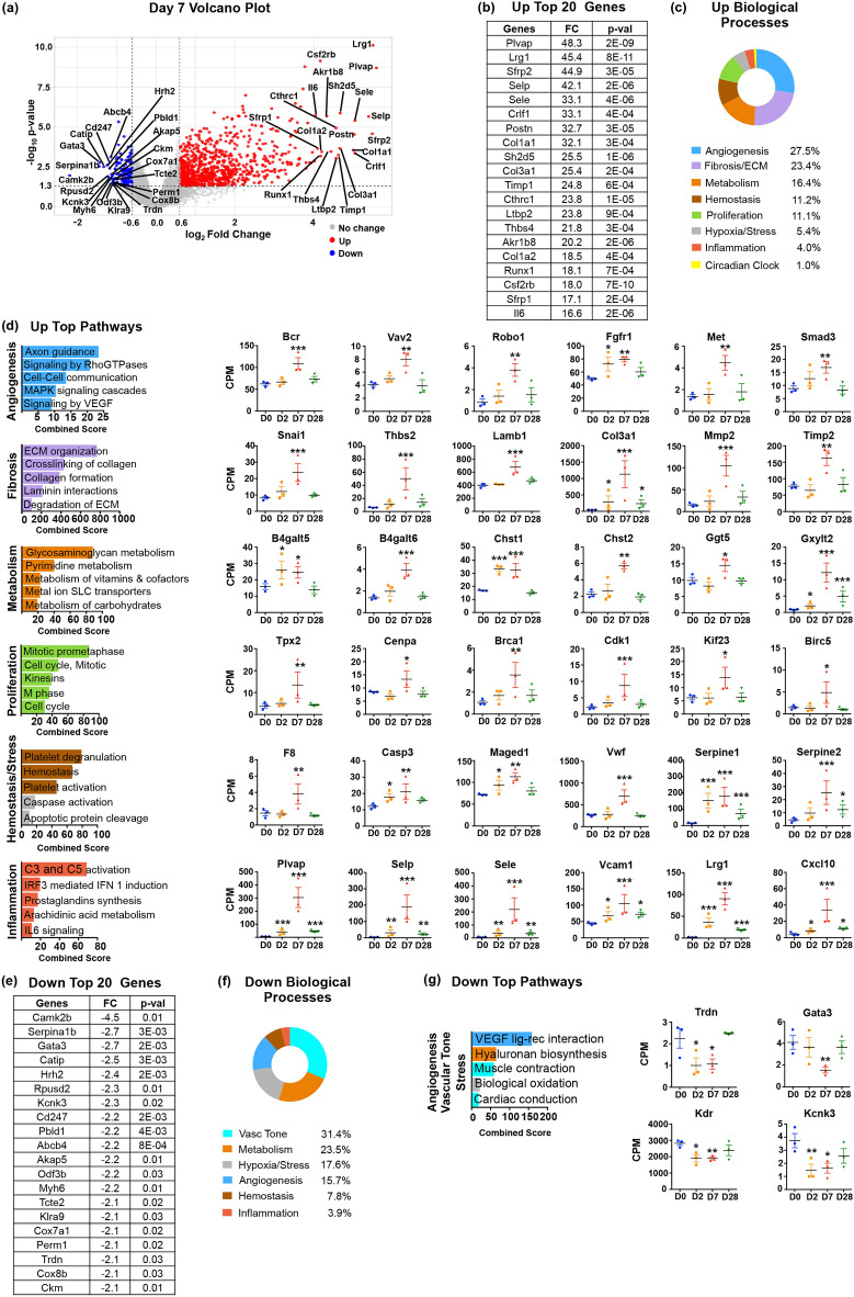
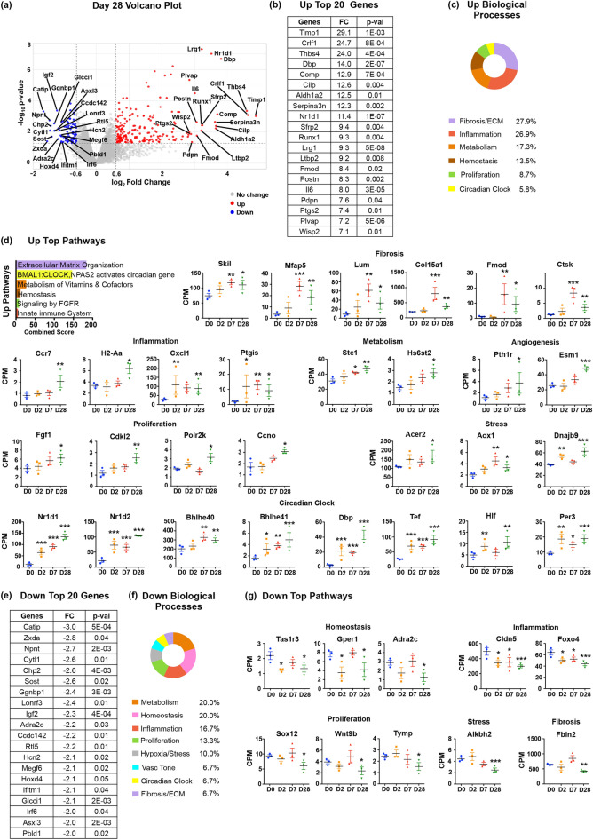
Endothelial cells (ECs) have essential roles in cardiac tissue repair after myocardial infarction (MI). To establish stage-specific and long-term effects of the ischemic injury on cardiac ECs, we analyzed their transcriptome at landmark time points after MI in mice. We found that early EC response at Day 2 post-MI centered on metabolic changes, acquisition of proinflammatory phenotypes, initiation of the S phase of cell cycle, and activation of stress-response pathways, followed by progression to mitosis (M/G2 phase) and acquisition of proangiogenic and mesenchymal properties during scar formation at Day 7. In contrast, genes involved in vascular physiology and maintenance of vascular tone were suppressed. Importantly, ECs did not return to pre-injury phenotypes after repair has been completed but maintained inflammatory, fibrotic and thrombotic characteristics and lost circadian rhythmicity. We discovered that the highest induced transcript is the mammalian-specific Sh2d5 gene that promoted migration and invasion of ECs through Rac1 GTPase. Our results revealed a synchronized, temporal activation of disease phenotypes, metabolic pathways, and proliferation in quiescent ECs after MI, indicating that precisely-timed interventions are necessary to optimize cardiac tissue repair and improve outcomes. Furthermore, long-term effects of acute ischemic injury on ECs may contribute to vascular dysfunction and development of heart failure.

摘要

内皮细胞(ECs)在心肌梗死后的心肌组织修复中具有重要作用。为了确定缺血性损伤对心脏 ECs 的阶段性和长期影响,我们在小鼠心梗后关键时间点分析了它们的转录组。我们发现,心梗后第 2 天的早期 EC 反应集中在代谢变化、获得促炎表型、细胞周期 S 期启动以及应激反应途径激活上,随后在第 7 天进入有丝分裂(M/G2 期)并获得促血管生成和间充质特性,形成疤痕。相比之下,参与血管生理和维持血管张力的基因受到抑制。重要的是,EC 在修复完成后并未恢复到损伤前的表型,而是保持炎症、纤维化和血栓形成的特征,并丧失了昼夜节律性。我们发现,诱导表达最高的基因是哺乳动物特异性的 Sh2d5 基因,它通过 Rac1 GTPase 促进 EC 的迁移和侵袭。我们的研究结果揭示了心梗后静息 EC 中疾病表型、代谢途径和增殖的同步、时间性激活,表明需要精确计时的干预措施来优化心肌组织修复并改善预后。此外,急性缺血性损伤对 ECs 的长期影响可能导致血管功能障碍和心力衰竭的发生。

https://cdn.ncbi.nlm.nih.gov/pmc/blobs/dac6/11063162/3e7e06ea1ef7/41598_2024_59155_Fig5_HTML.jpg
https://cdn.ncbi.nlm.nih.gov/pmc/blobs/dac6/11063162/6f1b824b88ab/41598_2024_59155_Fig1_HTML.jpg
https://cdn.ncbi.nlm.nih.gov/pmc/blobs/dac6/11063162/286507cb02a1/41598_2024_59155_Fig2_HTML.jpg
https://cdn.ncbi.nlm.nih.gov/pmc/blobs/dac6/11063162/0751f8748e77/41598_2024_59155_Fig3_HTML.jpg
https://cdn.ncbi.nlm.nih.gov/pmc/blobs/dac6/11063162/bde8e6696b55/41598_2024_59155_Fig4_HTML.jpg
https://cdn.ncbi.nlm.nih.gov/pmc/blobs/dac6/11063162/3e7e06ea1ef7/41598_2024_59155_Fig5_HTML.jpg

相似文献

1
Transcriptome analysis of cardiac endothelial cells after myocardial infarction reveals temporal changes and long-term deficits.心肌梗死后心脏内皮细胞转录组分析揭示了时间变化和长期缺陷。
Sci Rep. 2024 May 1;14(1):9991. doi: 10.1038/s41598-024-59155-8.
2
Single-cell transcriptome analyses reveal novel targets modulating cardiac neovascularization by resident endothelial cells following myocardial infarction.单细胞转录组分析揭示了心肌梗死后驻留内皮细胞调节心脏新生血管的新靶点。
Eur Heart J. 2019 Aug 7;40(30):2507-2520. doi: 10.1093/eurheartj/ehz305.
3
M1-like macrophage-derived exosomes suppress angiogenesis and exacerbate cardiac dysfunction in a myocardial infarction microenvironment.M1 样巨噬细胞衍生的外泌体在心肌梗死微环境中抑制血管生成并加重心脏功能障碍。
Basic Res Cardiol. 2020 Feb 28;115(2):22. doi: 10.1007/s00395-020-0781-7.
4
Vascular endothelial S1pr1 ameliorates adverse cardiac remodelling via stimulating reparative macrophage proliferation after myocardial infarction.血管内皮细胞的S1pr1通过刺激心肌梗死后的修复性巨噬细胞增殖来改善不良心脏重塑。
Cardiovasc Res. 2021 Jan 21;117(2):585-599. doi: 10.1093/cvr/cvaa046.
5
Cardiac endothelial cells express Wilms' tumor-1: Wt1 expression in the developing, adult and infarcted heart.心脏内皮细胞表达维尔姆斯瘤-1:Wt1 在心脏发育、成年和梗死中的表达。
J Mol Cell Cardiol. 2015 Apr;81:127-35. doi: 10.1016/j.yjmcc.2015.02.007. Epub 2015 Feb 11.
6
In vitro and in vivo proliferation, differentiation and migration of cardiac endothelial progenitor cells (SCA1+/CD31+ side-population cells).心脏内皮祖细胞(SCA1+/CD31+侧群细胞)的体外和体内增殖、分化和迁移。
J Thromb Haemost. 2011 Aug;9(8):1628-37. doi: 10.1111/j.1538-7836.2011.04375.x.
7
Nerve growth factor promotes cardiac repair following myocardial infarction.神经生长因子促进心肌梗死后的心脏修复。
Circ Res. 2010 Apr 16;106(7):1275-84. doi: 10.1161/CIRCRESAHA.109.210088. Epub 2010 Apr 1.
8
Lgr4 Governs a Pro-Inflammatory Program in Macrophages to Antagonize Post-Infarction Cardiac Repair.Lgr4 调控巨噬细胞中的促炎程序以拮抗心肌梗死后的心脏修复。
Circ Res. 2020 Sep 25;127(8):953-973. doi: 10.1161/CIRCRESAHA.119.315807. Epub 2020 Jun 30.
9
Mapping macrophage polarization over the myocardial infarction time continuum.描绘心肌梗死时间连续体上的巨噬细胞极化。
Basic Res Cardiol. 2018 Jun 4;113(4):26. doi: 10.1007/s00395-018-0686-x.
10
Angiogenesis after acute myocardial infarction.急性心肌梗死后的血管新生。
Cardiovasc Res. 2021 Apr 23;117(5):1257-1273. doi: 10.1093/cvr/cvaa287.

引用本文的文献

1
Unraveling the Complex Cellular Repair Mechanisms Following Myocardial Infarction.解析心肌梗死后复杂的细胞修复机制
Int J Mol Sci. 2025 Jun 23;26(13):6002. doi: 10.3390/ijms26136002.
2
Transcriptional signatures of endothelial cells shape immune responses in cardiopulmonary health and disease.内皮细胞的转录特征塑造心肺健康与疾病中的免疫反应。
JCI Insight. 2025 May 22;10(10). doi: 10.1172/jci.insight.191059.
3
Post-myocardial Infarction Cardiac Remodeling: Multidimensional Mechanisms and Clinical Prospects of Stem Cell Therapy.

本文引用的文献

1
Heart Disease and Stroke Statistics-2023 Update: A Report From the American Heart Association.《心脏病与卒中统计数据-2023 更新:美国心脏协会报告》。
Circulation. 2023 Feb 21;147(8):e93-e621. doi: 10.1161/CIR.0000000000001123. Epub 2023 Jan 25.
2
Signaling pathways and targeted therapy for myocardial infarction.心肌梗死的信号通路和靶向治疗。
Signal Transduct Target Ther. 2022 Mar 10;7(1):78. doi: 10.1038/s41392-022-00925-z.
3
Challenging the Paradigm: Anti-Inflammatory Interleukins and Angiogenesis.挑战传统观念:抗炎细胞因子与血管生成。
心肌梗死后心脏重塑:干细胞治疗的多维机制与临床前景
Stem Cell Rev Rep. 2025 May 5. doi: 10.1007/s12015-025-10888-7.
4
Transcriptional Signatures of the Right Ventricle in End-Stage Heart Failure.终末期心力衰竭时右心室的转录特征
Pulm Circ. 2025 Apr 24;15(2):e70090. doi: 10.1002/pul2.70090. eCollection 2025 Apr.
5
Deficiency of Caveolin-1 Aggravates Cardiac Infarction Injury by Disturbing the Endothelial Homeostasis.小窝蛋白-1缺乏通过扰乱内皮稳态加重心肌梗死损伤。
Int J Med Sci. 2025 Jan 21;22(4):920-932. doi: 10.7150/ijms.101074. eCollection 2025.
6
Empagliflozin in Acute Myocardial Infarction Reduces No-Reflow and Preserves Cardiac Function by Preventing Endothelial Damage.恩格列净用于急性心肌梗死可通过预防内皮损伤减轻无复流现象并保护心脏功能。
JACC Basic Transl Sci. 2024 Aug 30;10(1):43-61. doi: 10.1016/j.jacbts.2024.08.003. eCollection 2025 Jan.
Cells. 2022 Feb 8;11(3):587. doi: 10.3390/cells11030587.
4
Percutaneous coronary intervention with drug-eluting stents versus coronary artery bypass grafting in left main coronary artery disease: an individual patient data meta-analysis.经皮冠状动脉介入治疗联合药物洗脱支架与冠状动脉旁路移植术治疗左主干冠状动脉疾病的比较:一项个体患者数据分析荟萃研究。
Lancet. 2021 Dec 18;398(10318):2247-2257. doi: 10.1016/S0140-6736(21)02334-5. Epub 2021 Nov 15.
5
Role of Irisin in Myocardial Infarction, Heart Failure, and Cardiac Hypertrophy.鸢尾素在心肌梗死、心力衰竭和心肌肥厚中的作用。
Cells. 2021 Aug 16;10(8):2103. doi: 10.3390/cells10082103.
6
Preclinical models of myocardial infarction: from mechanism to translation.心肌梗死的临床前模型:从机制到转化。
Br J Pharmacol. 2022 Mar;179(5):770-791. doi: 10.1111/bph.15595. Epub 2021 Aug 11.
7
LTBP4 in Health and Disease.LTBP4 在健康和疾病中的作用。
Genes (Basel). 2021 May 23;12(6):795. doi: 10.3390/genes12060795.
8
The vasculature: a therapeutic target in heart failure?血管系统:心力衰竭的治疗靶点?
Cardiovasc Res. 2022 Jan 7;118(1):53-64. doi: 10.1093/cvr/cvab047.
9
Single cell sequencing reveals endothelial plasticity with transient mesenchymal activation after myocardial infarction.单细胞测序揭示心肌梗死后短暂间充质激活的内皮可塑性。
Nat Commun. 2021 Jan 29;12(1):681. doi: 10.1038/s41467-021-20905-1.
10
Cardiac endothelial cells maintain open chromatin and expression of cardiomyocyte myofibrillar genes.心脏内皮细胞维持开放染色质和心肌细胞肌原纤维基因的表达。
Elife. 2020 Dec 14;9:e55730. doi: 10.7554/eLife.55730.