Lin Brian, Shah Viral S, Chernoff Chaim, Sun Jiawei, Shipkovenska Gergana G, Vinarsky Vladimir, Waghray Avinash, Xu Jiajie, Leduc Andrew D, Hintschich Constantin A, Surve Manalee Vishnu, Xu Yanxin, Capen Diane E, Villoria Jorge, Dou Zhixun, Hariri Lida P, Rajagopal Jayaraj
Center for Regenerative Medicine, Massachusetts General Hospital, Boston, MA, USA.
Harvard Stem Cell Institute, Cambridge, MA, USA.
Nature. 2024 May;629(8013):869-877. doi: 10.1038/s41586-024-07377-1. Epub 2024 May 1.
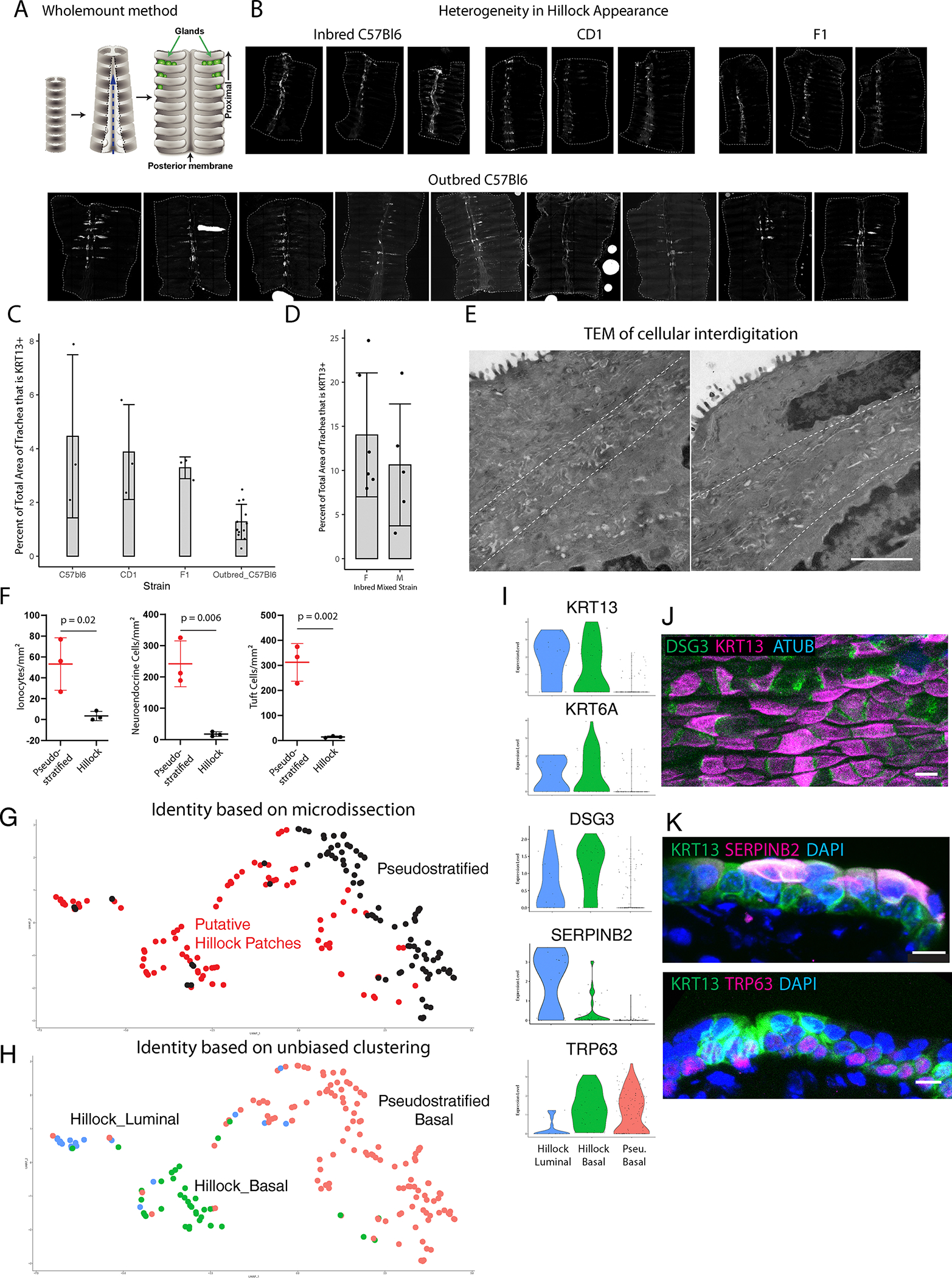
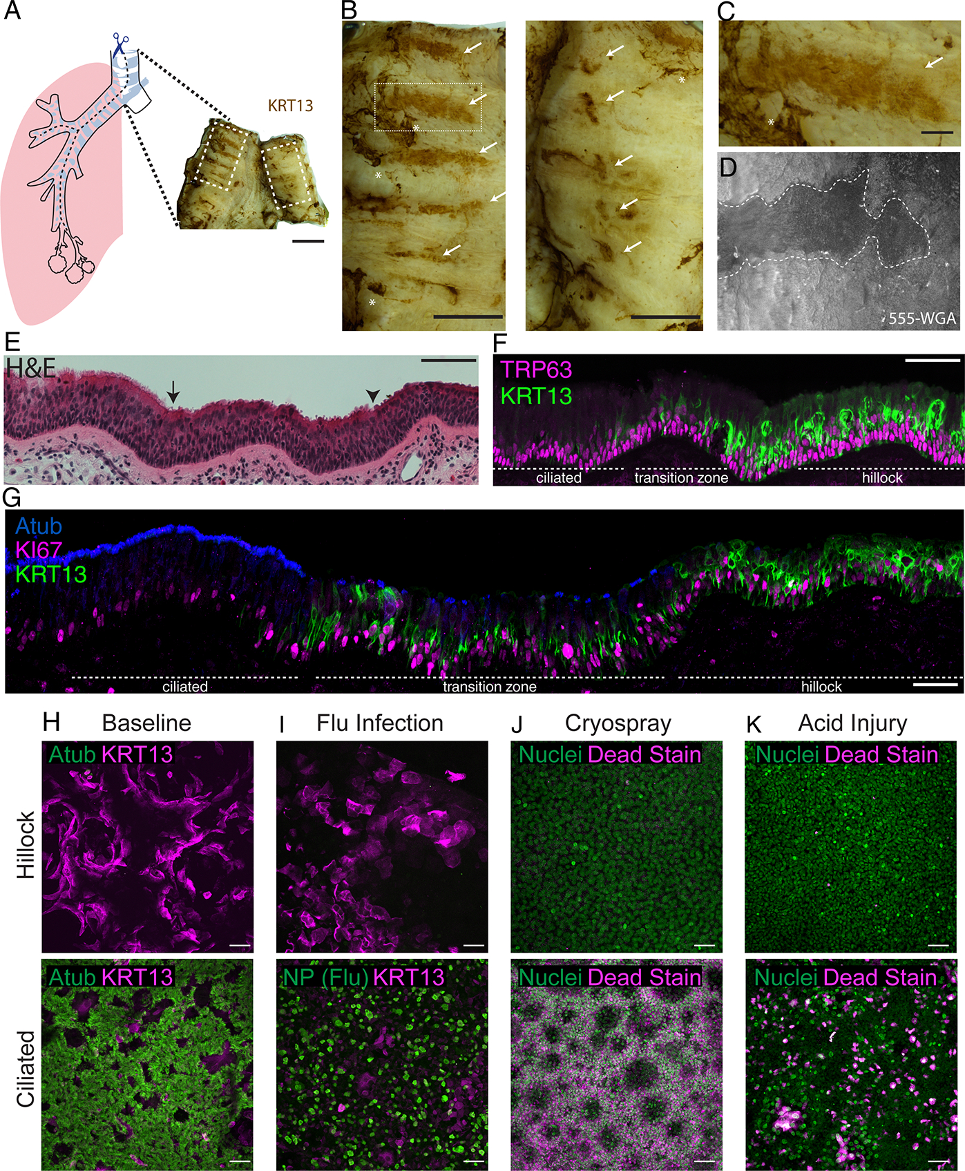
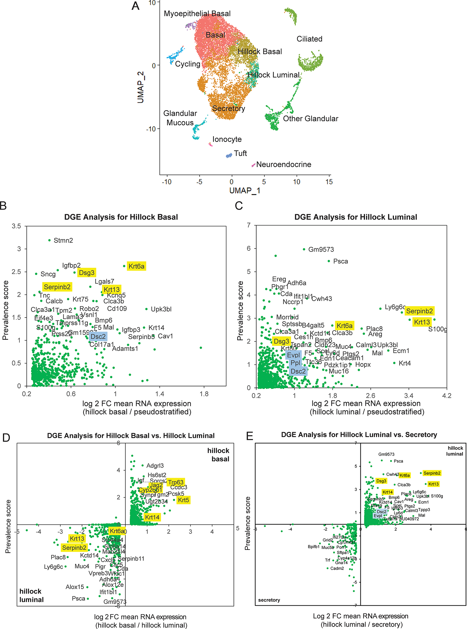
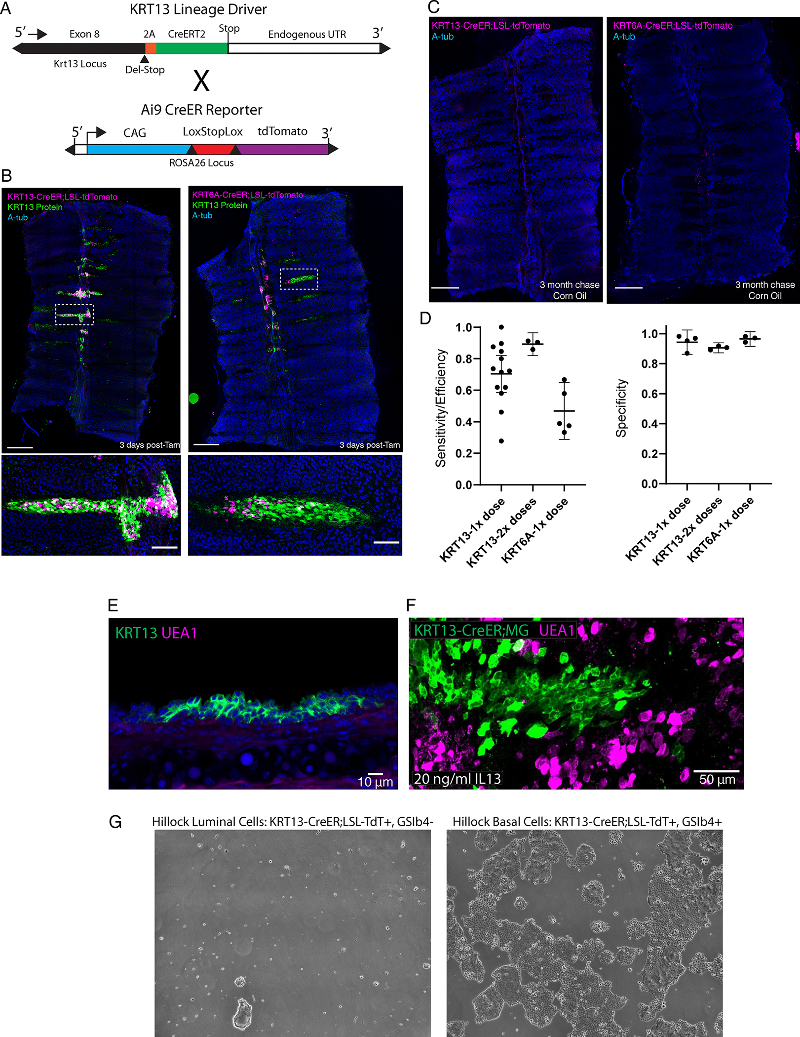
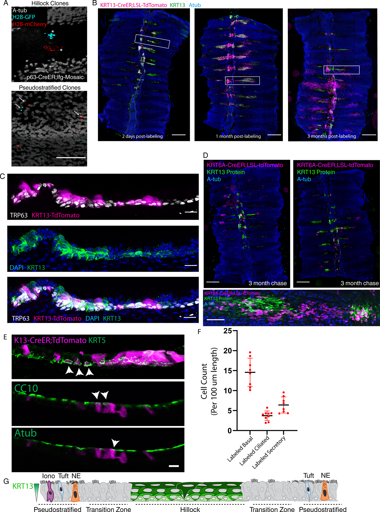
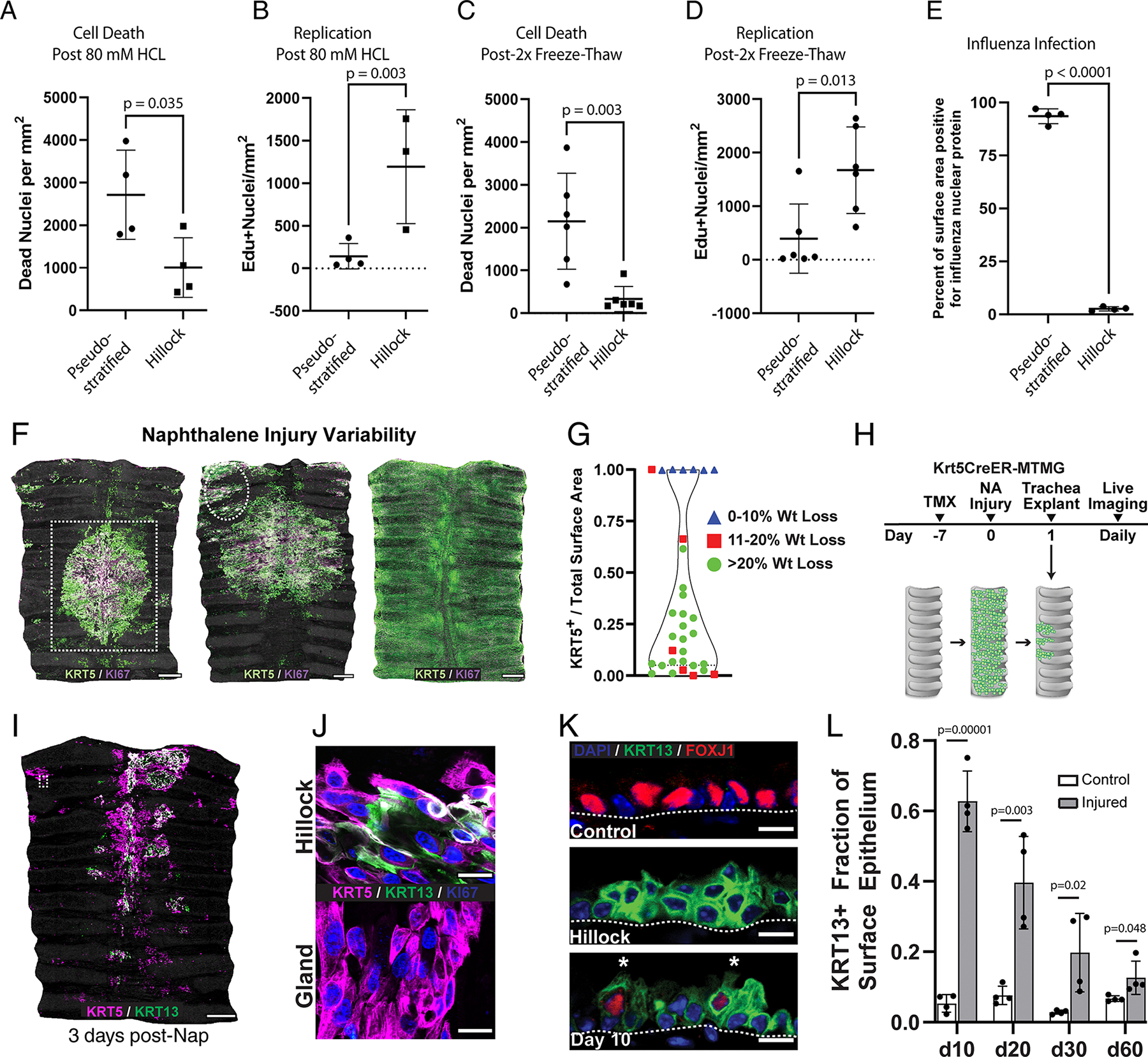
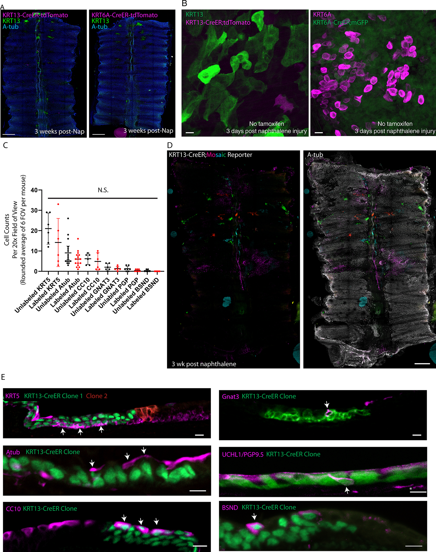
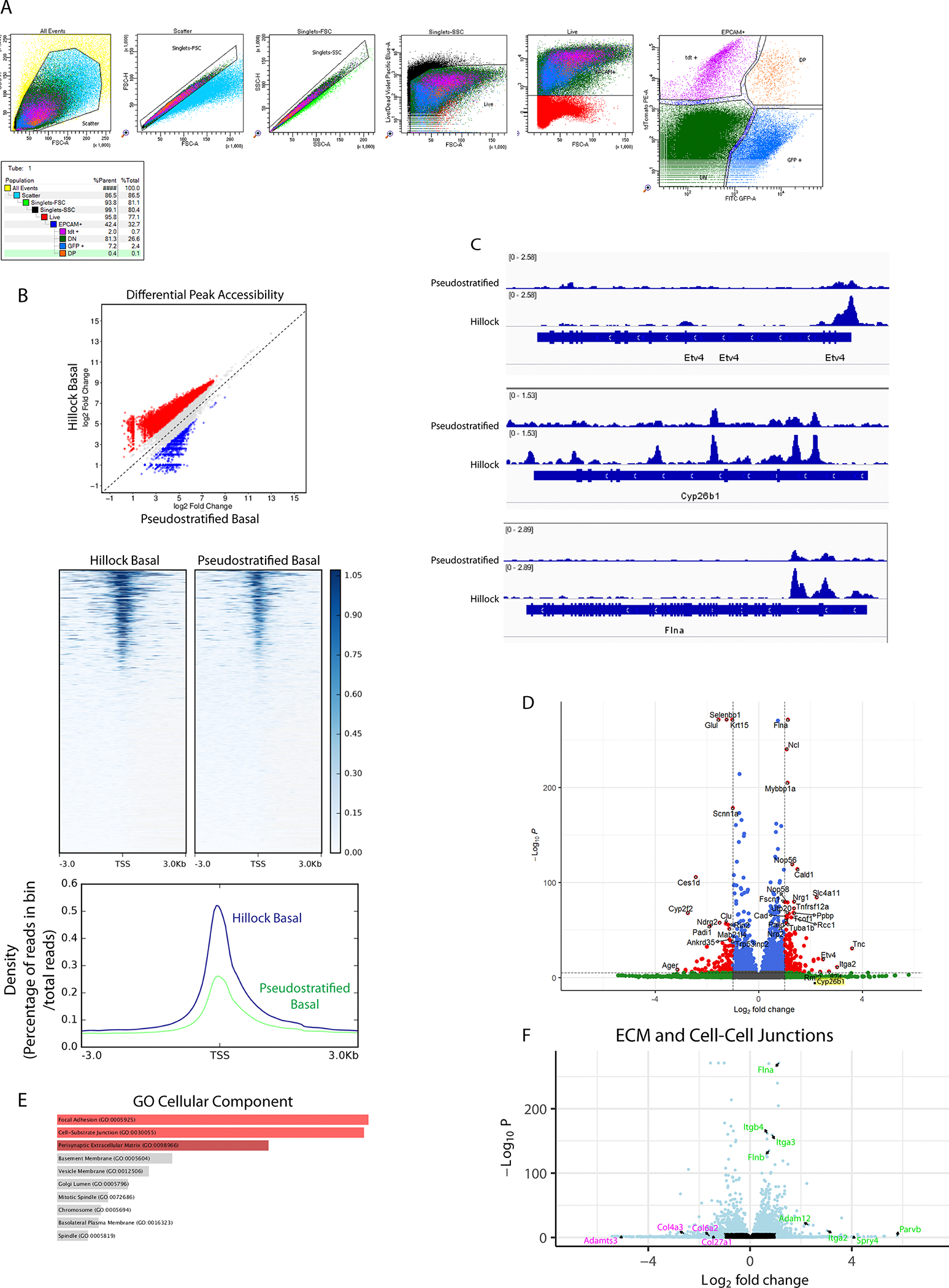
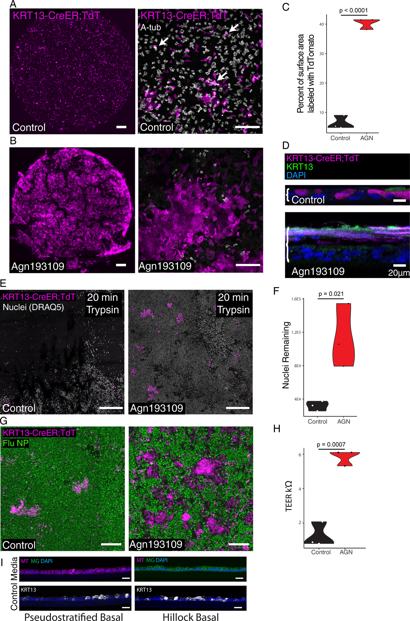
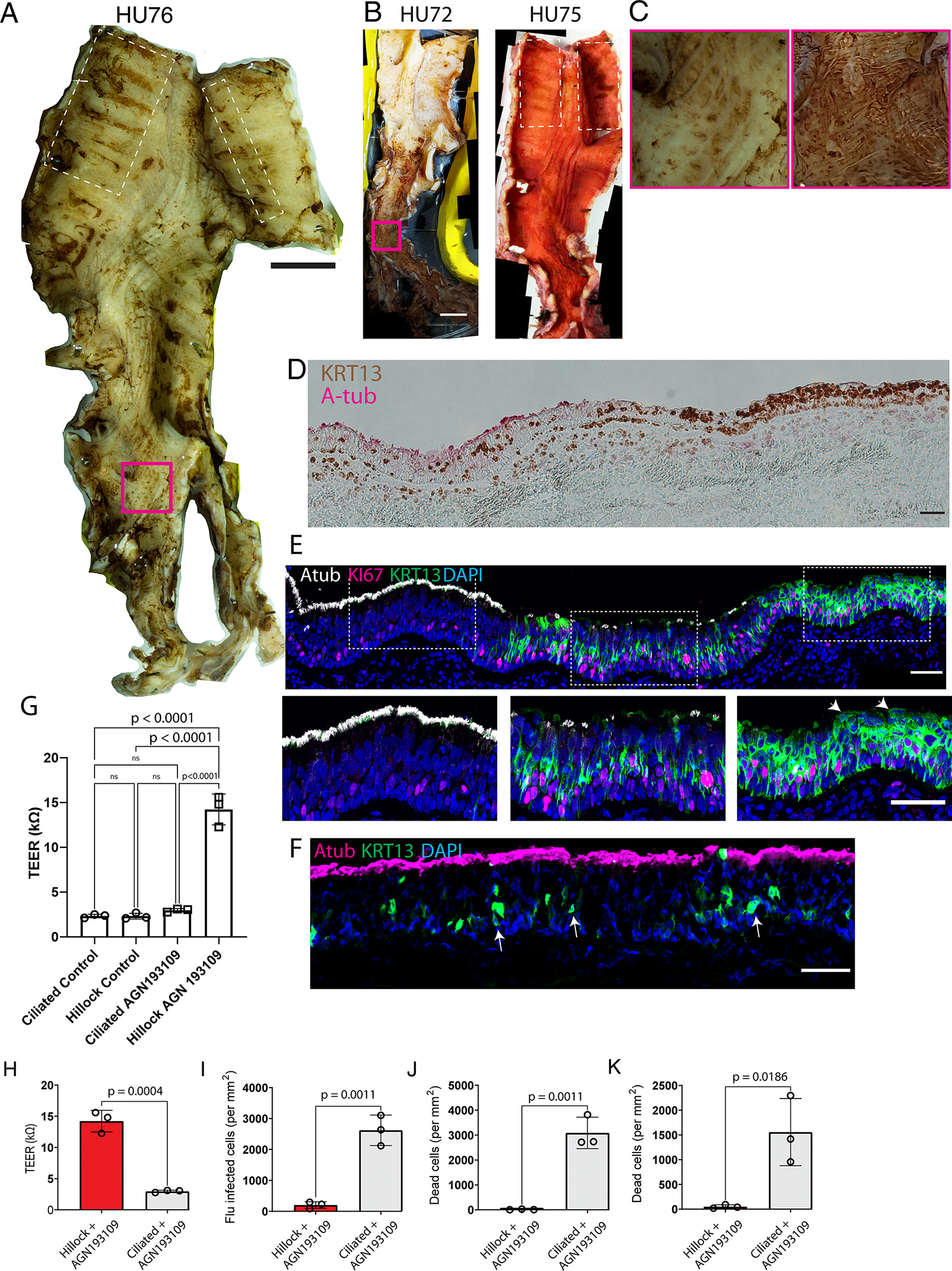
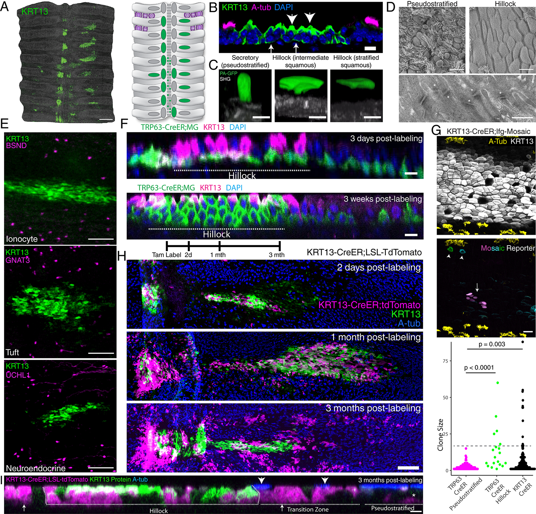
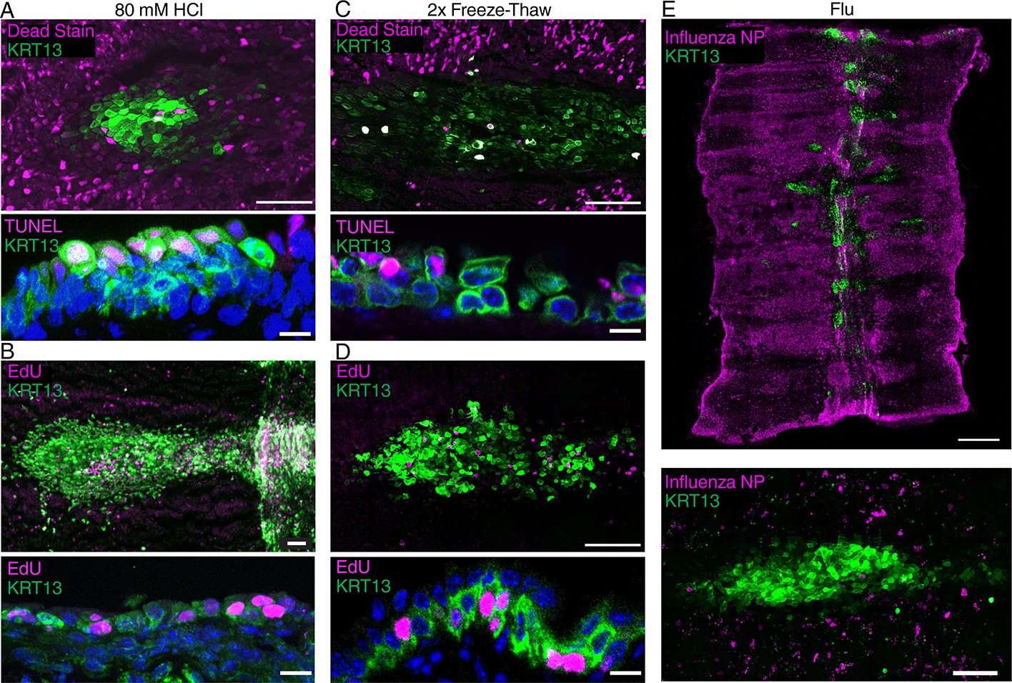
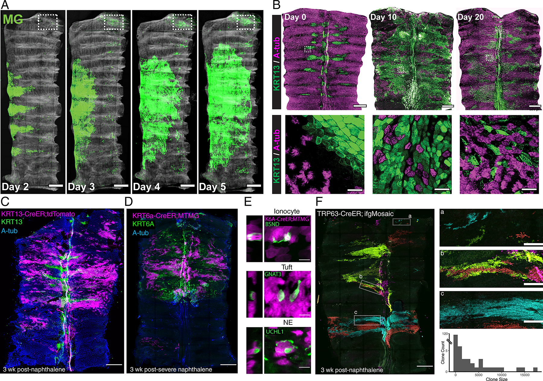
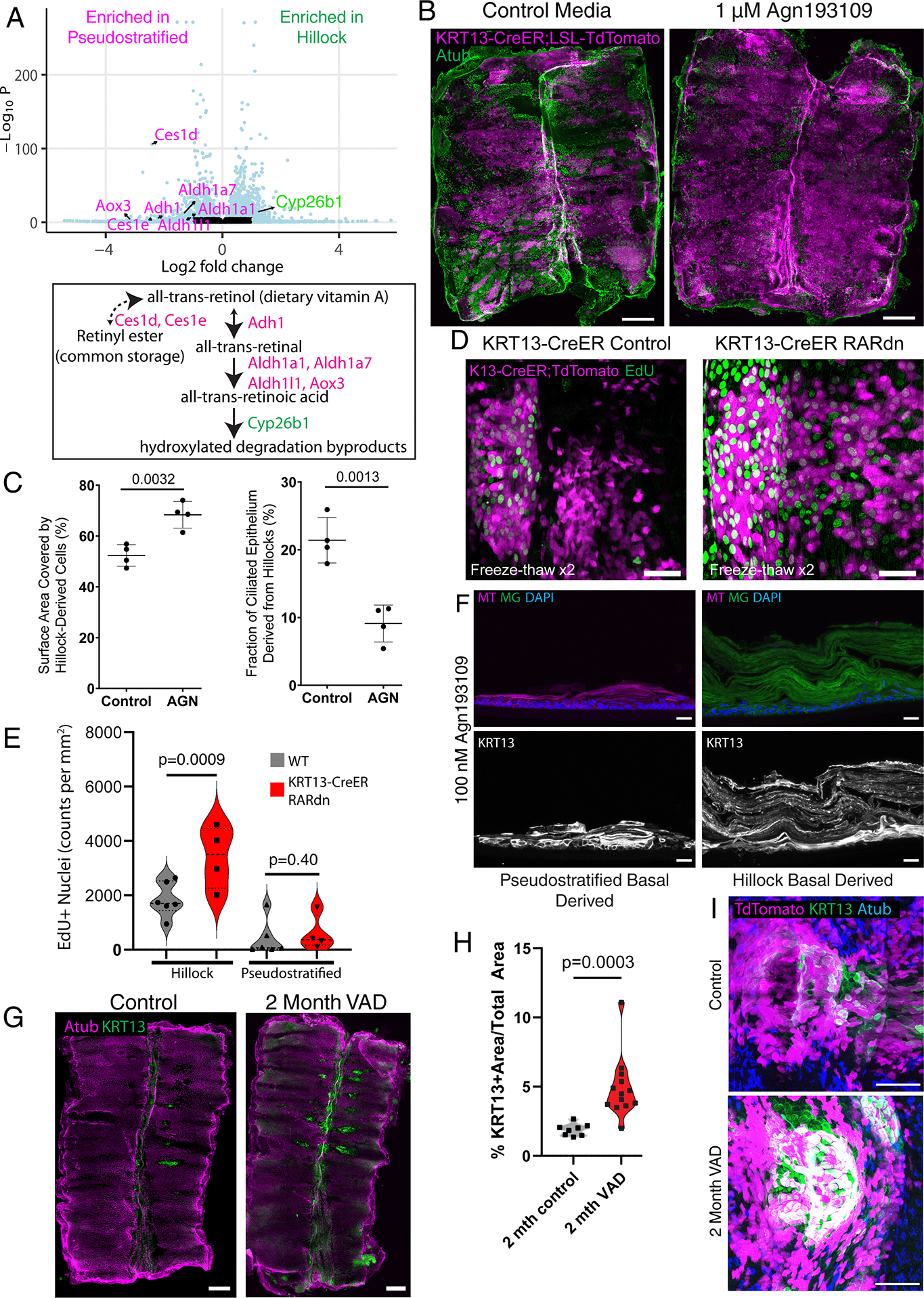
Airway hillocks are stratified epithelial structures of unknown function. Hillocks persist for months and have a unique population of basal stem cells that express genes associated with barrier function and cell adhesion. Hillock basal stem cells continually replenish overlying squamous barrier cells. They exhibit dramatically higher turnover than the abundant, largely quiescent classic pseudostratified airway epithelium. Hillocks resist a remarkably broad spectrum of injuries, including toxins, infection, acid and physical injury because hillock squamous cells shield underlying hillock basal stem cells from injury. Hillock basal stem cells are capable of massive clonal expansion that is sufficient to resurface denuded airway, and eventually regenerate normal airway epithelium with each of its six component cell types. Hillock basal stem cells preferentially stratify and keratinize in the setting of retinoic acid signalling inhibition, a known cause of squamous metaplasia. Here we show that mouse hillock expansion is the cause of vitamin A deficiency-induced squamous metaplasia. Finally, we identify human hillocks whose basal stem cells generate functional squamous barrier structures in culture. The existence of hillocks reframes our understanding of airway epithelial regeneration. Furthermore, we show that hillocks are one origin of 'squamous metaplasia', which is long thought to be a precursor of lung cancer.
气道小丘是功能未知的分层上皮结构。小丘可持续存在数月,具有独特的基底干细胞群体,这些细胞表达与屏障功能和细胞黏附相关的基因。小丘基底干细胞持续补充覆盖其上的鳞状屏障细胞。它们的更新速度比丰富的、大多处于静止状态的典型假复层气道上皮细胞快得多。小丘能抵抗范围广泛的损伤,包括毒素、感染、酸和物理损伤,因为小丘鳞状细胞可保护其下方的小丘基底干细胞免受损伤。小丘基底干细胞能够进行大量克隆扩增,足以重新覆盖剥脱的气道,并最终再生出具有六种组成细胞类型的正常气道上皮。在视黄酸信号抑制的情况下,小丘基底干细胞优先分层并角质化,视黄酸信号抑制是已知的鳞状化生原因。在这里,我们表明小鼠小丘扩增是维生素A缺乏诱导的鳞状化生的原因。最后,我们鉴定出了人类小丘,其基底干细胞在培养中可生成功能性鳞状屏障结构。小丘的存在重塑了我们对气道上皮再生的理解。此外,我们表明小丘是“鳞状化生”的一个起源,长期以来人们一直认为鳞状化生是肺癌的前兆。