• 文献检索
  • 文档翻译
  • 深度研究
  • 学术资讯
  • Suppr Zotero 插件Zotero 插件
  • 邀请有礼
  • 套餐&价格
  • 历史记录
应用&插件
Suppr Zotero 插件Zotero 插件浏览器插件Mac 客户端Windows 客户端微信小程序
定价
高级版会员购买积分包购买API积分包
服务
文献检索文档翻译深度研究API 文档MCP 服务
关于我们
关于 Suppr公司介绍联系我们用户协议隐私条款
关注我们

Suppr 超能文献

核心技术专利:CN118964589B侵权必究
粤ICP备2023148730 号-1Suppr @ 2026

文献检索

告别复杂PubMed语法,用中文像聊天一样搜索,搜遍4000万医学文献。AI智能推荐,让科研检索更轻松。

立即免费搜索

文件翻译

保留排版,准确专业,支持PDF/Word/PPT等文件格式,支持 12+语言互译。

免费翻译文档

深度研究

AI帮你快速写综述,25分钟生成高质量综述,智能提取关键信息,辅助科研写作。

立即免费体验

三维融合成像评估用于颅内动脉瘤栓塞的低轮廓可视化腔内支撑支架的贴合情况。

Three-dimensional fusion imaging to assess apposition of low-profile visualized intraluminal support stent for intracranial aneurysm coiling.

作者信息

Kato Naoki, Ishibashi Toshihiro, Otani Katharina, Abe Yukiko, Sano Tohru, Nagayama Gota, Fuga Michiyasu, Hataoka Shunsuke, Kan Issei, Murayama Yuichi

机构信息

Department of Neurosurgery, The Jikei University School of Medicine Tokyo, Tokyo, Japan.

Siemens Healthcare K.K., Tokyo, Japan.

出版信息

World Neurosurg X. 2024 Apr 21;23:100381. doi: 10.1016/j.wnsx.2024.100381. eCollection 2024 Jul.

DOI:10.1016/j.wnsx.2024.100381
PMID:38706708
原文链接:https://pmc.ncbi.nlm.nih.gov/articles/PMC11066469/
Abstract

OBJECTIVE

To investigate on three-dimensional (3D) fusion images the apposition of low-profile visualized intraluminal support (LVIS) stents in intracranial aneurysms after treatment and assess inter-rater reliability.

MATERIALS AND METHODS

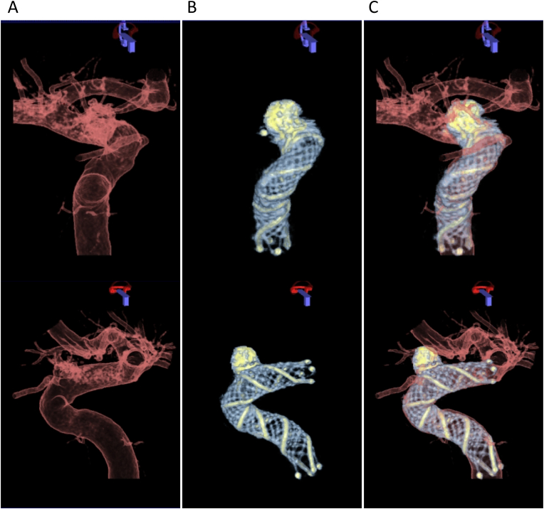
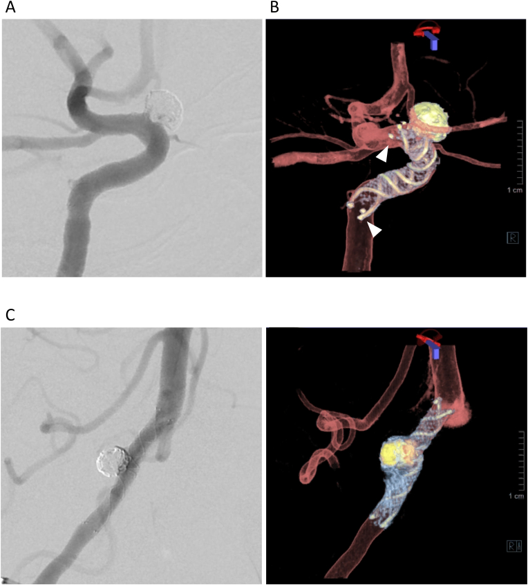
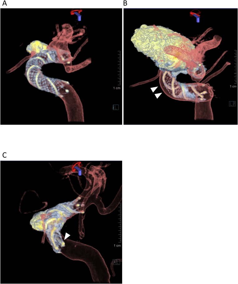
Records of all patients with unruptured intracranial aneurysms who were treated with the LVIS stent were retrospectively accessed and included in this study. Two neurosurgeons evaluated the presence of malapposition between the vessel walls and the stent trunk (crescent sign) and the vessel wall and the stent edges (edge malappostion) on 3D fusion images. These images were high-resolution cone-beam computed tomography images of the LVIS stent fused with 3D-digital subtraction angiography images of the vessels. Associations between malapposition and aneurysm location were assessed by Fisher's exact test, and inter-rater agreement was estimated using Cohen's kappa statistic.

RESULTS

Forty consecutive patients were included. In all patients, 3D fusion imaging successfully visualized the tantalum helical strands and the closed-cell structure of the nitinol material of the low-profile visualized intraluminal support. A crescent sign was observed in 27.5 % and edge malapposition in 47.5 % of the patients. Malapposition was not significantly associated with location ( = 0.23 crescent sign,  = 0.07 edge malapposition). Almost perfect ( = 0.88) and substantial ( = 0.76) agreements between the two raters were found for the detection of crescent signs and edge appositions, respectively.

CONCLUSIONS

3D fusion imaging provided clear visualization of the LVIS stent and parent arteries, and could detect malapposition with excellent inter-rater reliability. This technique may provide valuable guidance for surgeons in determining postoperative management.

摘要

目的

通过三维(3D)融合图像研究低轮廓可视化腔内支撑(LVIS)支架在颅内动脉瘤治疗后的贴合情况,并评估评分者间的可靠性。

材料与方法

回顾性查阅所有接受LVIS支架治疗的未破裂颅内动脉瘤患者的记录,并纳入本研究。两名神经外科医生在3D融合图像上评估血管壁与支架主干之间(新月征)以及血管壁与支架边缘之间(边缘贴合不良)是否存在贴合不良。这些图像是LVIS支架的高分辨率锥形束计算机断层扫描图像与血管的3D数字减影血管造影图像融合而成。通过Fisher精确检验评估贴合不良与动脉瘤位置之间的关联,并使用Cohen卡方统计量估计评分者间的一致性。

结果

连续纳入40例患者。在所有患者中,3D融合成像成功显示了低轮廓可视化腔内支撑的钽螺旋丝和镍钛诺材料的闭孔结构。27.5%的患者观察到新月征,47.5%的患者观察到边缘贴合不良。贴合不良与位置无显著相关性(新月征P = 0.23,边缘贴合不良P = 0.07)。两名评分者在检测新月征和边缘贴合方面的一致性分别为几乎完美(κ = 0.88)和高度一致(κ = 0.76)。

结论

3D融合成像能够清晰显示LVIS支架和供血动脉,并能以优异的评分者间可靠性检测到贴合不良。该技术可为外科医生确定术后管理提供有价值的指导。

https://cdn.ncbi.nlm.nih.gov/pmc/blobs/4d35/11066469/c56b27b78a1e/gr3.jpg
https://cdn.ncbi.nlm.nih.gov/pmc/blobs/4d35/11066469/e36ed1d08b48/gr1.jpg
https://cdn.ncbi.nlm.nih.gov/pmc/blobs/4d35/11066469/05c5e0acae2e/gr2.jpg
https://cdn.ncbi.nlm.nih.gov/pmc/blobs/4d35/11066469/c56b27b78a1e/gr3.jpg
https://cdn.ncbi.nlm.nih.gov/pmc/blobs/4d35/11066469/e36ed1d08b48/gr1.jpg
https://cdn.ncbi.nlm.nih.gov/pmc/blobs/4d35/11066469/05c5e0acae2e/gr2.jpg
https://cdn.ncbi.nlm.nih.gov/pmc/blobs/4d35/11066469/c56b27b78a1e/gr3.jpg

相似文献

1
Three-dimensional fusion imaging to assess apposition of low-profile visualized intraluminal support stent for intracranial aneurysm coiling.三维融合成像评估用于颅内动脉瘤栓塞的低轮廓可视化腔内支撑支架的贴合情况。
World Neurosurg X. 2024 Apr 21;23:100381. doi: 10.1016/j.wnsx.2024.100381. eCollection 2024 Jul.
2
Visualization of stent apposition after stent-assisted coiling of intracranial aneurysms using high resolution 3D fusion images acquired by C-arm CT.应用 C 臂 CT 获得的高分辨率 3D 融合图像对颅内动脉瘤支架辅助弹簧圈栓塞术后支架贴壁情况进行可视化。
J Neurointerv Surg. 2020 Feb;12(2):192-196. doi: 10.1136/neurintsurg-2019-014966. Epub 2019 Aug 12.
3
Flow Diverter Apposition in Patients with Large or Giant Intracranial Aneurysms Evaluated on Three-Dimensional Fusion Images Acquired by High-Resolution Cone-Beam Computed Tomography and Digital Subtraction Angiography.高分辨率锥形束 CT 和数字减影血管造影三维融合图像评估的大型或巨大颅内动脉瘤患者的血流导向装置贴附。
World Neurosurg. 2021 Mar;147:e388-e395. doi: 10.1016/j.wneu.2020.12.068. Epub 2021 Jan 7.
4
225 intracranial aneurysms treated with the Low-profile Visualized Intraluminal Support (LVIS) stent: a single-center retrospective study.采用低轮廓可视化腔内支撑(LVIS)支架治疗的225例颅内动脉瘤:一项单中心回顾性研究。
Neurol Res. 2018 Jun;40(6):445-451. doi: 10.1080/01616412.2018.1457608. Epub 2018 Apr 23.
5
Enterprise stents versus low-profile visualized intraluminal support stents for stent-assisted coiling of unruptured paraclinoid aneurysms.企业支架与低轮廓可视化腔内支撑支架用于未破裂床突旁动脉瘤的支架辅助弹簧圈栓塞术的比较
Technol Health Care. 2023;31(5):1855-1865. doi: 10.3233/THC-220697.
6
Hemodynamic differences by increasing low profile visualized intraluminal support (LVIS) stent local compaction across intracranial aneurysm orifice.通过增加低轮廓可视化腔内支撑(LVIS)支架在颅内动脉瘤口的局部压缩来改变血流动力学差异。
Interv Neuroradiol. 2020 Oct;26(5):557-565. doi: 10.1177/1591019920952903. Epub 2020 Aug 23.
7
Long-term outcomes of Y-stent-assisted coil embolization using Low-profile Visualized Intraluminal Support Junior (LVIS Jr) for intracranial bifurcation aneurysms.使用低剖面可视化腔内支撑支架 Junior(LVIS Jr)辅助线圈栓塞治疗颅内分叉动脉瘤的长期结果。
Clin Neurol Neurosurg. 2022 Jun;217:107275. doi: 10.1016/j.clineuro.2022.107275. Epub 2022 May 2.
8
A new low-profile visualized intraluminal support (LVIS) device, LVIS Blue: laboratory comparison between old and new LVIS.一种新型低调可视化腔内支撑(LVIS)装置,LVIS Blue:新旧LVIS的实验室比较
Neurol Res. 2018 Jan;40(1):78-85. doi: 10.1080/01616412.2017.1398881. Epub 2017 Nov 5.
9
Overlapped Stenting Combined with Coiling for Blood Blister-Like Aneurysms: Comparison of Low-Profile Visualized Intraluminal Support (LVIS) Stent and Non-LVIS Stent.重叠支架置入联合弹簧圈栓塞治疗血泡样动脉瘤:低轮廓可视化腔内支撑(LVIS)支架与非LVIS支架的比较
World Neurosurg. 2017 Aug;104:729-735. doi: 10.1016/j.wneu.2017.03.092. Epub 2017 Mar 27.
10
LVIS Jr. stent for treatment of intracranial aneurysms with parent vessel diameter of 2.5 mm or less.LVIS Jr.支架用于治疗载瘤血管直径为2.5毫米或更小的颅内动脉瘤。
Interv Neuroradiol. 2018 Jun;24(3):246-253. doi: 10.1177/1591019918759307. Epub 2018 Feb 20.

本文引用的文献

1
Multi-centric European post-market follow-up study of the Neuroform Atlas Stent System: primary results.多中心欧洲上市后随访研究Neuroform Atlas 支架系统:初步结果。
J Neurointerv Surg. 2022 Jul;14(7):694-698. doi: 10.1136/neurintsurg-2021-017849. Epub 2021 Sep 2.
2
Flow Diverter Apposition in Patients with Large or Giant Intracranial Aneurysms Evaluated on Three-Dimensional Fusion Images Acquired by High-Resolution Cone-Beam Computed Tomography and Digital Subtraction Angiography.高分辨率锥形束 CT 和数字减影血管造影三维融合图像评估的大型或巨大颅内动脉瘤患者的血流导向装置贴附。
World Neurosurg. 2021 Mar;147:e388-e395. doi: 10.1016/j.wneu.2020.12.068. Epub 2021 Jan 7.
3
Incomplete stent apposition of low-profile visualized intraluminal support stents in the treatment of cerebral aneurysms.
低轮廓可视化腔内支撑支架在治疗脑动脉瘤中支架贴壁不完整的情况。
J Neurointerv Surg. 2020 Jun;12(6):591-597. doi: 10.1136/neurintsurg-2019-015505. Epub 2019 Dec 9.
4
Combination of high-resolution cone beam computed tomography and metal artefact reduction software: a new image fusion technique for evaluating intracranial stent apposition after aneurysm treatment.高分辨率锥形束计算机断层扫描与金属伪影减少软件的结合:一种评估动脉瘤治疗后颅内支架贴壁情况的新型图像融合技术。
BMJ Case Rep. 2019 Sep 17;12(9):e230687. doi: 10.1136/bcr-2019-230687.
5
Visualization of stent apposition after stent-assisted coiling of intracranial aneurysms using high resolution 3D fusion images acquired by C-arm CT.应用 C 臂 CT 获得的高分辨率 3D 融合图像对颅内动脉瘤支架辅助弹簧圈栓塞术后支架贴壁情况进行可视化。
J Neurointerv Surg. 2020 Feb;12(2):192-196. doi: 10.1136/neurintsurg-2019-014966. Epub 2019 Aug 12.
6
Quantitative Analysis of Conebeam CT for Delineating Stents in Stent-Assisted Coil Embolization.锥形束 CT 定量分析在支架辅助线圈栓塞中对支架的描绘。
AJNR Am J Neuroradiol. 2018 Mar;39(3):488-493. doi: 10.3174/ajnr.A5533. Epub 2018 Feb 1.
7
Critical role of angiographic acquisition modality and reconstruction on morphometric and haemodynamic analysis of intracranial aneurysms.血管造影采集方式和重建对颅内动脉瘤形态和血流动力学分析的关键作用。
J Neurointerv Surg. 2018 Sep;10(9):911-915. doi: 10.1136/neurintsurg-2017-013677. Epub 2018 Jan 19.
8
Safety and effectiveness of the Low Profile Visualized Intraluminal Support (LVIS and LVIS Jr) devices in the endovascular treatment of intracranial aneurysms: results of the TRAIL multicenter observational study.颅内动脉瘤血管内治疗中使用低轮廓可视化腔内支撑装置(LVIS 和 LVIS Jr)的安全性和有效性:TRAIL 多中心观察性研究结果。
J Neurointerv Surg. 2018 Jul;10(7):675-681. doi: 10.1136/neurintsurg-2017-013375. Epub 2017 Nov 24.
9
Microguidewire Looping to Traverse Stented Parent Arteries of Intracranial Aneurysms.微导丝成袢技术穿越颅内动脉瘤载瘤动脉支架
J Korean Neurosurg Soc. 2017 Mar;60(2):262-268. doi: 10.3340/jkns.2016.0707.009. Epub 2017 Mar 1.
10
In vitro comparison of intracranial stent visibility using various concentrations of gadolinium contrast agent under 1.5 T and 3 T MR angiography.在 1.5T 和 3T MRI 血管造影下使用不同浓度钆造影剂对颅内支架可视性的体外比较。
J Neurointerv Surg. 2017 Apr;9(4):399-404. doi: 10.1136/neurintsurg-2016-012490. Epub 2016 Aug 10.