• 文献检索
  • 文档翻译
  • 深度研究
  • 学术资讯
  • Suppr Zotero 插件Zotero 插件
  • 邀请有礼
  • 套餐&价格
  • 历史记录
应用&插件
Suppr Zotero 插件Zotero 插件浏览器插件Mac 客户端Windows 客户端微信小程序
定价
高级版会员购买积分包购买API积分包
服务
文献检索文档翻译深度研究API 文档MCP 服务
关于我们
关于 Suppr公司介绍联系我们用户协议隐私条款
关注我们

Suppr 超能文献

核心技术专利:CN118964589B侵权必究
粤ICP备2023148730 号-1Suppr @ 2026

文献检索

告别复杂PubMed语法,用中文像聊天一样搜索,搜遍4000万医学文献。AI智能推荐,让科研检索更轻松。

立即免费搜索

文件翻译

保留排版,准确专业,支持PDF/Word/PPT等文件格式,支持 12+语言互译。

免费翻译文档

深度研究

AI帮你快速写综述,25分钟生成高质量综述,智能提取关键信息,辅助科研写作。

立即免费体验

大鼠胚胎性腺的组织发生

The histogenesis of the gonad in rat embryos.

作者信息

Satoh M

机构信息

Department of Obstetrics and Gynecology, Faculty of Medicine, University of Tokyo, Japan.

出版信息

J Anat. 1985 Dec;143:17-37.

PMID:3870724
原文链接:https://pmc.ncbi.nlm.nih.gov/articles/PMC1166420/
Abstract

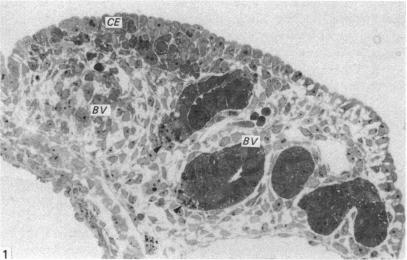
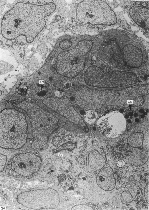
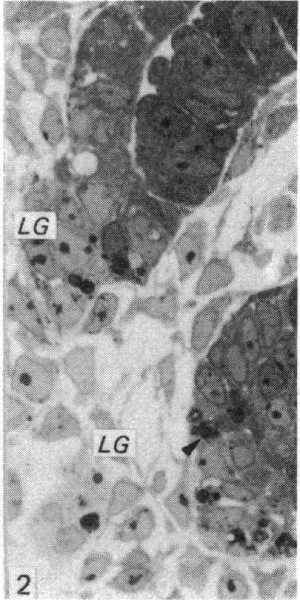
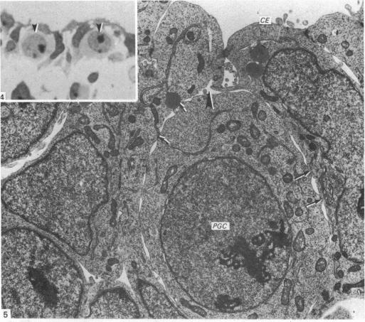
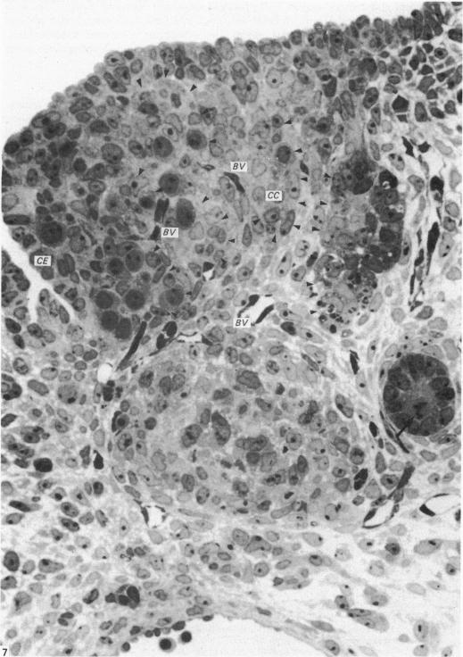
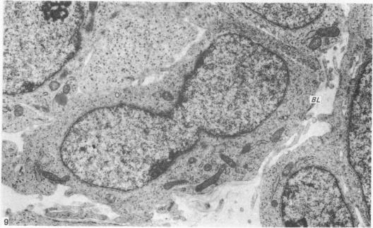
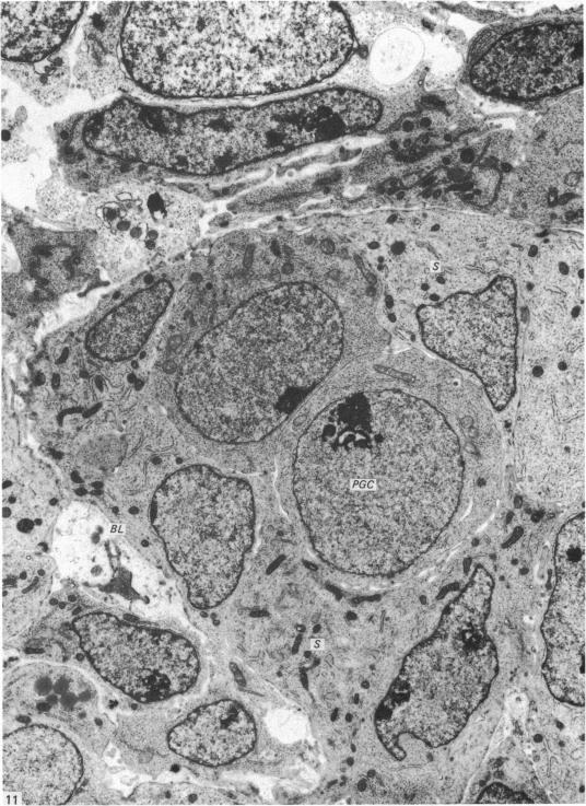
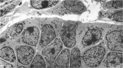
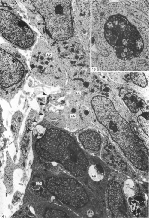
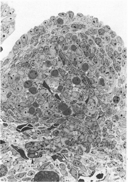
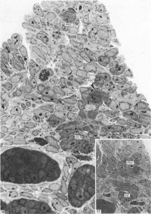
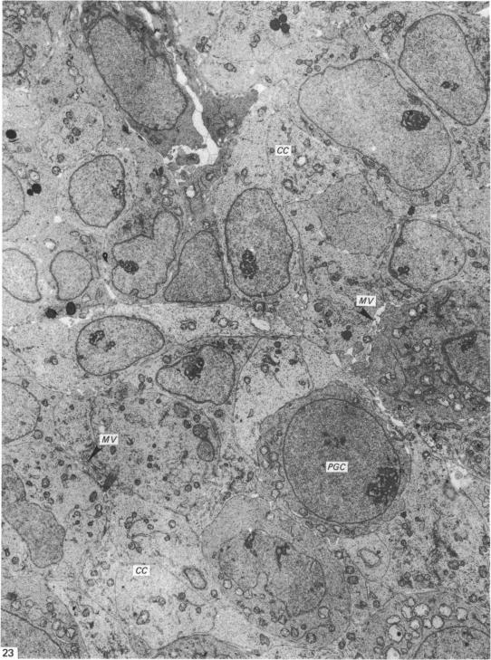
Gonadogenesis was investigated using Wistar rat embryos at 12-14 days after fertilisation. In indifferent gonads, a mesonephric tubule which bifurcates from the mesonephric duct at the upper end of the gonadal anlage ramifies into six to eight branches, the distal portions of which are contiguous, in contact with, or in close proximity to, the coelomic epithelium of the gonadal ridge. After the primordial germ cells reach the gonadal ridge, the overlying epithelium proliferates and clear cells appear in the distal portion of each mesonephric tubule, proliferating and forming cord-like structures. The primordial germ cells appear to enter these cell cords by an amoeboid type of movement. The basal lamina covering the cell cord partially disappears near the germ cells. The germ cells within the cord migrate toward the proximal portion of the cell cord and proliferate in great profusion. From the present observations, it can be concluded that the gonad is mainly formed of clear cell cords originating in mesonephric tubules into which germ cells have entered. The original mesenchymal cells and blood vessels form the interstitial tissue of the gonad. The rete testis and rete ovarii are of mesonephric tubule origin.

摘要

在受精后12至14天,使用Wistar大鼠胚胎研究性腺发生。在未分化性腺中,一条中肾管在性腺原基上端从中肾管分支出来,分成六到八个分支,其远端部分相邻、接触或紧邻性腺嵴的体腔上皮。原始生殖细胞到达性腺嵴后,覆盖其上的上皮细胞增殖,每个中肾管远端出现透明细胞,这些透明细胞增殖并形成索状结构。原始生殖细胞似乎通过阿米巴样运动进入这些细胞索。覆盖细胞索的基膜在生殖细胞附近部分消失。细胞索内的生殖细胞向细胞索近端迁移并大量增殖。从目前的观察结果可以得出结论,性腺主要由起源于中肾管的透明细胞索组成,生殖细胞已进入这些中肾管。原始间充质细胞和血管形成性腺的间质组织。睾丸网和卵巢网起源于中肾管。

https://cdn.ncbi.nlm.nih.gov/pmc/blobs/49c8/1166420/aab7efc98afe/janat00190-0040-a.jpg
https://cdn.ncbi.nlm.nih.gov/pmc/blobs/49c8/1166420/a9523b1f0676/janat00190-0026-a.jpg
https://cdn.ncbi.nlm.nih.gov/pmc/blobs/49c8/1166420/54a5bb1aa1bb/janat00190-0026-b.jpg
https://cdn.ncbi.nlm.nih.gov/pmc/blobs/49c8/1166420/f620ff49610e/janat00190-0026-c.jpg
https://cdn.ncbi.nlm.nih.gov/pmc/blobs/49c8/1166420/a5203cb9c5bd/janat00190-0027-a.jpg
https://cdn.ncbi.nlm.nih.gov/pmc/blobs/49c8/1166420/e5981dd3c7b6/janat00190-0027-b.jpg
https://cdn.ncbi.nlm.nih.gov/pmc/blobs/49c8/1166420/6fef713f0e58/janat00190-0029-a.jpg
https://cdn.ncbi.nlm.nih.gov/pmc/blobs/49c8/1166420/f5580c8f0c73/janat00190-0030-a.jpg
https://cdn.ncbi.nlm.nih.gov/pmc/blobs/49c8/1166420/657d0be3696a/janat00190-0030-b.jpg
https://cdn.ncbi.nlm.nih.gov/pmc/blobs/49c8/1166420/5ce14ed3f668/janat00190-0031-a.jpg
https://cdn.ncbi.nlm.nih.gov/pmc/blobs/49c8/1166420/cde3a54e159d/janat00190-0032-a.jpg
https://cdn.ncbi.nlm.nih.gov/pmc/blobs/49c8/1166420/df1c559cb94e/janat00190-0033-a.jpg
https://cdn.ncbi.nlm.nih.gov/pmc/blobs/49c8/1166420/d0ebde4890c1/janat00190-0033-b.jpg
https://cdn.ncbi.nlm.nih.gov/pmc/blobs/49c8/1166420/70fa375a820b/janat00190-0034-a.jpg
https://cdn.ncbi.nlm.nih.gov/pmc/blobs/49c8/1166420/f00c2d8a574c/janat00190-0035-a.jpg
https://cdn.ncbi.nlm.nih.gov/pmc/blobs/49c8/1166420/0b5e22fbc8e5/janat00190-0036-a.jpg
https://cdn.ncbi.nlm.nih.gov/pmc/blobs/49c8/1166420/006e24076f5b/janat00190-0038-a.jpg
https://cdn.ncbi.nlm.nih.gov/pmc/blobs/49c8/1166420/2884bc9ab4e5/janat00190-0038-b.jpg
https://cdn.ncbi.nlm.nih.gov/pmc/blobs/49c8/1166420/870d38909fd4/janat00190-0038-c.jpg
https://cdn.ncbi.nlm.nih.gov/pmc/blobs/49c8/1166420/7385b009619f/janat00190-0038-d.jpg
https://cdn.ncbi.nlm.nih.gov/pmc/blobs/49c8/1166420/93a743fb7eb4/janat00190-0039-a.jpg
https://cdn.ncbi.nlm.nih.gov/pmc/blobs/49c8/1166420/aab7efc98afe/janat00190-0040-a.jpg

相似文献

1
The histogenesis of the gonad in rat embryos.大鼠胚胎性腺的组织发生
J Anat. 1985 Dec;143:17-37.
2
Histogenesis and organogenesis of the gonad in human embryos.人类胚胎性腺的组织发生和器官发生
J Anat. 1991 Aug;177:85-107.
3
Ultrastructure of the early ovary and testis in pig embryos.猪胚胎早期卵巢和睾丸的超微结构
Am J Anat. 1975 Sep;144(1):89-111. doi: 10.1002/aja.1001440106.
4
Sertoli cells of the mouse testis originate from the coelomic epithelium.小鼠睾丸的支持细胞起源于体腔上皮。
Dev Biol. 1998 Nov 15;203(2):323-33. doi: 10.1006/dbio.1998.9068.
5
Ultrastructure of fetal human gonad before sexual differentiation and during early testicular and ovarian development.性分化前及早期睾丸和卵巢发育期间的人类胎儿性腺超微结构
J Submicrosc Cytol Pathol. 1990 Jul;22(3):389-400.
6
Testicular differentiation in mammals under normal and experimental conditions.正常及实验条件下哺乳动物的睾丸分化
J Electron Microsc Tech. 1991 Oct;19(2):158-71. doi: 10.1002/jemt.1060190204.
7
Expression of type I and III collagen during morphogenesis of fetal rat testis and ovary.I型和III型胶原蛋白在胎鼠睾丸和卵巢形态发生过程中的表达。
Anat Rec. 1987 Sep;219(1):91-101. doi: 10.1002/ar.1092190115.
8
The genus Acipenser as a model for vertebrate urogenital development: ultrastructure of nephrostomial tubule formation and of initial gonadogenesis.
Ann Anat. 2002 Sep;184(5):443-54. doi: 10.1016/S0940-9602(02)80077-2.
9
Germ cell depletion does not alter the morphogenesis of the fetal testis or ovary in the red-eared slider turtle (Trachemys scripta).生殖细胞耗竭不会改变红耳龟(滑龟)胎儿睾丸或卵巢的形态发生。
J Exp Zool B Mol Dev Evol. 2007 May 15;308(3):236-41. doi: 10.1002/jez.b.21136.
10
Primordial germ cells and gonadal development in the golden hamster.金黄仓鼠的原始生殖细胞与性腺发育
J Anat. 1989 Jun;164:155-63.

引用本文的文献

1
Integration of mouse ovary morphogenesis with developmental dynamics of the oviduct, ovarian ligaments, and rete ovarii.将小鼠卵巢形态发生与输卵管、卵巢韧带和卵巢固有韧带的发育动力学相结合。
Elife. 2022 Sep 27;11:e81088. doi: 10.7554/eLife.81088.
2
Concerted morphogenesis of genital ridges and nephric ducts in the mouse captured through whole-embryo imaging.通过胚胎整体成像捕捉到的小鼠生殖器嵴和肾管的协同形态发生。
Development. 2021 Sep 15;148(18). doi: 10.1242/dev.199208. Epub 2021 May 18.
3
On cell contribution to gonadal soma formation in quail-chick chimeras during the indifferent stage of gonadal development.

本文引用的文献

1
The use of lead citrate at high pH as an electron-opaque stain in electron microscopy.在电子显微镜检查中,将高pH值的柠檬酸铅用作电子不透明染色剂。
J Cell Biol. 1963 Apr;17(1):208-12. doi: 10.1083/jcb.17.1.208.
2
Development of the human ovary--a study using histochemical technics.
Obstet Gynecol. 1961 Aug;18:152-81.
3
The presence of a common embryonic blastema for ovarian and testicular parenchymal (follicular, interstitial and tubular) cells in cattle Bos taurus.
Z Zellforsch Mikrosk Anat. 1966;74(4):505-28. doi: 10.1007/BF00496841.
4
关于性腺发育中性未分化阶段鹌鹑-鸡嵌合体中细胞对性腺体细胞形成的贡献。
Anat Embryol (Berl). 1989;179(3):237-42. doi: 10.1007/BF00326588.
4
Origin of somatic cells and histogenesis in the primordial gonad of the Japanese tree frog Rhacophorus arboreus.日本树蛙(Rhacophorus arboreus)原始性腺中体细胞的起源和组织发生
Anat Embryol (Berl). 1989;180(2):165-73. doi: 10.1007/BF00309768.
5
Primordial germ cells and gonadal development in the golden hamster.金黄仓鼠的原始生殖细胞与性腺发育
J Anat. 1989 Jun;164:155-63.
6
Histogenesis and organogenesis of the gonad in human embryos.人类胚胎性腺的组织发生和器官发生
J Anat. 1991 Aug;177:85-107.
Follicle formation in the immature mouse ovary: the role of the rete ovarii.未成熟小鼠卵巢中的卵泡形成:卵巢网的作用。
J Anat. 1973 Nov;116(Pt 2):207-17.
5
Ultrastructure of the indifferent gonad in male and female pig embryos.
Tissue Cell. 1976;8(1):163-74. doi: 10.1016/0040-8166(76)90028-8.
6
Rat gonadal and ovarioan organogenesis with and without germ cells. An ultrastructural study.
Dev Biol. 1975 May;44(1):1-21. doi: 10.1016/0012-1606(75)90372-3.