• 文献检索
  • 文档翻译
  • 深度研究
  • 学术资讯
  • Suppr Zotero 插件Zotero 插件
  • 邀请有礼
  • 套餐&价格
  • 历史记录
应用&插件
Suppr Zotero 插件Zotero 插件浏览器插件Mac 客户端Windows 客户端微信小程序
定价
高级版会员购买积分包购买API积分包
服务
文献检索文档翻译深度研究API 文档MCP 服务
关于我们
关于 Suppr公司介绍联系我们用户协议隐私条款
关注我们

Suppr 超能文献

核心技术专利:CN118964589B侵权必究
粤ICP备2023148730 号-1Suppr @ 2026

文献检索

告别复杂PubMed语法,用中文像聊天一样搜索,搜遍4000万医学文献。AI智能推荐,让科研检索更轻松。

立即免费搜索

文件翻译

保留排版,准确专业,支持PDF/Word/PPT等文件格式,支持 12+语言互译。

免费翻译文档

深度研究

AI帮你快速写综述,25分钟生成高质量综述,智能提取关键信息,辅助科研写作。

立即免费体验

人类胚胎性腺的组织发生和器官发生

Histogenesis and organogenesis of the gonad in human embryos.

作者信息

Satoh M

机构信息

Department of Obstetrics and Gynecology, Faculty of Medicine, University of Tokyo, Japan.

出版信息

J Anat. 1991 Aug;177:85-107.

PMID:1769902
原文链接:https://pmc.ncbi.nlm.nih.gov/articles/PMC1260417/
Abstract

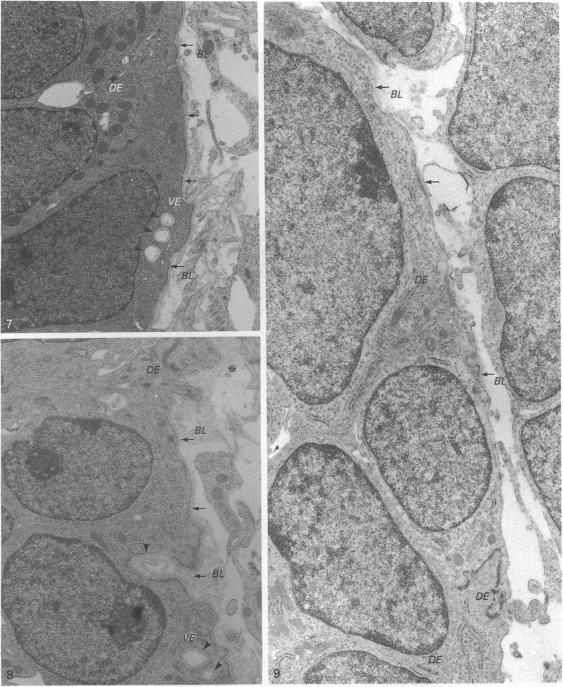
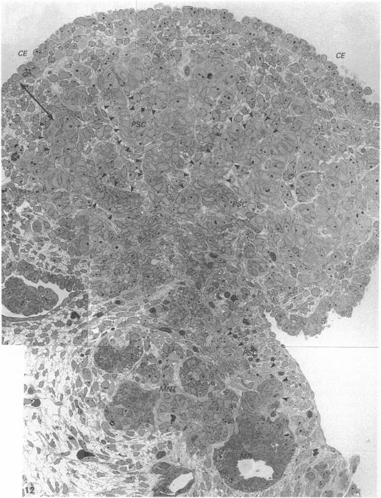
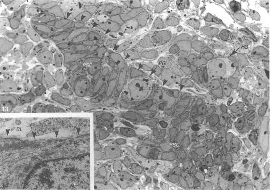
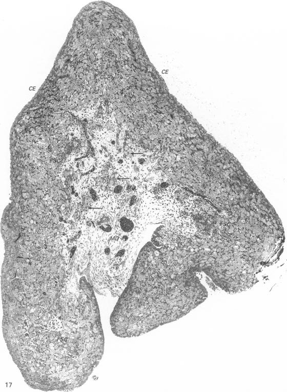
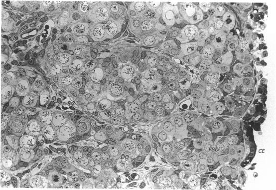
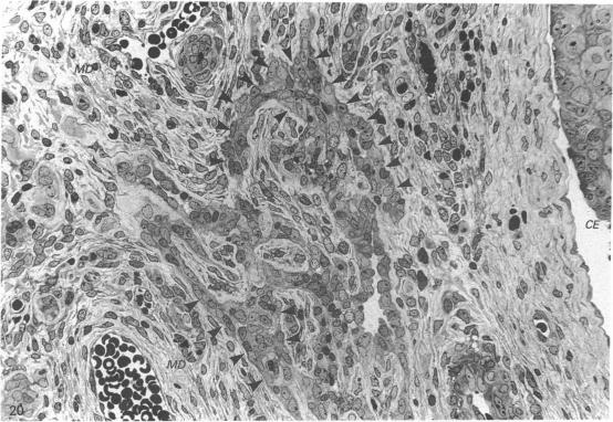
The histogenesis and organogenesis of the human gonad in 12 embryos and 6 fetuses of ovulational ages 5 to 18 weeks was investigated by histological and ultrastructural examination, including observation of almost complete serial Epon-embedded sections of entire gonads of 10 embryos. This investigation revealed that the main constituent cells of the gonads are derived from the mesonephros, and that the coelomic epithelium is not involved in the formation of the main component at any stage. With the migration of the primordial germ cells into the gonadal ridge, the coelomic epithelium becomes stratified to form a moderate protrusion of the gonad into the coelomic cavity and the coelomic epithelial cells develop into short pillars which form cord-like structures, the so-called primary sex cords. Shortly afterwards, concomitantly with the development into the subsequent prominent protrusion of the gonad into the coelomic cavity, cells emerging from the mesonephros are incorporated into the gonad to form 'primordial sex cords'. At this stage, a stratified, pile-like arrangement of coelomic epithelium flattens into monolaminar or oligolaminar structures. In the testis, the 'primordial sex cords' differentiate into seminiferous sex cords by elaborating a surrounding basal lamina. In the ovary, these 'primordial sex cords' become displaced towards the peripheral regions of the gonad by the enlargement of these cords, as well as by the formation of the interstitium, or so-called medulla, at the base of the ovary; they differentiate into 'folliculogenous sex cords' which give rise to follicular cells.

摘要

通过组织学和超微结构检查,对12例胚龄为5至18周的胚胎及6例胎儿的人性腺进行了组织发生和器官发生的研究,包括观察10例胚胎整个性腺几乎完整的系列环氧树脂包埋切片。该研究表明,性腺的主要组成细胞来源于中肾,体腔上皮在任何阶段均不参与主要成分的形成。随着原始生殖细胞迁移至性腺嵴,体腔上皮分层,形成性腺向体腔的适度突出,体腔上皮细胞发育成短柱状,形成索状结构,即所谓的初级性索。此后不久,伴随着性腺向体腔后续突出的发育,从中肾出现的细胞并入性腺形成“原始性索”。在此阶段,分层的、堆积状排列的体腔上皮扁平为单层或多层结构。在睾丸中,“原始性索”通过形成周围的基膜分化为生精性索。在卵巢中,这些“原始性索”因这些索的增大以及卵巢底部间质或所谓髓质的形成而向性腺周边区域移位;它们分化为产生卵泡细胞的“卵泡源性性索”。

https://cdn.ncbi.nlm.nih.gov/pmc/blobs/88ff/1260417/3f8dbf7d189f/janat00155-0108-a.jpg
https://cdn.ncbi.nlm.nih.gov/pmc/blobs/88ff/1260417/21f90dc8b705/janat00155-0093-a.jpg
https://cdn.ncbi.nlm.nih.gov/pmc/blobs/88ff/1260417/301923c53e90/janat00155-0094-a.jpg
https://cdn.ncbi.nlm.nih.gov/pmc/blobs/88ff/1260417/32d1307bbc33/janat00155-0094-b.jpg
https://cdn.ncbi.nlm.nih.gov/pmc/blobs/88ff/1260417/b700980a2e51/janat00155-0096-a.jpg
https://cdn.ncbi.nlm.nih.gov/pmc/blobs/88ff/1260417/067c74ec1425/janat00155-0096-b.jpg
https://cdn.ncbi.nlm.nih.gov/pmc/blobs/88ff/1260417/96dd76f40f14/janat00155-0097-a.jpg
https://cdn.ncbi.nlm.nih.gov/pmc/blobs/88ff/1260417/597de481b816/janat00155-0098-a.jpg
https://cdn.ncbi.nlm.nih.gov/pmc/blobs/88ff/1260417/785f4f8a11b9/janat00155-0099-a.jpg
https://cdn.ncbi.nlm.nih.gov/pmc/blobs/88ff/1260417/93da1544535c/janat00155-0100-a.jpg
https://cdn.ncbi.nlm.nih.gov/pmc/blobs/88ff/1260417/f149e7eca968/janat00155-0101-a.jpg
https://cdn.ncbi.nlm.nih.gov/pmc/blobs/88ff/1260417/4d2c20a2f2cc/janat00155-0102-a.jpg
https://cdn.ncbi.nlm.nih.gov/pmc/blobs/88ff/1260417/553b6b1e5918/janat00155-0103-a.jpg
https://cdn.ncbi.nlm.nih.gov/pmc/blobs/88ff/1260417/3ce469bb0602/janat00155-0104-a.jpg
https://cdn.ncbi.nlm.nih.gov/pmc/blobs/88ff/1260417/2cb127ebe374/janat00155-0105-a.jpg
https://cdn.ncbi.nlm.nih.gov/pmc/blobs/88ff/1260417/2e2c8de3cdf8/janat00155-0105-b.jpg
https://cdn.ncbi.nlm.nih.gov/pmc/blobs/88ff/1260417/3f8dbf7d189f/janat00155-0108-a.jpg

相似文献

1
Histogenesis and organogenesis of the gonad in human embryos.人类胚胎性腺的组织发生和器官发生
J Anat. 1991 Aug;177:85-107.
2
Sertoli cells of the mouse testis originate from the coelomic epithelium.小鼠睾丸的支持细胞起源于体腔上皮。
Dev Biol. 1998 Nov 15;203(2):323-33. doi: 10.1006/dbio.1998.9068.
3
[Histologic study of the gonads in ovarian differentiation during organogenesis].[器官发生过程中卵巢分化期性腺的组织学研究]
Arch Anat Cytol Pathol. 1989;37(5-6):201-7.
4
Ultrastructure of the early ovary and testis in pig embryos.猪胚胎早期卵巢和睾丸的超微结构
Am J Anat. 1975 Sep;144(1):89-111. doi: 10.1002/aja.1001440106.
5
The histogenesis of the gonad in rat embryos.大鼠胚胎性腺的组织发生
J Anat. 1985 Dec;143:17-37.
6
Germ cell depletion does not alter the morphogenesis of the fetal testis or ovary in the red-eared slider turtle (Trachemys scripta).生殖细胞耗竭不会改变红耳龟(滑龟)胎儿睾丸或卵巢的形态发生。
J Exp Zool B Mol Dev Evol. 2007 May 15;308(3):236-41. doi: 10.1002/jez.b.21136.
7
Testicular differentiation in mammals under normal and experimental conditions.正常及实验条件下哺乳动物的睾丸分化
J Electron Microsc Tech. 1991 Oct;19(2):158-71. doi: 10.1002/jemt.1060190204.
8
Ultrastructure of fetal human gonad before sexual differentiation and during early testicular and ovarian development.性分化前及早期睾丸和卵巢发育期间的人类胎儿性腺超微结构
J Submicrosc Cytol Pathol. 1990 Jul;22(3):389-400.
9
[Coelomic gonadal epithelia during the period of human embryonic morphologic sex differentiation].
Arkh Anat Gistol Embriol. 1979 Jun;76(6):84-92.
10
Ultrastructural events in horse gonadal morphogenesis.马性腺形态发生中的超微结构事件。
J Reprod Fertil Suppl. 1979(27):479-85.

引用本文的文献

1
Construction of human pluripotent stem cell-derived testicular organoids and their use as humanized testis models for evaluating the effects of semaglutide.人多能干细胞来源的睾丸类器官的构建及其作为评估司美格鲁肽效果的人源化睾丸模型的应用。
Theranostics. 2025 Jan 27;15(6):2597-2623. doi: 10.7150/thno.104523. eCollection 2025.
2
Relevance of Molecular Pathology for the Diagnosis of Sex Cord-Stromal Tumors of the Ovary: A Narrative Review.分子病理学在卵巢性索间质肿瘤诊断中的相关性:一篇叙述性综述
Cancers (Basel). 2023 Dec 15;15(24):5864. doi: 10.3390/cancers15245864.
3
CRISPR/Cas9-mediated activation of NR5A1 steers female human embryonic stem cell-derived bipotential gonadal-like cells towards a steroidogenic cell fate.

本文引用的文献

1
The use of lead citrate at high pH as an electron-opaque stain in electron microscopy.在电子显微镜检查中,将高pH值的柠檬酸铅用作电子不透明染色剂。
J Cell Biol. 1963 Apr;17(1):208-12. doi: 10.1083/jcb.17.1.208.
2
Development of the human ovary--a study using histochemical technics.
Obstet Gynecol. 1961 Aug;18:152-81.
3
A comparative study of the development of the fetal testis and ovary in the monkey (Macaca fascicularis).
Reprod Nutr Dev (1980). 1980;20(5A):1439-59. doi: 10.1051/rnd:19800804.
4
CRISPR/Cas9 介导的 NR5A1 激活将人胚胎干细胞来源的双潜能性腺样细胞向类固醇生成细胞命运定向。
J Ovarian Res. 2023 Sep 20;16(1):194. doi: 10.1186/s13048-023-01264-5.
4
Single-cell analysis of the developing human ovary defines distinct insights into ovarian somatic and germline progenitors.单细胞分析人类卵巢发育过程,深入了解卵巢体细胞和生殖细胞祖细胞。
Dev Cell. 2023 Oct 23;58(20):2097-2111.e3. doi: 10.1016/j.devcel.2023.07.014. Epub 2023 Aug 14.
5
Notch signaling sculpts the stem cell niche.Notch信号通路塑造干细胞微环境。
Front Cell Dev Biol. 2022 Dec 20;10:1027222. doi: 10.3389/fcell.2022.1027222. eCollection 2022.
6
Modeling Human Gonad Development in Organoids.类器官中人类性腺发育的建模。
Tissue Eng Regen Med. 2022 Dec;19(6):1185-1206. doi: 10.1007/s13770-022-00492-y. Epub 2022 Nov 9.
7
Comparative anatomy on the development of sperm transporting pathway between the testis and mesonephros.睾丸与中肾之间精子运输途径发育的比较解剖学
Histochem Cell Biol. 2022 Mar;157(3):321-332. doi: 10.1007/s00418-021-02057-x. Epub 2022 Jan 6.
8
A rare case of polyorchidism in a 40-year-old man. A case report.一名40岁男性患多睾症的罕见病例。病例报告。
Ann Med Surg (Lond). 2021 May 12;66:102389. doi: 10.1016/j.amsu.2021.102389. eCollection 2021 Jun.
9
Concerted morphogenesis of genital ridges and nephric ducts in the mouse captured through whole-embryo imaging.通过胚胎整体成像捕捉到的小鼠生殖器嵴和肾管的协同形态发生。
Development. 2021 Sep 15;148(18). doi: 10.1242/dev.199208. Epub 2021 May 18.
10
An In Vitro Differentiation Protocol for Human Embryonic Bipotential Gonad and Testis Cell Development.人类胚胎双向性性腺和睾丸细胞体外分化方案。
Stem Cell Reports. 2020 Dec 8;15(6):1377-1391. doi: 10.1016/j.stemcr.2020.10.009. Epub 2020 Nov 19.
Development of the early human ovary and role of the mesonephros in the differentiation of the cortex.早期人类卵巢的发育及中肾在皮质分化中的作用。
Anat Embryol (Berl). 1982;165(2):253-80. doi: 10.1007/BF00305481.
5
Follicle formation in the immature mouse ovary: the role of the rete ovarii.未成熟小鼠卵巢中的卵泡形成:卵巢网的作用。
J Anat. 1973 Nov;116(Pt 2):207-17.
6
The histogenesis of the gonad in rat embryos.大鼠胚胎性腺的组织发生
J Anat. 1985 Dec;143:17-37.
7
The development of the sexually indifferent gonad in the prosimian, Galago crassicaudatus crassicaudatus.原猴类粗尾婴猴(Galago crassicaudatus crassicaudatus)中性腺未分化期的发育
Am J Anat. 1988 Jan;181(1):89-105. doi: 10.1002/aja.1001810110.
8
Ultrastructure of the indifferent gonad in male and female pig embryos.
Tissue Cell. 1976;8(1):163-74. doi: 10.1016/0040-8166(76)90028-8.