From the Department of Physics, School of Natural Sciences (J.T., M.S., T.D., F.P., D.P., F.S.), Munich Institute of Biomedical Engineering (J.T., M.S., T.D., T.L., F.P., D.P., F.S.), Department of Diagnostic and Interventional Radiology, School of Medicine, Klinikum rechts der Isar (J.T., M.S., T.D., F.P., D.P.), Institute for Advanced Study (J.T., F.P., D.P.), and Computational Imaging and Inverse Problems, Department of Computer Science, School of Computation, Information, and Technology (T.L.), Technical University of Munich, Boltzmannstrasse 11, 85748 Garching, Germany.
Radiol Artif Intell. 2024 Jul;6(4):e230275. doi: 10.1148/ryai.230275.
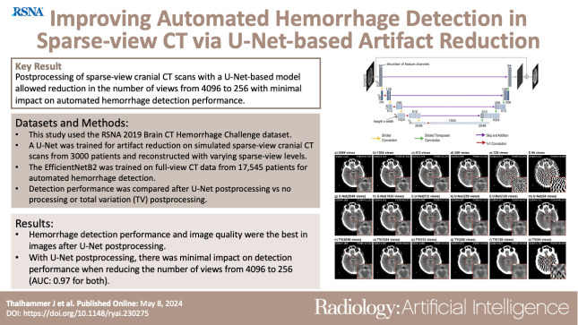
Purpose To explore the potential benefits of deep learning-based artifact reduction in sparse-view cranial CT scans and its impact on automated hemorrhage detection. Materials and Methods In this retrospective study, a U-Net was trained for artifact reduction on simulated sparse-view cranial CT scans in 3000 patients, obtained from a public dataset and reconstructed with varying sparse-view levels. Additionally, EfficientNet-B2 was trained on full-view CT data from 17 545 patients for automated hemorrhage detection. Detection performance was evaluated using the area under the receiver operating characteristic curve (AUC), with differences assessed using the DeLong test, along with confusion matrices. A total variation (TV) postprocessing approach, commonly applied to sparse-view CT, served as the basis for comparison. A Bonferroni-corrected significance level of .001/6 = .00017 was used to accommodate for multiple hypotheses testing. Results Images with U-Net postprocessing were better than unprocessed and TV-processed images with respect to image quality and automated hemorrhage detection. With U-Net postprocessing, the number of views could be reduced from 4096 (AUC: 0.97 [95% CI: 0.97, 0.98]) to 512 (0.97 [95% CI: 0.97, 0.98], < .00017) and to 256 views (0.97 [95% CI: 0.96, 0.97], < .00017) with a minimal decrease in hemorrhage detection performance. This was accompanied by mean structural similarity index measure increases of 0.0210 (95% CI: 0.0210, 0.0211) and 0.0560 (95% CI: 0.0559, 0.0560) relative to unprocessed images. Conclusion U-Net-based artifact reduction substantially enhanced automated hemorrhage detection in sparse-view cranial CT scans. CT, Head/Neck, Hemorrhage, Diagnosis, Supervised Learning © RSNA, 2024.
目的 探讨基于深度学习的稀疏视野颅脑 CT 扫描伪影减少技术的潜在优势及其对自动出血检测的影响。
材料与方法 本回顾性研究在一个公共数据集的 3000 例患者中模拟稀疏视野颅脑 CT 扫描,使用 U-Net 对伪影进行减少处理,并使用不同稀疏视图水平进行重建。此外,使用 17545 例患者的全视野 CT 数据训练 EfficientNet-B2 进行自动出血检测。使用受试者工作特征曲线下面积(AUC)评估检测性能,使用 DeLong 检验评估差异,并使用混淆矩阵。总变差(TV)后处理方法是稀疏视图 CT 的常用方法,作为比较的基础。使用 Bonferroni 校正的.001/6 =.00017 显著性水平来容纳多个假设检验。
结果 与未处理和 TV 处理图像相比,使用 U-Net 后处理的图像在图像质量和自动出血检测方面都更好。使用 U-Net 后处理,视图数量可从 4096 减少到 512(AUC:0.97 [95%CI:0.97,0.98], <.00017)和 256 视图(0.97 [95%CI:0.96,0.97], <.00017),而出血检测性能的轻微下降。这伴随着平均结构相似性指数测量值的增加,相对于未处理图像分别增加了 0.0210(95%CI:0.0210,0.0211)和 0.0560(95%CI:0.0559,0.0560)。
结论 U-Net 基于伪影减少技术可显著提高稀疏视野颅脑 CT 扫描的自动出血检测性能。