Becker Taylor, Struble Roger D, Rappaport Charles
Department of Internal Medicine, University of Iowa, C123 GH, 200 Hawkins Dr., Iowa City, IA, 52242, USA.
Department of Internal Medicine, Division of Pulmonary, Critical Care, and Occupational Medicine, University of Iowa, Iowa City, USA.
Ultrasound J. 2024 May 8;16(1):27. doi: 10.1186/s13089-024-00357-6.
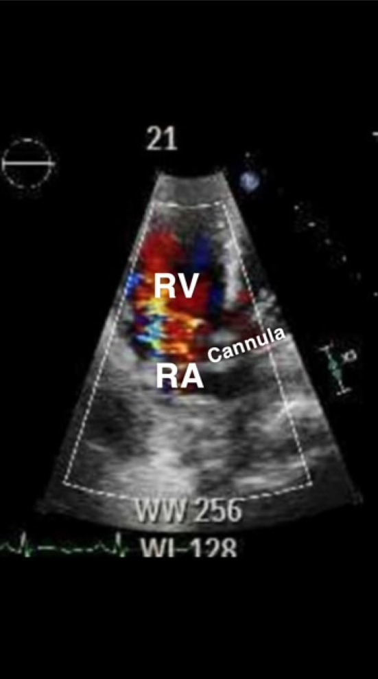
Point-of-care ultrasound (POCUS) has become a mainstay in the evaluation of critically ill patients in the intensive care unit (ICU). ECMO patients are susceptible to complications during prolonged ICU stay, including cannula malposition, which has deleterious consequences. Although the literature surrounding utility of ultrasound on ECMO patients is expansive, direct comparison between radiographic imaging versus ultrasound for identification of cannula malposition is lacking.
The authors identified four patients with cannula malposition discovered through POCUS that was missed on routine radiographic imaging. Identification and correction of malposition changed their ECMO course.
This case series is the first in literature demonstrating that ultrasound may be superior to radiographic images for ECMO cannula malposition. Further investigation into this subject is warranted.
床旁超声(POCUS)已成为重症监护病房(ICU)中危重症患者评估的主要手段。体外膜肺氧合(ECMO)患者在ICU长期住院期间易发生并发症,包括插管位置不当,这会产生有害后果。尽管关于超声在ECMO患者中的应用的文献很多,但缺乏影像学检查与超声在识别插管位置不当方面的直接比较。
作者发现4例通过POCUS发现插管位置不当的患者,而常规影像学检查未发现。识别并纠正位置不当改变了他们的ECMO治疗过程。
该病例系列是文献中首个证明超声在识别ECMO插管位置不当方面可能优于影像学检查的。对此主题进行进一步研究是有必要的。