Department of Obstetrics and Gynecology, Madigan Army Medical Center, Tacoma, WA, United States of America.
Department of Obstetrics and Gynecology, Division of Gynecologic Oncology, Baylor College of Medicine, Houston, TX, United States of America.
PLoS One. 2024 May 10;19(5):e0292978. doi: 10.1371/journal.pone.0292978. eCollection 2024.
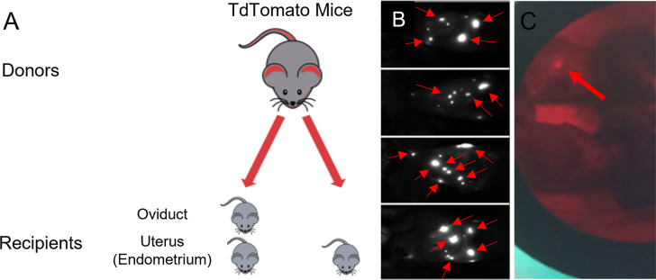
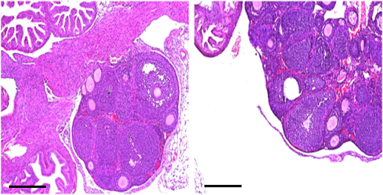
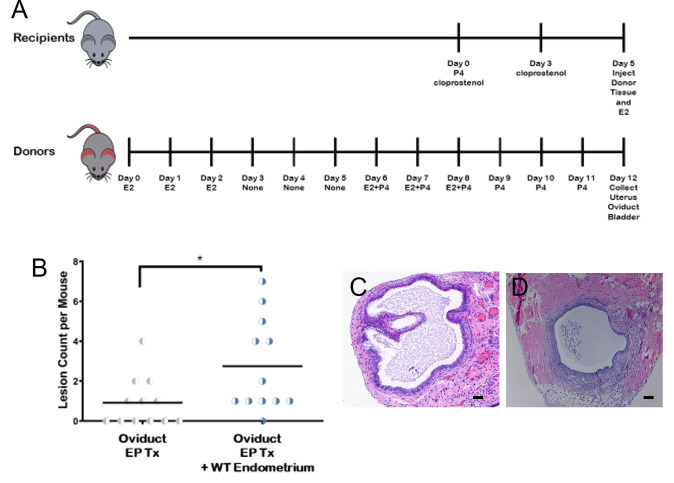
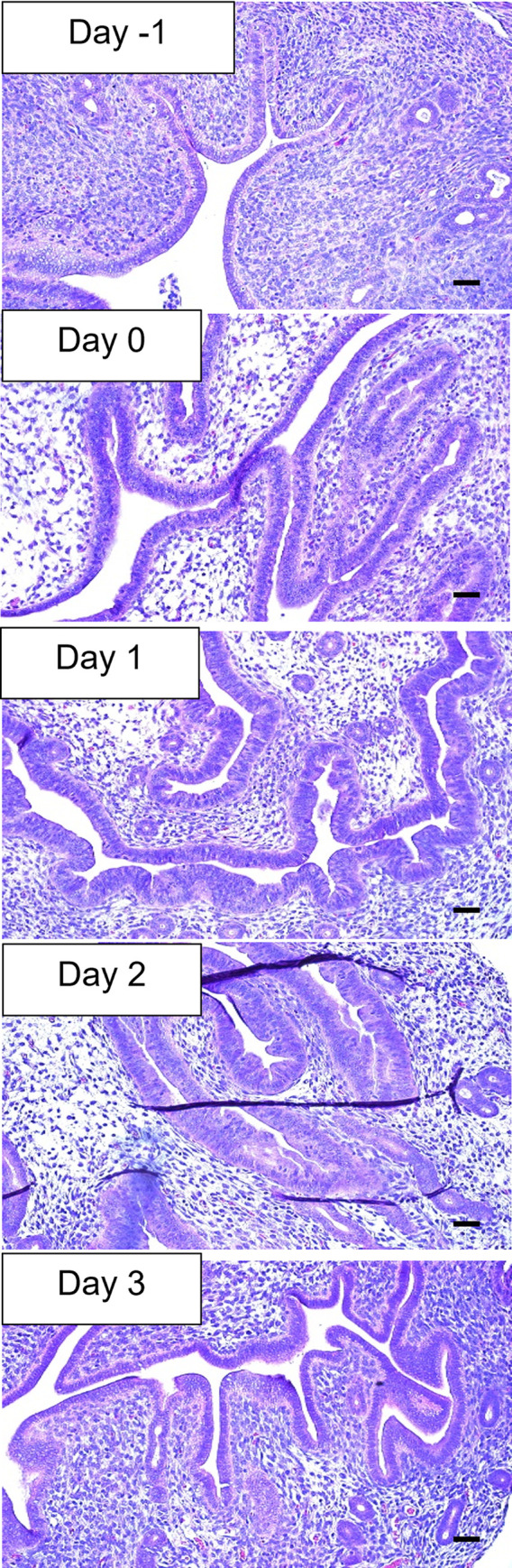
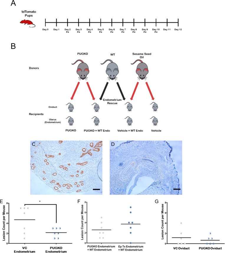
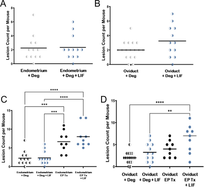
Endosalpingiosis (ES) and endometriosis (EM) refer to the growth of tubal and endometrial epithelium respectively, outside of their site of origin. We hypothesize that uterine secretome factors drive ectopic growth. To test this, we developed a mouse model of ES and EM using tdTomato (tdT) transgenic fluorescent mice as donors. To block implantation factors, progesterone knockout (PKO) tdT mice were created. Fluorescent lesions were present after oviduct implantation with and without WT endometrium. Implantation was increased (p<0.05) when tdt oviductal tissue was implanted with endometrium compared to oviductal tissue alone. Implantation was reduced (p<0.0005) in animals implanted with minced tdT oviductal tissue with PKO tdT endometrium compared to WT endometrium. Finally, oviductal tissues was incubated with and without a known implantation factor, leukemia inhibitory factor (LIF) prior to and during implantation. LIF promoted lesion implantation. In conclusion, endometrial derived implantation factors, such as LIF, are necessary to initiate ectopic tissue growth. We have developed an animal model of ectopic growth of gynecologic tissues in a WT mouse which will potentially allow for development of new prevention and treatment modalities.
内移性输卵管上皮内瘤(ES)和子宫内膜异位症(EM)分别指输卵管和子宫内膜上皮在其起源部位以外的生长。我们假设子宫分泌因子驱动异位生长。为了验证这一点,我们使用 tdTomato(tdT)转基因荧光小鼠作为供体开发了 ES 和 EM 的小鼠模型。为了阻断着床因子,创建了孕酮敲除(PKO)tdT 小鼠。在有无 WT 子宫内膜的情况下,输卵管植入后均出现荧光病变。与仅植入输卵管组织相比,当将 tdt 输卵管组织与子宫内膜一起植入时,植入增加(p<0.05)。与植入 WT 子宫内膜相比,植入 PKO tdT 子宫内膜的切碎 tdT 输卵管组织的动物的植入减少(p<0.0005)。最后,在植入前和植入期间,将输卵管组织与和不与已知的着床因子白血病抑制因子(LIF)孵育。LIF 促进了病变的植入。总之,子宫内膜来源的着床因子,如 LIF,对于启动异位组织生长是必要的。我们已经开发出一种在 WT 小鼠中异位生长妇科组织的动物模型,这可能为开发新的预防和治疗方法提供了可能。