• 文献检索
  • 文档翻译
  • 深度研究
  • 学术资讯
  • Suppr Zotero 插件Zotero 插件
  • 邀请有礼
  • 套餐&价格
  • 历史记录
应用&插件
Suppr Zotero 插件Zotero 插件浏览器插件Mac 客户端Windows 客户端微信小程序
定价
高级版会员购买积分包购买API积分包
服务
文献检索文档翻译深度研究API 文档MCP 服务
关于我们
关于 Suppr公司介绍联系我们用户协议隐私条款
关注我们

Suppr 超能文献

核心技术专利:CN118964589B侵权必究
粤ICP备2023148730 号-1Suppr @ 2026

文献检索

告别复杂PubMed语法,用中文像聊天一样搜索,搜遍4000万医学文献。AI智能推荐,让科研检索更轻松。

立即免费搜索

文件翻译

保留排版,准确专业,支持PDF/Word/PPT等文件格式,支持 12+语言互译。

免费翻译文档

深度研究

AI帮你快速写综述,25分钟生成高质量综述,智能提取关键信息,辅助科研写作。

立即免费体验

3.0T CMR 中主动心脏植入物引起的图像失真的三维评估。

Three-dimensional assessment of image distortion induced by active cardiac implants in 3.0T CMR.

机构信息

Department of Internal Medicine I, Cardiology, University Hospital Wuerzburg, Oberduerbacher Strasse 6a, 97080, Wuerzburg, Germany.

German Heart Center Munich, Electrophysiology, Munich, Germany.

出版信息

Sci Rep. 2024 May 15;14(1):11130. doi: 10.1038/s41598-024-61283-0.

DOI:10.1038/s41598-024-61283-0
PMID:38750100
原文链接:https://pmc.ncbi.nlm.nih.gov/articles/PMC11096309/
Abstract

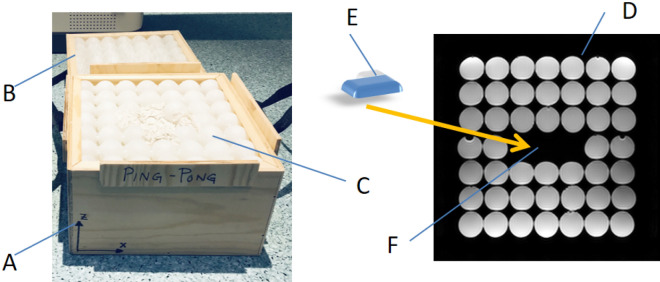
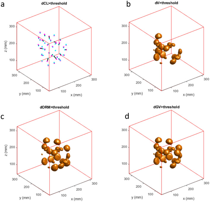
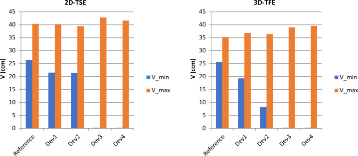
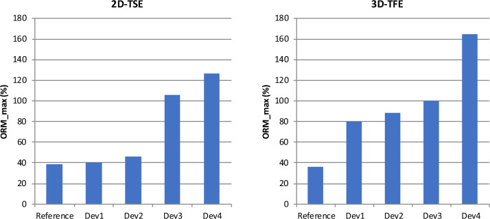
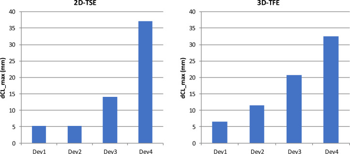
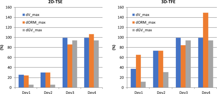
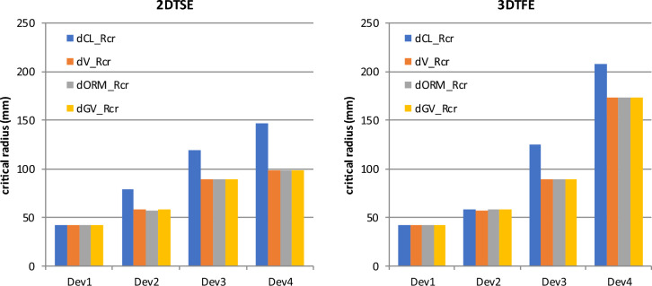
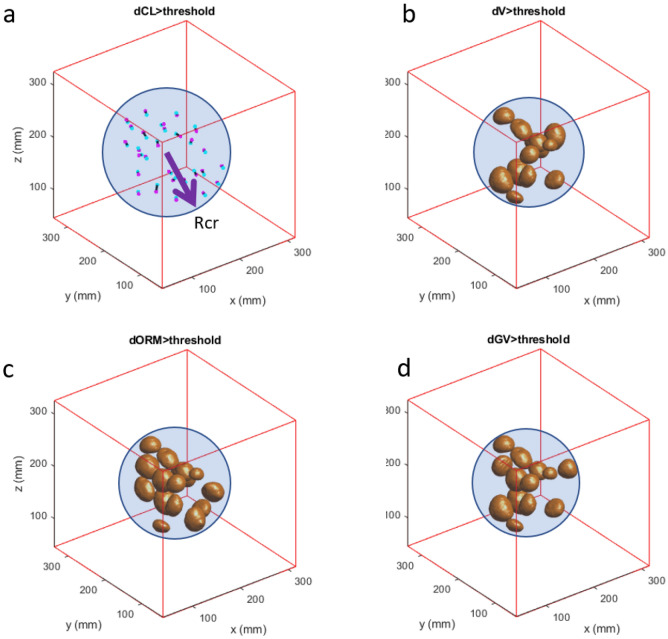
CMR at 3.0T in the presence of active cardiac implants remains a challenge due to susceptibility artifacts. Beyond a signal void that cancels image information, magnetic field inhomogeneities may cause distorted appearances of anatomical structures. Understanding influencing factors and the extent of distortion are a first step towards optimizing the image quality of CMR with active implants at 3.0T. All measurements were obtained at a clinical 3.0T scanner. An in-house designed phantom with a 3D cartesian grid of water filled spheres was used to analyze the distortion caused by four representative active cardiac devices (cardiac loop recorder, pacemaker, 2 ICDs). For imaging a gradient echo (3D-TFE) sequence and a turbo spin echo (2D-TSE) sequence were used. The work defines metrics to quantify the different features of distortion such as changes in size, location and signal intensity. It introduces a specialized segmentation technique based on a reaction-diffusion-equation. The distortion features are dependent on the amount of magnetic material in the active implants and showed a significant increase when measured with the 3D TFE compared to the 2D TSE. This work presents a quantitative approach for the evaluation of image distortion at 3.0T caused by active cardiac implants and serves as foundation for both further optimization of sequences and devices but also for planning of imaging procedures.

摘要

在存在心脏活性植入物的情况下,由于磁化率伪影,3.0T 的心脏磁共振成像仍然是一个挑战。除了信号缺失导致图像信息丢失之外,磁场不均匀性可能会导致解剖结构的外观失真。了解影响因素和失真程度是优化 3.0T 心脏活性植入物磁共振图像质量的第一步。所有测量均在临床 3.0T 扫描仪上进行。使用内部设计的带有三维笛卡尔网格充满水的球体的体模来分析四个代表性的心脏活性设备(心脏环记录仪、起搏器、2 个 ICD)引起的失真。对于成像,使用梯度回波(3D-TFE)序列和涡轮自旋回波(2D-TSE)序列。该工作定义了用于量化失真的不同特征的指标,例如大小、位置和信号强度的变化。它介绍了一种基于反应扩散方程的专门分割技术。失真特征取决于心脏活性植入物中的磁性材料的量,并且与 2D-TSE 相比,使用 3D-TFE 测量时会显著增加。该工作提出了一种用于评估 3.0T 心脏活性植入物引起的图像失真的定量方法,既可以为序列和设备的进一步优化提供基础,也可以为成像程序的规划提供基础。

https://cdn.ncbi.nlm.nih.gov/pmc/blobs/d7e3/11096309/ab47f9f7529a/41598_2024_61283_Fig8_HTML.jpg
https://cdn.ncbi.nlm.nih.gov/pmc/blobs/d7e3/11096309/95a149fc2bb7/41598_2024_61283_Fig1_HTML.jpg
https://cdn.ncbi.nlm.nih.gov/pmc/blobs/d7e3/11096309/d2a629e7ca8e/41598_2024_61283_Fig2_HTML.jpg
https://cdn.ncbi.nlm.nih.gov/pmc/blobs/d7e3/11096309/8610649a9c41/41598_2024_61283_Fig3_HTML.jpg
https://cdn.ncbi.nlm.nih.gov/pmc/blobs/d7e3/11096309/bffb522cc744/41598_2024_61283_Fig4_HTML.jpg
https://cdn.ncbi.nlm.nih.gov/pmc/blobs/d7e3/11096309/a82f3cf1579d/41598_2024_61283_Fig5_HTML.jpg
https://cdn.ncbi.nlm.nih.gov/pmc/blobs/d7e3/11096309/d3f5df67e2f1/41598_2024_61283_Fig6_HTML.jpg
https://cdn.ncbi.nlm.nih.gov/pmc/blobs/d7e3/11096309/55f8d3f1f341/41598_2024_61283_Fig7_HTML.jpg
https://cdn.ncbi.nlm.nih.gov/pmc/blobs/d7e3/11096309/ab47f9f7529a/41598_2024_61283_Fig8_HTML.jpg

相似文献

1
Three-dimensional assessment of image distortion induced by active cardiac implants in 3.0T CMR.3.0T CMR 中主动心脏植入物引起的图像失真的三维评估。
Sci Rep. 2024 May 15;14(1):11130. doi: 10.1038/s41598-024-61283-0.
2
Signal voids of active cardiac implants at 3.0 T CMR.3.0T心脏磁共振成像中活性心脏植入物的信号缺失
Sci Rep. 2022 Apr 15;12(1):6285. doi: 10.1038/s41598-022-09690-z.
3
Qualitative and quantitative assessment of wrist MRI at 3.0T: comparison between isotropic 3D turbo spin echo and isotropic 3D fast field echo and 2D turbo spin echo.3.0T腕部MRI的定性和定量评估:各向同性3D快速自旋回波、各向同性3D快速场回波与2D快速自旋回波之间的比较
Acta Radiol. 2013 Apr 1;54(3):284-91. doi: 10.1258/ar.2012.120475. Epub 2013 Jan 14.
4
Advantages of a T1-Weighted Gradient-Recalled Echo (GRE) Sequence With a Radial 3D Sampling Approach Versus 2D Turbo Spin-Echo and Cartesian 3D GRE Sequences in Head and Neck MRI.头部和颈部 MRI 中 T1 加权梯度回波(GRE)序列与 2D 涡轮自旋回波和笛卡尔 3D GRE 序列的径向 3D 采样方法相比的优势。
AJR Am J Roentgenol. 2020 Apr;214(4):747-753. doi: 10.2214/AJR.19.21579. Epub 2020 Jan 8.
5
Multispectral 3D phase-encoded turbo spin-echo for imaging near metal: Limitations and possibilities demonstrated by simulations and phantom experiments.用于金属附近成像的多光谱3D相位编码涡轮自旋回波:通过模拟和体模实验展示的局限性与可能性
Magn Reson Imaging. 2017 Jun;39:31-43. doi: 10.1016/j.mri.2017.01.016. Epub 2017 Jan 25.
6
Artifact-robust diffusion-weighted whole-body imaging with background suppression at 3 T using improved turbo spin-echo diffusion-weighted imaging.利用改进的快速自旋回波扩散加权成像在3T下进行具有背景抑制的伪影稳健型扩散加权全身成像。
Br J Radiol. 2019 Feb;92(1094):20180489. doi: 10.1259/bjr.20180489. Epub 2018 Nov 14.
7
Geometrically undistorted MRI in the presence of field inhomogeneities using compressed sensing accelerated broadband 3D phase encoded turbo spin-echo imaging.在存在场不均匀性的情况下,使用压缩感知加速宽带3D相位编码涡轮自旋回波成像的几何无畸变磁共振成像。
Phys Med Biol. 2015 Jan 21;60(2):615-31. doi: 10.1088/0031-9155/60/2/615. Epub 2014 Dec 30.
8
Super-Resolution Magnetic Resonance Imaging of the Knee Using 2-Dimensional Turbo Spin Echo Imaging.使用二维涡轮自旋回波成像的膝关节超分辨率磁共振成像。
Invest Radiol. 2020 Aug;55(8):481-493. doi: 10.1097/RLI.0000000000000676.
9
Three-dimensional isotropic T2-weighted cervical MRI at 3T: comparison with two-dimensional T2-weighted sequences.3T 三维各向同性 T2 加权颈椎 MRI:与二维 T2 加权序列的比较。
Clin Radiol. 2012 Feb;67(2):106-13. doi: 10.1016/j.crad.2011.06.011. Epub 2011 Dec 3.
10
Magnetic resonance imaging of hyaline cartilage defects at 1.5T and 3.0T: comparison of medium T2-weighted fast spin echo, T1-weighted two-dimensional and three-dimensional gradient echo pulse sequences.1.5T和3.0T下透明软骨缺损的磁共振成像:中等T2加权快速自旋回波、T1加权二维和三维梯度回波脉冲序列的比较
Acta Radiol. 2005 Feb;46(1):67-73. doi: 10.1080/02841850510012625.

本文引用的文献

1
Impact of wideband cardiac magnetic resonance on diagnosis, decision-making and outcomes in patients with implantable cardioverter defibrillators.宽频带心脏磁共振对植入式心脏复律除颤器患者的诊断、决策和预后的影响。
Eur Heart J Cardiovasc Imaging. 2023 Jan 23;24(2):181-189. doi: 10.1093/ehjci/jeac227.
2
Signal voids of active cardiac implants at 3.0 T CMR.3.0T心脏磁共振成像中活性心脏植入物的信号缺失
Sci Rep. 2022 Apr 15;12(1):6285. doi: 10.1038/s41598-022-09690-z.
3
A modular phantom and software to characterize 3D geometric distortion in MRI.
一种用于 MRI 三维几何变形特征分析的模块化体模和软件。
Phys Med Biol. 2020 Sep 28;65(19):195008. doi: 10.1088/1361-6560/ab9c64.
4
Evaluation of the right heart using cardiovascular magnetic resonance imaging in patients with cardiac devices.在植入心脏装置的患者中使用心血管磁共振成像评估右心。
Int J Cardiol. 2020 Oct 1;316:266-271. doi: 10.1016/j.ijcard.2020.05.021. Epub 2020 May 7.
5
Standardized image interpretation and post-processing in cardiovascular magnetic resonance - 2020 update : Society for Cardiovascular Magnetic Resonance (SCMR): Board of Trustees Task Force on Standardized Post-Processing.心血管磁共振标准化图像解读和后处理 - 2020 年更新:心血管磁共振学会(SCMR):标准化后处理董事会信托基金工作组。
J Cardiovasc Magn Reson. 2020 Mar 12;22(1):19. doi: 10.1186/s12968-020-00610-6.
6
MRI in Patients with Cardiac Implantable Electronic Devices.磁共振成像在心脏植入式电子设备患者中的应用。
Radiology. 2018 Nov;289(2):281-292. doi: 10.1148/radiol.2018180285. Epub 2018 Sep 25.
7
Cardiovascular magnetic resonance imaging in patients with cardiac implantable electronic devices: a device-dependent imaging strategy for improved image quality.心脏植入式电子设备患者的心血管磁共振成像:一种基于设备的成像策略,可提高图像质量。
Eur Heart J Cardiovasc Imaging. 2018 Sep 1;19(9):1051-1061. doi: 10.1093/ehjci/jex243.
8
A Decade of Information on the Use of Cardiac Implantable Electronic Devices and Interventional Electrophysiological Procedures in the European Society of Cardiology Countries: 2017 Report from the European Heart Rhythm Association.欧洲心脏病学会国家中心使用心脏植入式电子设备和介入性电生理程序的十年信息:欧洲心律协会 2017 年报告。
Europace. 2017 Aug 1;19(suppl_2):ii1-ii90. doi: 10.1093/europace/eux258.
9
MR Imaging in Patients with Cardiac Pacemakers and Implantable Cardioverter Defibrillators.心脏起搏器和植入式心脏复律除颤器患者的磁共振成像
Rofo. 2017 Mar;189(3):204-217. doi: 10.1055/s-0043-102029. Epub 2017 Feb 15.
10
Imaging near orthopedic hardware.骨科植入物附近的成像
J Magn Reson Imaging. 2017 Jul;46(1):24-39. doi: 10.1002/jmri.25577. Epub 2017 Feb 2.