• 文献检索
  • 文档翻译
  • 深度研究
  • 学术资讯
  • Suppr Zotero 插件Zotero 插件
  • 邀请有礼
  • 套餐&价格
  • 历史记录
应用&插件
Suppr Zotero 插件Zotero 插件浏览器插件Mac 客户端Windows 客户端微信小程序
定价
高级版会员购买积分包购买API积分包
服务
文献检索文档翻译深度研究API 文档MCP 服务
关于我们
关于 Suppr公司介绍联系我们用户协议隐私条款
关注我们

Suppr 超能文献

核心技术专利:CN118964589B侵权必究
粤ICP备2023148730 号-1Suppr @ 2026

文献检索

告别复杂PubMed语法,用中文像聊天一样搜索,搜遍4000万医学文献。AI智能推荐,让科研检索更轻松。

立即免费搜索

文件翻译

保留排版,准确专业,支持PDF/Word/PPT等文件格式,支持 12+语言互译。

免费翻译文档

深度研究

AI帮你快速写综述,25分钟生成高质量综述,智能提取关键信息,辅助科研写作。

立即免费体验

比较有效成像方式检测胃肿瘤:一项随机 3 臂 II 期试验。

Comparison of Effective Imaging Modalities for Detecting Gastric Neoplasms: A Randomized 3-Arm Phase II Trial.

机构信息

Department of Gastroenterology and Endoscopy, National Cancer Center Hospital East, Kashiwa, Japan.

Endoscopy Division, National Cancer Center Hospital, Tokyo, Japan.

出版信息

Am J Gastroenterol. 2024 Oct 1;119(10):2010-2018. doi: 10.14309/ajg.0000000000002871. Epub 2024 May 16.

DOI:10.14309/ajg.0000000000002871
PMID:38752623
原文链接:https://pmc.ncbi.nlm.nih.gov/articles/PMC11446510/
Abstract

INTRODUCTION

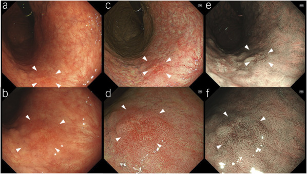
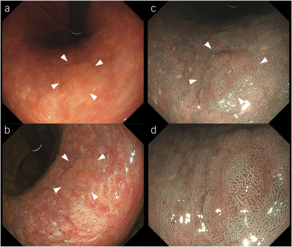
The early detection of gastric neoplasms (GNs) leads to favorable treatment outcomes. The latest endoscopic system, EVIS X1, includes third-generation narrow-band imaging (3G-NBI), texture and color enhancement imaging (TXI), and high-definition white-light imaging (WLI). Therefore, this randomized phase II trial aimed to identify the most promising imaging modality for GN detection using 3G-NBI and TXI.

METHODS

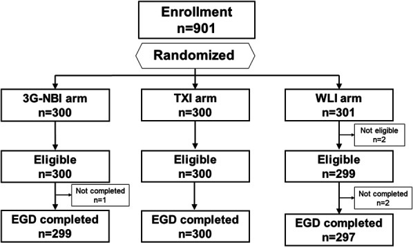
Patients with scheduled surveillance endoscopy after a history of esophageal cancer or GN or preoperative endoscopy for known esophageal cancer or GN were randomly assigned to the 3G-NBI, TXI, or WLI groups. Endoscopic observations were performed to detect new GN lesions, and all suspected lesions were biopsied. The primary endpoint was the GN detection rate during primary observation. Secondary endpoints were the rate of missed GNs, early gastric cancer detection rate, and positive predictive value for a GN diagnosis. The decision rule had a higher GN detection rate between 3G-NBI and TXI, outperforming WLI by >1.0%.

RESULTS

Finally, 901 patients were enrolled and assigned to the 3G-NBI, TXI, and WLI groups (300, 300, and 301 patients, respectively). GN detection rates in the 3G-NBI, TXI, and WLI groups were 7.3, 5.0, and 5.6%, respectively. The rates of missed GNs were 1.0, 0.7, and 1.0%, the detection rates of early gastric cancer were 5.7, 4.0, and 5.6%, and the positive predictive values for the diagnosis of GN were 36.5, 21.3, and 36.8% in the 3G-NBI, TXI, and WLI groups, respectively.

DISCUSSION

Compared with TXI and WLI, 3G-NBI is a more promising modality for GN detection.

摘要

简介

早期发现胃肿瘤(GNs)可带来良好的治疗效果。最新的内镜系统 EVIS X1 包括第三代窄带成像(3G-NBI)、纹理和颜色增强成像(TXI)以及高清白光成像(WLI)。因此,本项随机 II 期试验旨在通过 3G-NBI 和 TXI 来确定最有前途的 GN 检测成像方式。

方法

患有食管癌或 GN 病史的患者或已知患有食管癌或 GN 的患者在术前接受内镜检查,这些患者被随机分配至 3G-NBI、TXI 或 WLI 组。进行内镜观察以检测新的 GN 病变,所有疑似病变均进行活检。主要终点是初次观察时的 GN 检出率。次要终点是 GN 漏诊率、早期胃癌检出率和 GN 诊断的阳性预测值。决策规则要求 3G-NBI 和 TXI 的 GN 检出率更高,优于 WLI 的幅度>1.0%。

结果

最终纳入 901 例患者,分别分配至 3G-NBI、TXI 和 WLI 组(每组 300、300 和 301 例)。3G-NBI、TXI 和 WLI 组的 GN 检出率分别为 7.3%、5.0%和 5.6%。GN 漏诊率分别为 1.0%、0.7%和 1.0%,早期胃癌检出率分别为 5.7%、4.0%和 5.6%,GN 诊断的阳性预测值分别为 36.5%、21.3%和 36.8%。

讨论

与 TXI 和 WLI 相比,3G-NBI 是一种更有前途的 GN 检测方式。

https://cdn.ncbi.nlm.nih.gov/pmc/blobs/a0d2/11446510/69cb6158c6ce/acg-119-2010-g003.jpg
https://cdn.ncbi.nlm.nih.gov/pmc/blobs/a0d2/11446510/09c591bae62c/acg-119-2010-g001.jpg
https://cdn.ncbi.nlm.nih.gov/pmc/blobs/a0d2/11446510/2964638adf51/acg-119-2010-g002.jpg
https://cdn.ncbi.nlm.nih.gov/pmc/blobs/a0d2/11446510/69cb6158c6ce/acg-119-2010-g003.jpg
https://cdn.ncbi.nlm.nih.gov/pmc/blobs/a0d2/11446510/09c591bae62c/acg-119-2010-g001.jpg
https://cdn.ncbi.nlm.nih.gov/pmc/blobs/a0d2/11446510/2964638adf51/acg-119-2010-g002.jpg
https://cdn.ncbi.nlm.nih.gov/pmc/blobs/a0d2/11446510/69cb6158c6ce/acg-119-2010-g003.jpg

相似文献

1
Comparison of Effective Imaging Modalities for Detecting Gastric Neoplasms: A Randomized 3-Arm Phase II Trial.比较有效成像方式检测胃肿瘤:一项随机 3 臂 II 期试验。
Am J Gastroenterol. 2024 Oct 1;119(10):2010-2018. doi: 10.14309/ajg.0000000000002871. Epub 2024 May 16.
2
The Efficacy of Texture and Color Enhancement Imaging Observation in the Detection of Colorectal Lesions: A Multicenter, Randomized Controlled Trial (deTXIon Study).纹理和颜色增强成像观察在结直肠病变检测中的疗效:一项多中心随机对照试验(deTXIon研究)
Gastroenterology. 2025 Aug;169(2):337-345.e2. doi: 10.1053/j.gastro.2025.03.007. Epub 2025 Mar 18.
3
Second Examination of the Right Colon Using Narrow-Band Imaging Increases Adenoma Detection Rates in the Right Colon: A Multicenter, Randomized Controlled Trial.使用窄带成像技术对右半结肠进行第二次检查可提高右半结肠腺瘤的检出率:一项多中心随机对照试验
J Gastroenterol Hepatol. 2025 Jul;40(7):1667-1674. doi: 10.1111/jgh.16964. Epub 2025 Apr 14.
4
Efficacy of high-vision transnasal endoscopy using texture and colour enhancement imaging and narrow-band imaging to evaluate gastritis: a randomized controlled trial.高分辨率经鼻内镜联合纹理增强及窄带成像技术评估胃炎的疗效:一项随机对照试验。
Ann Med. 2022 Dec;54(1):1004-1013. doi: 10.1080/07853890.2022.2063372.
5
An Additional 30-s Observation of the Right-Sided Colon Using a Novel Endoscopic System with Texture and Color Enhancement Imaging Decreases Polyp Miss Rates: A Multicenter Study.使用具有纹理和颜色增强成像的新型内镜系统对右侧结肠进行额外30秒观察可降低息肉漏检率:一项多中心研究
Diagnostics (Basel). 2025 Jul 11;15(14):1759. doi: 10.3390/diagnostics15141759.
6
White-Light Imaging and Image-Enhanced Endoscopy With Magnifying Endoscopy for the Optical Diagnosis of Superficial Nonampullary Duodenal Epithelial Tumors: A Systematic Review and Meta-Analysis.白光成像及联合放大内镜的图像增强内镜用于浅表非壶腹十二指肠上皮肿瘤的光学诊断:一项系统评价和Meta分析
J Gastroenterol Hepatol. 2025 Feb;40(2):379-386. doi: 10.1111/jgh.16822. Epub 2024 Nov 25.
7
Texture and Colour Enhancement Imaging versus White Light Endoscopy for Detection of Dysplasia within Barrett's Oesophagus: A Pilot Study.纹理与颜色增强成像对比白光内镜检查用于检测巴雷特食管内发育异常的初步研究
Digestion. 2025 Jun 16:1-9. doi: 10.1159/000546637.
8
Virtual NBI image synthesis using stable diffusion for enhanced recognition of early gastric cancer: a technical validation study.使用稳定扩散进行虚拟窄带成像(NBI)图像合成以增强早期胃癌识别:一项技术验证研究
Ann Med. 2025 Dec;57(1):2523565. doi: 10.1080/07853890.2025.2523565. Epub 2025 Jun 28.
9
Narrow band imaging diagnosis of bladder cancer: systematic review and meta-analysis.窄带成像诊断膀胱癌:系统评价和荟萃分析。
BJU Int. 2012 Dec;110(11 Pt B):E680-7. doi: 10.1111/j.1464-410X.2012.11500.x. Epub 2012 Sep 18.
10
Usefulness of third-generation narrow band imaging and texture and color enhancement imaging in improving visibility of superficial early gastric cancer: A study using color difference.第三代窄带成像及纹理与色彩增强成像在提高早期浅表性胃癌可视性方面的效用:一项色差研究
DEN Open. 2022 Nov 24;3(1):e186. doi: 10.1002/deo2.186. eCollection 2023 Apr.

引用本文的文献

1
Image-enhanced endoscopy in upper GI tract: State-of-the-art review.上消化道的图像增强内镜检查:最新综述
Indian J Gastroenterol. 2025 Sep 2. doi: 10.1007/s12664-025-01850-z.
2
Assessing the effectiveness of texture and color enhancement imaging versus white-light endoscopy in detecting gastrointestinal lesions: A systematic review and meta-analysis.评估纹理和颜色增强成像与白光内镜检查在检测胃肠道病变中的有效性:一项系统评价和荟萃分析。
DEN Open. 2025 Apr 30;6(1):e70128. doi: 10.1002/deo2.70128. eCollection 2026 Apr.
3
Effect of texture and color enhancement imaging on the visibility of gastric tumors.

本文引用的文献

1
Metachronous primary gastric cancer after endoscopic resection in patients with esophageal squamous cell carcinoma.食管鳞癌内镜切除术后胃的异时性原发性癌。
Gastric Cancer. 2023 Nov;26(6):988-1001. doi: 10.1007/s10120-023-01413-1. Epub 2023 Jun 27.
2
Endoscopic diagnosis of gastritis using white light imaging and texture and color enhancement imaging.使用白光成像以及纹理和颜色增强成像进行胃炎的内镜诊断。
Endosc Int Open. 2023 Feb 2;11(2):E136-E141. doi: 10.1055/a-2005-7486. eCollection 2023 Feb.
3
Visibility of early gastric cancers by texture and color enhancement imaging using a high-definition ultrathin transnasal endoscope.
纹理增强及色彩增强成像技术对胃肿瘤可视性的影响。
Sci Rep. 2024 Aug 18;14(1):19125. doi: 10.1038/s41598-024-70236-6.
使用高清超微经鼻内窥镜的纹理和颜色增强成像技术对早期胃癌的可视性。
Sci Rep. 2023 Feb 3;13(1):1994. doi: 10.1038/s41598-023-29284-7.
4
Confusion and prospects for carcinogenesis of gastric adenoma and dysplasia: What is the correct answer currently?胃腺瘤和异型增生的癌变困惑与展望:目前正确答案是什么?
World J Gastroenterol. 2022 Dec 28;28(48):6900-6908. doi: 10.3748/wjg.v28.i48.6900.
5
Usefulness of third-generation narrow band imaging and texture and color enhancement imaging in improving visibility of superficial early gastric cancer: A study using color difference.第三代窄带成像及纹理与色彩增强成像在提高早期浅表性胃癌可视性方面的效用:一项色差研究
DEN Open. 2022 Nov 24;3(1):e186. doi: 10.1002/deo2.186. eCollection 2023 Apr.
6
Efficacy of high-vision transnasal endoscopy using texture and colour enhancement imaging and narrow-band imaging to evaluate gastritis: a randomized controlled trial.高分辨率经鼻内镜联合纹理增强及窄带成像技术评估胃炎的疗效:一项随机对照试验。
Ann Med. 2022 Dec;54(1):1004-1013. doi: 10.1080/07853890.2022.2063372.
7
Visibility of early gastric cancer in texture and color enhancement imaging.早期胃癌在纹理和颜色增强成像中的可视性。
DEN Open. 2021 Aug 24;2(1):e46. doi: 10.1002/deo2.46. eCollection 2022 Apr.
8
Emerging texture and color enhancement imaging in early gastric cancer.早期胃癌的新兴纹理和色彩增强成像技术。
Dig Endosc. 2022 May;34(4):714-720. doi: 10.1111/den.14182. Epub 2021 Dec 1.
9
TXI: Texture and Color Enhancement Imaging for Endoscopic Image Enhancement.TXI:用于内窥镜图像增强的纹理和颜色增强成像。
J Healthc Eng. 2021 Apr 7;2021:5518948. doi: 10.1155/2021/5518948. eCollection 2021.
10
Efficacy of Texture and Color Enhancement Imaging in visualizing gastric mucosal atrophy and gastric neoplasms.纹理和色彩增强成像在胃黏膜萎缩和胃癌中的可视化效果。
Sci Rep. 2021 Mar 25;11(1):6910. doi: 10.1038/s41598-021-86296-x.