• 文献检索
  • 文档翻译
  • 深度研究
  • 学术资讯
  • Suppr Zotero 插件Zotero 插件
  • 邀请有礼
  • 套餐&价格
  • 历史记录
应用&插件
Suppr Zotero 插件Zotero 插件浏览器插件Mac 客户端Windows 客户端微信小程序
定价
高级版会员购买积分包购买API积分包
服务
文献检索文档翻译深度研究API 文档MCP 服务
关于我们
关于 Suppr公司介绍联系我们用户协议隐私条款
关注我们

Suppr 超能文献

核心技术专利:CN118964589B侵权必究
粤ICP备2023148730 号-1Suppr @ 2026

文献检索

告别复杂PubMed语法,用中文像聊天一样搜索,搜遍4000万医学文献。AI智能推荐,让科研检索更轻松。

立即免费搜索

文件翻译

保留排版,准确专业,支持PDF/Word/PPT等文件格式,支持 12+语言互译。

免费翻译文档

深度研究

AI帮你快速写综述,25分钟生成高质量综述,智能提取关键信息,辅助科研写作。

立即免费体验

纹理和色彩增强成像在胃黏膜萎缩和胃癌中的可视化效果。

Efficacy of Texture and Color Enhancement Imaging in visualizing gastric mucosal atrophy and gastric neoplasms.

机构信息

Department of Gastroenterology, Graduate School of Medicine, Chiba University, Inohana 1-8-1, Chiba-City, 260-8670, Japan.

Department of Medical Oncology, Chiba University, Chiba, Japan.

出版信息

Sci Rep. 2021 Mar 25;11(1):6910. doi: 10.1038/s41598-021-86296-x.

DOI:10.1038/s41598-021-86296-x
PMID:33767278
原文链接:https://pmc.ncbi.nlm.nih.gov/articles/PMC7994634/
Abstract

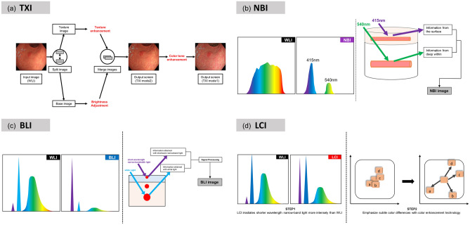
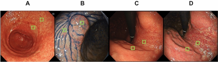
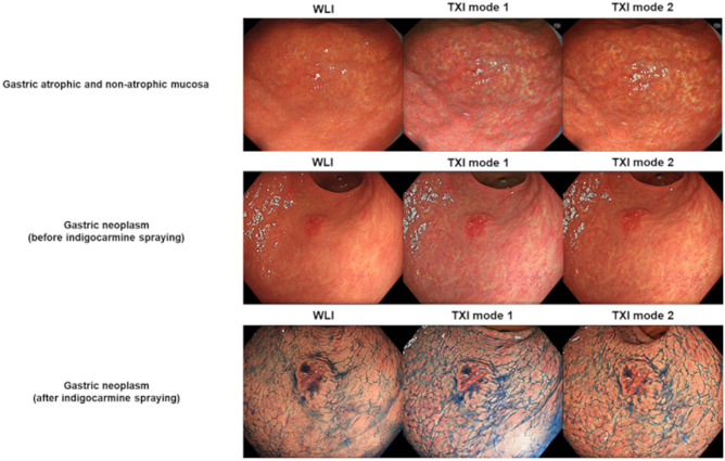
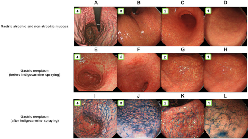
In 2020, Olympus Medical Systems Corporation introduced the Texture and Color Enhancement Imaging (TXI) as a new image-enhanced endoscopy. This study aimed to evaluate the visibility of neoplasms and mucosal atrophy in the upper gastrointestinal tract through TXI. We evaluated 72 and 60 images of 12 gastric neoplasms and 20 gastric atrophic/nonatrophic mucosa, respectively. The visibility of gastric mucosal atrophy and gastric neoplasm was assessed by six endoscopists using a previously reported visibility scale (1 = poor to 4 = excellent). Color differences between gastric mucosal atrophy and nonatrophic mucosa and between gastric neoplasm and adjacent areas were assessed using the International Commission on Illumination Lab* color space system. The visibility of mucosal atrophy and gastric neoplasm was significantly improved in TXI mode 1 compared with that in white-light imaging (WLI) (visibility score: 3.8 ± 0.5 vs. 2.8 ± 0.9, p < 0.01 for mucosal atrophy; visibility score: 2.8 ± 1.0 vs. 2.0 ± 0.9, p < 0.01 for gastric neoplasm). Regarding gastric atrophic and nonatrophic mucosae, TXI mode 1 had a significantly greater color difference than WLI (color differences: 14.2 ± 8.0 vs. 8.7 ± 4.2, respectively, p < 0.01). TXI may be a useful observation modality in the endoscopic screening of the upper gastrointestinal tract.

摘要

2020 年,奥林巴斯医疗系统公司推出了纹理和色彩增强成像(TXI)作为一种新的图像增强内镜。本研究旨在通过 TXI 评估上消化道肿瘤和黏膜萎缩的可见性。我们分别评估了 12 个胃肿瘤的 72 个和 20 个胃萎缩/非萎缩黏膜的 60 个图像。六位内镜医生使用之前报道的可视性评分(1=差到 4=优秀)评估胃黏膜萎缩和胃肿瘤的可视性。使用国际照明委员会 Lab*颜色空间系统评估胃黏膜萎缩和非萎缩黏膜以及胃肿瘤和相邻区域之间的颜色差异。与白光成像(WLI)相比,TXI 模式 1 显著提高了黏膜萎缩和胃肿瘤的可视性(可视性评分:3.8±0.5 对 2.8±0.9,p<0.01;可视性评分:2.8±1.0 对 2.0±0.9,p<0.01)。对于胃萎缩和非萎缩黏膜,TXI 模式 1 与 WLI 相比具有显著更大的颜色差异(颜色差异:14.2±8.0 对 8.7±4.2,均 p<0.01)。TXI 可能是上消化道内镜筛查的有用观察方式。

https://cdn.ncbi.nlm.nih.gov/pmc/blobs/ce50/7994634/33e7d132302b/41598_2021_86296_Fig4_HTML.jpg
https://cdn.ncbi.nlm.nih.gov/pmc/blobs/ce50/7994634/a189ea7f90eb/41598_2021_86296_Fig1_HTML.jpg
https://cdn.ncbi.nlm.nih.gov/pmc/blobs/ce50/7994634/4e9236b4d913/41598_2021_86296_Fig2_HTML.jpg
https://cdn.ncbi.nlm.nih.gov/pmc/blobs/ce50/7994634/924fafc3265f/41598_2021_86296_Fig3_HTML.jpg
https://cdn.ncbi.nlm.nih.gov/pmc/blobs/ce50/7994634/33e7d132302b/41598_2021_86296_Fig4_HTML.jpg

相似文献

1
Efficacy of Texture and Color Enhancement Imaging in visualizing gastric mucosal atrophy and gastric neoplasms.纹理和色彩增强成像在胃黏膜萎缩和胃癌中的可视化效果。
Sci Rep. 2021 Mar 25;11(1):6910. doi: 10.1038/s41598-021-86296-x.
2
Effect of texture and color enhancement imaging on the visibility of gastric tumors.纹理增强及色彩增强成像技术对胃肿瘤可视性的影响。
Sci Rep. 2024 Aug 18;14(1):19125. doi: 10.1038/s41598-024-70236-6.
3
Texture and color enhancement imaging improves the visibility of gastric neoplasms: clinical trial with image catalogue assessment using conventional and newly developed endoscopes.纹理和色彩增强成像提高胃肿瘤的可视性:使用传统和新开发的内镜进行图像目录评估的临床试验。
BMC Gastroenterol. 2023 Nov 13;23(1):389. doi: 10.1186/s12876-023-03030-9.
4
Visibility of early gastric cancers by texture and color enhancement imaging using a high-definition ultrathin transnasal endoscope.使用高清超微经鼻内窥镜的纹理和颜色增强成像技术对早期胃癌的可视性。
Sci Rep. 2023 Feb 3;13(1):1994. doi: 10.1038/s41598-023-29284-7.
5
Efficacy of high-vision transnasal endoscopy using texture and colour enhancement imaging and narrow-band imaging to evaluate gastritis: a randomized controlled trial.高分辨率经鼻内镜联合纹理增强及窄带成像技术评估胃炎的疗效:一项随机对照试验。
Ann Med. 2022 Dec;54(1):1004-1013. doi: 10.1080/07853890.2022.2063372.
6
Using texture and colour enhancement imaging to evaluate gastrointestinal diseases in clinical practice: a review.利用纹理和色彩增强成像技术评估临床胃肠道疾病:综述。
Ann Med. 2022 Dec;54(1):3315-3332. doi: 10.1080/07853890.2022.2147992.
7
Visibility of early gastric cancer in texture and color enhancement imaging.早期胃癌在纹理和颜色增强成像中的可视性。
DEN Open. 2021 Aug 24;2(1):e46. doi: 10.1002/deo2.46. eCollection 2022 Apr.
8
Comparison of Texture and Color Enhancement Imaging with White Light Imaging in 52 Patients with Short-Segment Barrett's Esophagus.52 例短节段 Barrett 食管患者的纹理及色彩增强成像与白光成像的对比。
Med Sci Monit. 2023 Jun 13;29:e940249. doi: 10.12659/MSM.940249.
9
Texture and Color Enhancement Imaging Increases Color Changes and Improves Visibility for Squamous Cell Carcinoma Suspicious Lesions in the Pharynx and Esophagus.纹理与颜色增强成像增加了颜色变化并提高了咽部和食管鳞状细胞癌可疑病变的可视性。
Diagnostics (Basel). 2021 Oct 23;11(11):1971. doi: 10.3390/diagnostics11111971.
10
Topic highlight on texture and color enhancement imaging in gastrointestinal diseases.胃肠道疾病的纹理和颜色增强成像专题亮点。
World J Gastroenterol. 2024 Apr 14;30(14):1934-1940. doi: 10.3748/wjg.v30.i14.1934.

引用本文的文献

1
Visibility Evaluation of Fundic Gland Polyp Associated With Proton Pump Inhibitor in Texture and Color Enhancement Imaging.质子泵抑制剂相关性胃底腺息肉在纹理和颜色增强成像中的可视性评估
DEN Open. 2025 May 22;6(1):e70147. doi: 10.1002/deo2.70147. eCollection 2026 Apr.
2
Red dichromatic imaging enhances submucosal visibility during endoscopic submucosal dissection: Pilot study.红色双色成像在内镜黏膜下剥离术中增强黏膜下可视性:初步研究。
Endosc Int Open. 2025 May 16;13:a25923546. doi: 10.1055/a-2592-3546. eCollection 2025.
3
Assessing the effectiveness of texture and color enhancement imaging versus white-light endoscopy in detecting gastrointestinal lesions: A systematic review and meta-analysis.

本文引用的文献

1
Linked Color Imaging Can Improve Detection Rate of Early Gastric Cancer in a High-Risk Population: A Multi-Center Randomized Controlled Clinical Trial.链接显色内镜成像技术可提高高危人群早期胃癌检出率:一项多中心随机对照临床试验。
Dig Dis Sci. 2021 Apr;66(4):1212-1219. doi: 10.1007/s10620-020-06289-0. Epub 2020 May 4.
2
RGB Pixel Brightness Characteristics of Linked Color Imaging in Early Gastric Cancer: A Pilot Study.早期胃癌中联合彩色成像的RGB像素亮度特征:一项初步研究
Gastroenterol Res Pract. 2020 Mar 31;2020:2105874. doi: 10.1155/2020/2105874. eCollection 2020.
3
Early gastric cancer detection in high-risk patients: a multicentre randomised controlled trial on the effect of second-generation narrow band imaging.
评估纹理和颜色增强成像与白光内镜检查在检测胃肠道病变中的有效性:一项系统评价和荟萃分析。
DEN Open. 2025 Apr 30;6(1):e70128. doi: 10.1002/deo2.70128. eCollection 2026 Apr.
4
Visibility evaluation of gastric epithelial neoplasm of fundic gland mucosa lineage using texture and color enhancement imaging.利用纹理和颜色增强成像对胃底腺黏膜谱系胃上皮肿瘤进行可视性评估。
DEN Open. 2025 Apr 8;5(1):e70110. doi: 10.1002/deo2.70110. eCollection 2025 Apr.
5
Image-enhanced endoscopy in upper gastrointestinal disease: focusing on texture and color enhancement imaging and red dichromatic imaging.上消化道疾病的图像增强内镜检查:聚焦于纹理和颜色增强成像以及红色双色成像。
Clin Endosc. 2025 Mar;58(2):163-180. doi: 10.5946/ce.2024.159. Epub 2024 Nov 6.
6
Novel endoscopic techniques for the diagnosis of gastric infection: a systematic review and network meta-analysis.用于诊断胃部感染的新型内镜技术:系统评价与网状Meta分析
Front Microbiol. 2024 Aug 26;15:1377541. doi: 10.3389/fmicb.2024.1377541. eCollection 2024.
7
Effect of texture and color enhancement imaging on the visibility of gastric tumors.纹理增强及色彩增强成像技术对胃肿瘤可视性的影响。
Sci Rep. 2024 Aug 18;14(1):19125. doi: 10.1038/s41598-024-70236-6.
8
Efficacy of texture and color enhancement imaging for the visibility and diagnostic accuracy of non-polypoid colorectal lesions.纹理和颜色增强成像对非息肉样结直肠病变可视性及诊断准确性的效果
DEN Open. 2024 May 29;5(1):e380. doi: 10.1002/deo2.380. eCollection 2025 Apr.
9
Comparison of Prognosis and Metachronous Gastric Tumor Rates After Endoscopic Submucosal Dissection Between Gastric Neoplasm of Fundic Gland Type Neoplasms and Conventional Gastric Adenocarcinoma.胃底腺型肿瘤与传统胃腺癌在内镜黏膜下剥离术后的预后及异时性胃肿瘤发生率比较
Cureus. 2024 Apr 17;16(4):e58467. doi: 10.7759/cureus.58467. eCollection 2024 Apr.
10
Comparison of Effective Imaging Modalities for Detecting Gastric Neoplasms: A Randomized 3-Arm Phase II Trial.比较有效成像方式检测胃肿瘤:一项随机 3 臂 II 期试验。
Am J Gastroenterol. 2024 Oct 1;119(10):2010-2018. doi: 10.14309/ajg.0000000000002871. Epub 2024 May 16.
高危人群胃癌早期检测:第二代窄带成像效果的多中心随机对照试验。
Gut. 2021 Jan;70(1):67-75. doi: 10.1136/gutjnl-2019-319631. Epub 2020 Apr 2.
4
Comparison of endoscopic visibility and miss rate for early gastric cancers after Helicobacter pylori eradication with white-light imaging versus linked color imaging.幽门螺杆菌根除后,白光成像与连接色彩成像对比对早期胃癌的内镜可视性和漏诊率的比较。
Dig Endosc. 2020 Jul;32(5):769-777. doi: 10.1111/den.13585. Epub 2019 Dec 26.
5
Linked color imaging identifies important risk factors associated with gastric cancer after successful eradication of Helicobacter pylori.链接颜色成像可识别成功根除幽门螺杆菌后与胃癌相关的重要危险因素。
Gastrointest Endosc. 2019 Nov;90(5):763-769. doi: 10.1016/j.gie.2019.06.043. Epub 2019 Jul 9.
6
Global cancer statistics 2018: GLOBOCAN estimates of incidence and mortality worldwide for 36 cancers in 185 countries.全球癌症统计数据 2018:GLOBOCAN 对全球 185 个国家/地区 36 种癌症的发病率和死亡率的估计。
CA Cancer J Clin. 2018 Nov;68(6):394-424. doi: 10.3322/caac.21492. Epub 2018 Sep 12.
7
Blue laser imaging-bright improves the real-time detection rate of early gastric cancer: a randomized controlled study.蓝激光成像-亮提高早期胃癌实时检出率:一项随机对照研究。
Gastrointest Endosc. 2019 Jan;89(1):47-57. doi: 10.1016/j.gie.2018.08.049. Epub 2018 Sep 4.
8
Improved Visibility of Barrett's Esophagus with Linked Color Imaging: Inter- and Intra-Rater Reliability and Quantitative Analysis.窄带光成像增强巴雷特食管的可视性:观察者间和观察者内的可靠性和定量分析。
Digestion. 2018;97(2):183-194. doi: 10.1159/000485459. Epub 2018 Jan 10.
9
Objective Endoscopic Analysis with Linked Color Imaging regarding Gastric Mucosal Atrophy: A Pilot Study.关于胃黏膜萎缩的内镜下联合彩色成像分析:一项初步研究。
Gastroenterol Res Pract. 2017;2017:5054237. doi: 10.1155/2017/5054237. Epub 2017 Nov 15.
10
Linked color imaging (LCI), a novel image-enhanced endoscopy technology, emphasizes the color of early gastric cancer.联动成像(LCI)是一种新型的图像增强内镜技术,它突出了早期胃癌的颜色。
Endosc Int Open. 2017 Oct;5(10):E1005-E1013. doi: 10.1055/s-0043-117881. Epub 2017 Oct 10.