Department of Gastroenterology and Hepatology, Tokyo Medical University Hospital, 6-7-1 Nishishinjuku, Shinjuku-ku, Tokyo, 160-0023, Japan.
Department of Gastroenterological Endoscopy, Tokyo Medical University Hospital, Tokyo, Japan.
Sci Rep. 2023 Feb 3;13(1):1994. doi: 10.1038/s41598-023-29284-7.
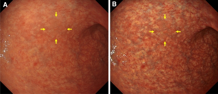
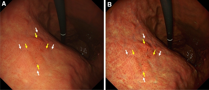
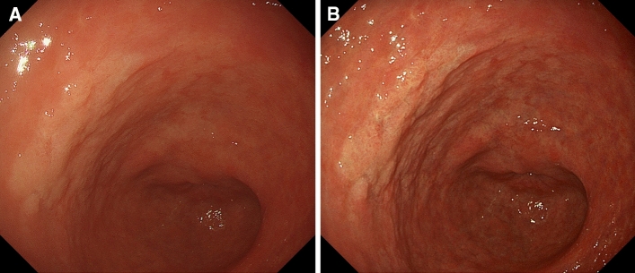
We evaluated whether texture and color enhancement imaging (TXI) using a high-definition ultrathin transnasal endoscope (UTE) improves the visibility of early gastric cancer (EGC) compared with white-light imaging (WLI). This study included 31 EGCs observed by TXI mode 2 using a high-definition UTE prior to endoscopic submucosal dissection. The first outcome was to compare the color differences based on Commission Internationale de l'Eclairage Lab* color space between EGCs and the surrounding mucosa by WLI and TXI using the UTE (objective appearance of EGC). The second outcome was to assess the visibility of EGCs by WLI and TXI using the UTE in an image evaluation test performed on 10 endoscopists (subjective appearance of EGC). Color differences between EGCs and non-neoplastic mucosa were significantly higher in TXI than in WLI in all EGCs (TXI: 16.0 ± 10.1 vs. WLI: 10.2 ± 5.5 [mean ± standard deviation], P < 0.001). Median visibility scores evaluated by 10 endoscopists using TXI were significantly higher than those evaluated using WLI (TXI: 4 [interquartile range, 4-4] vs. WLI: 4 [interquartile range, 3-4], P < 0.001). TXI using high-definition UTE improved both objective and subjective visibility of EGCs compared with WLI.
我们评估了使用高清超微型经鼻内镜(UTE)的纹理和色彩增强成像(TXI)是否比白光成像(WLI)提高了早期胃癌(EGC)的可视性。这项研究纳入了 31 例在进行内镜黏膜下剥离术之前通过 TXI 模式 2 使用高清 UTE 观察到的 EGC。第一个结果是通过 WLI 和 TXI 使用 UTE 比较 EGC 和周围黏膜之间基于国际照明委员会 Lab*颜色空间的颜色差异(EGC 的客观外观)。第二个结果是通过 WLI 和 TXI 使用 UTE 在 10 名内镜医师进行的图像评估测试中评估 EGC 的可视性(EGC 的主观外观)。在所有 EGC 中,TXI 下 EGC 与非肿瘤性黏膜之间的颜色差异明显高于 WLI(TXI:16.0±10.1 比 WLI:10.2±5.5[平均值±标准差],P<0.001)。使用 TXI 评估的 10 名内镜医师的中位数可视评分明显高于使用 WLI 评估的评分(TXI:4[四分位数范围,4-4]比 WLI:4[四分位数范围,3-4],P<0.001)。与 WLI 相比,使用高清 UTE 的 TXI 提高了 EGC 的客观和主观可视性。