Department of Radiology, Beijing Chaoyang Hospital, Capital Medical University, No. 8, Gongti South Road, Chaoyang District, Beijing, 100020, China.
Department of Medical Imaging, Peking University Shenzhen Hospital, Shenzhen, 518036, China.
Brain Imaging Behav. 2024 Oct;18(5):1001-1009. doi: 10.1007/s11682-024-00899-2. Epub 2024 May 24.
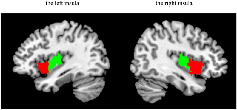
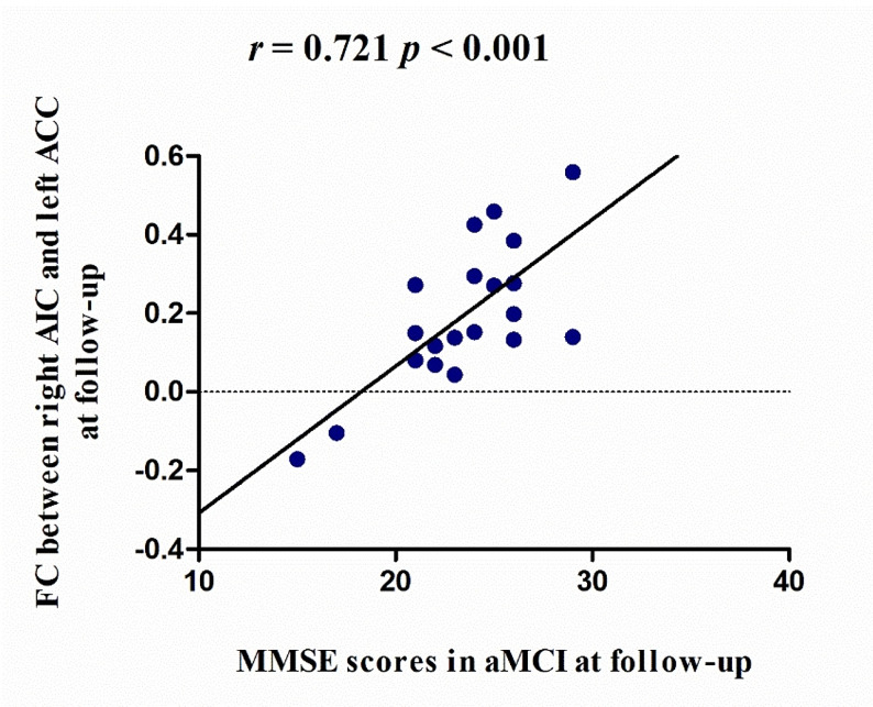
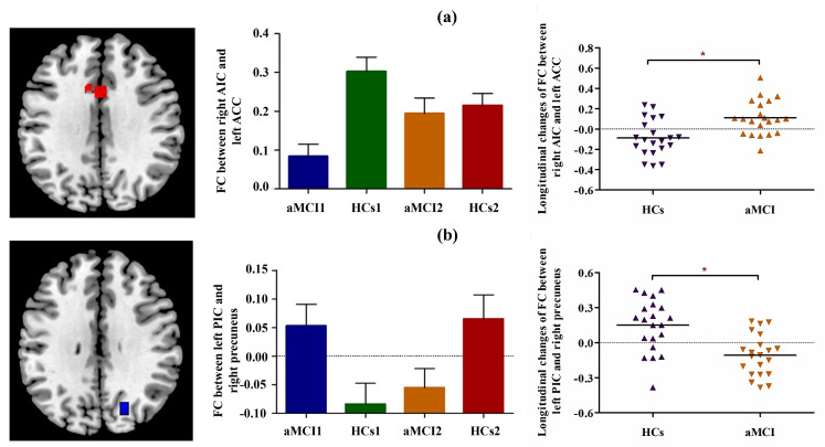
The insula, a crucial hub of the human brain network, can be divided into anterior and posterior regions. Previous studies have reported that different insula subregions play various roles in amnestic mild cognitive impairment (aMCI). However, the longitudinal changes in the functional connectivity (FC) of each insula subregion in aMCI patients over time remain unclear. Twenty aMCI patients and 20 healthy controls (HCs) were recruited and underwent resting-state functional magnetic resonance imaging (fMRI) scans and neuropsychological assessments at baseline and at the 15-month follow-up. FMRI data were preprocessed using SPM 12 and the CONN toolbox. Two-way analysis of covariance was used to compare longitudinal changes in the FC of each insula subregion with covariates including sex, age, education, follow-up interval, volume of gray matter, and global correlation (GCOR). Pearson's correlation was used to evaluate the relationship between insula subregional FC and neuropsychological performance in aMCI patients. In aMCI patients, the right anterior insula exhibited significantly increased FC with the left anterior cingulate cortex, whereas the left posterior insula exhibited decreased FC with the right precuneus compared with HCs. Furthermore, FC between the right anterior insula and left anterior cingulate cortex was significantly correlated with global cognition at follow-up. The current findings revealed different functional alterations in the insula subregions and provided new insights into the neurodegenerative process in aMCI patients.
脑岛是人类大脑网络的关键枢纽,可以分为前区和后区。先前的研究报告称,不同的脑岛亚区在遗忘型轻度认知障碍(aMCI)中发挥着不同的作用。然而,aMCI 患者脑岛各亚区功能连接(FC)随时间的纵向变化尚不清楚。本研究招募了 20 名 aMCI 患者和 20 名健康对照者(HCs),并在基线和 15 个月随访时进行了静息态功能磁共振成像(fMRI)扫描和神经心理学评估。FMRI 数据使用 SPM12 和 CONN 工具箱进行预处理。采用双因素协方差分析比较了各脑岛亚区 FC 的纵向变化,协变量包括性别、年龄、教育、随访间隔、灰质体积和全局相关(GCOR)。采用 Pearson 相关分析评估了 aMCI 患者脑岛亚区 FC 与神经心理学表现之间的关系。在 aMCI 患者中,与 HCs 相比,右侧前脑岛与左侧前扣带皮层的 FC 增加,而左侧后脑岛与右侧楔前叶的 FC 减少。此外,右侧前脑岛与左侧前扣带皮层的 FC 与随访时的整体认知显著相关。本研究结果揭示了脑岛亚区的不同功能改变,并为 aMCI 患者的神经退行性过程提供了新的见解。