Minton Dennis M, Ailiani Aditya R, Focht Michael D K, Kersh Mariana E, Li Amy, Elliehausen Christian J, Sonsalla Michelle M, Lamming Dudley W, Marolf Angela J, Santangelo Kelly S, Salmon Adam B, Konopka Adam R
Division of Geriatrics and Gerontology, Department of Medicine, University of Wisconsin-Madison, Madison, Wisconsin, USA.
Geriatric Research Education and Clinical Center, William S. Middleton Memorial Veterans Hospital, Madison, Wisconsin, USA.
bioRxiv. 2025 Mar 6:2024.05.14.594256. doi: 10.1101/2024.05.14.594256.
Genetic deletion of mTOR has protected against post-traumatic osteoarthritis (OA) in male mice, however, effects of pharmacological mTOR-inhibition are equivocal and have not been tested in aging models nor in female subjects. Therefore, the goal of this study was to determine if mTOR-inhibition by rapamycin can modify OA pathology in aging non-human primates and female mice.
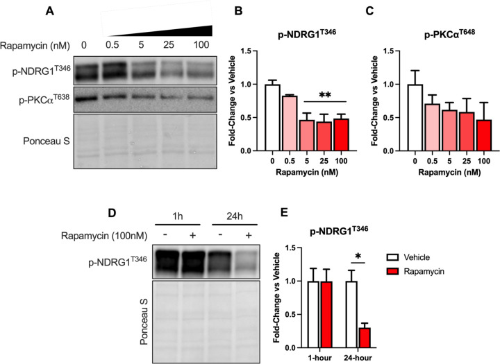
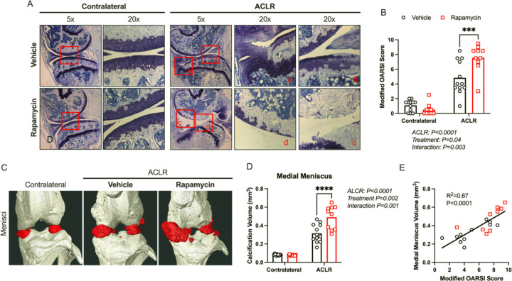
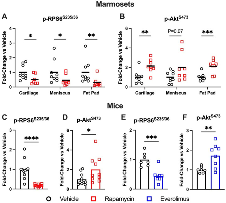
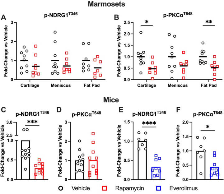
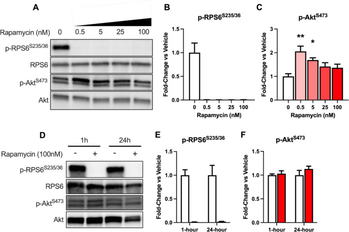
Common marmosets were administered oral rapamycin (1mg/kg/day) or vehicle starting near mid-life until death. Five-month-old, female C57BL/6J mice were treated with vehicle or rapamycin (IP, 2mg/kg, 3x/week) for 8-weeks following non-invasive ACL rupture. Knee OA pathology was assessed via microCT and histology. Phosphorylation of mTORC1 (p-RPS6) and mTORC2 (p-Akt, p-NDRG1, p-PKCα) substrates were evaluated via western blot in articular cartilage, meniscus, and/or infrapatellar fat pad. ATDC5 cells were cultured with rapamycin to determine time and dose effects on mTORC1/2 signaling.
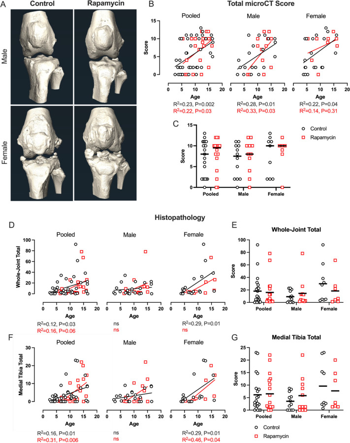
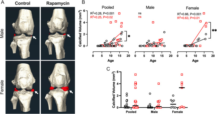
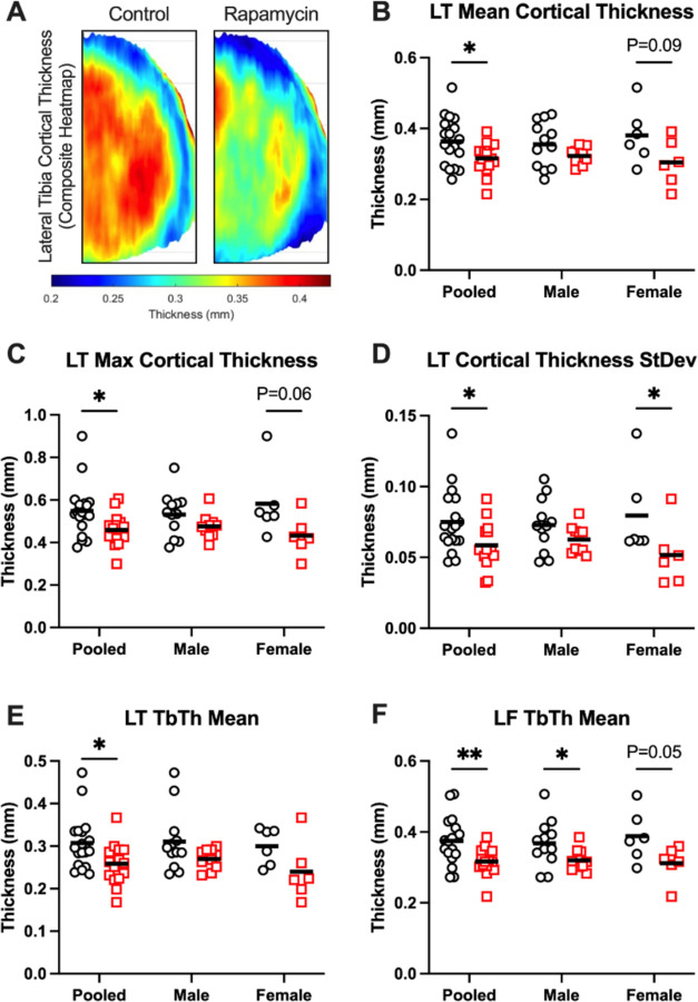
In marmosets, rapamycin did not impact age-related radiographic OA severity or cartilage pathology but increased medial meniscus calcification and lowered lateral tibia subchondral thickness, particularly in females. In female mice, rapamycin worsened ACLR-induced meniscus calcification and cartilage pathology. In marmoset and mouse joint tissues, rapamycin inhibited mTORC1 and increased p-Akt but not p-NDRG1 or p-PKCα. This mTOR signaling pattern was replicated in ATDC5 cells during exposure to low concentrations of rapamycin.
Rapamycin attenuated mTORC1 signaling with feedback activation of Akt in articular cartilage, meniscus, and/or infrapatellar fat pad and was accompanied by deleterious effects on meniscus calcification and/or cartilage pathology in female mice and common marmosets.
mTOR基因缺失可保护雄性小鼠免受创伤后骨关节炎(OA)的影响,然而,mTOR药理学抑制的效果并不明确,且尚未在衰老模型或雌性动物中进行测试。因此,本研究的目的是确定雷帕霉素抑制mTOR是否能改变衰老的非人灵长类动物和雌性小鼠的OA病理。
普通狨猴从接近中年开始直至死亡,口服雷帕霉素(1mg/kg/天)或赋形剂。5月龄雌性C57BL/6J小鼠在非侵入性前交叉韧带断裂后,用赋形剂或雷帕霉素(腹腔注射,2mg/kg,每周3次)治疗8周。通过显微CT和组织学评估膝关节OA病理。通过蛋白质免疫印迹法评估关节软骨、半月板和/或髌下脂肪垫中mTORC1(p-RPS6)和mTORC2(p-Akt、p-NDRG1、p-PKCα)底物的磷酸化。用雷帕霉素培养ATDC5细胞,以确定对mTORC1/2信号传导的时间和剂量效应。
在狨猴中,雷帕霉素不影响与年龄相关的放射学OA严重程度或软骨病理,但增加了内侧半月板钙化并降低了外侧胫骨软骨下厚度,尤其是在雌性中。在雌性小鼠中,雷帕霉素使前交叉韧带重建(ACLR)诱导的半月板钙化和软骨病理恶化。在狨猴和小鼠关节组织中,雷帕霉素抑制mTORC1并增加p-Akt,但不增加p-NDRG1或p-PKCα。在暴露于低浓度雷帕霉素期间,这种mTOR信号模式在ATDC5细胞中得到重现。
雷帕霉素减弱了关节软骨、半月板和/或髌下脂肪垫中mTORC1信号传导,并伴有Akt的反馈激活,同时对雌性小鼠和普通狨猴的半月板钙化和/或软骨病理产生有害影响。