Brown Meghan S, Mirhaidari Cyrus, Johnson Jordan, Larson Brandon M, Cook Chad, Shue Robert, Ventimiglia Anthony J, Cody Derek G
From Summa Health Department of Surgery, Akron, Ohio.
Northeast Ohio Medical University (NEOMED), Rootstown, Ohio.
Plast Reconstr Surg Glob Open. 2024 May 20;12(5):e5709. doi: 10.1097/GOX.0000000000005709. eCollection 2024 May.
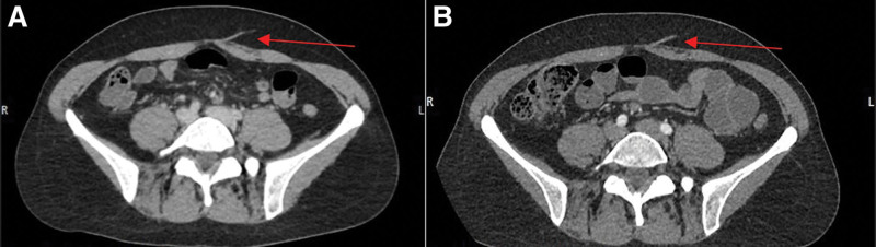
Due to variations in perforator vasculature, deep inferior epigastric artery perforator (DIEP) flap preoperative imaging can minimize operative time required to locate the most suitable perforators. Dedicated computed tomography angiography (CTA) has been the gold standard; however, many patients have already undergone a staging computed tomography (CT) per oncologic workup. The benefits from CTA may also be realized with a staging CT or CT with IV contrast.
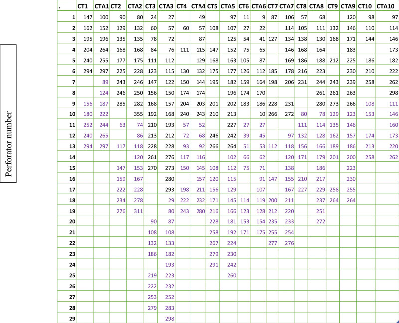
Ten patients who underwent DIEP flap reconstruction with staging CT and CTA within 3 years of one another were included in this study. Reviewers evaluated axial views of both imaging modalities separately to identify each visible perforator in reference to the pubic symphysis from the xiphoid to the pubic symphysis. An intraclass correlation coefficient (ICC) was used to determine agreement in location of perforators between the two imaging studies. Statistical analysis was performed using an ICC and Wilcoxon signed rank-tests.
The identified perforators within the patient cohort had an excellent correlation between their location on CT and CTA based upon ICC. The mean number of perforators identified in the CT group was 15.3 (SD 4.9) and in the CTA group was 18.8 (SD 6.4), which was not statistically different ( = 0.247).
CT has similar efficacy in identifying number of perforators and perforator location to dedicated CTA for preoperative planning in DIEP flaps. This has the potential for decreased patient contrast and ionizing radiation exposure as well as improved patient and healthcare resource utilization.
由于穿支血管存在变异,腹壁下深动脉穿支(DIEP)皮瓣术前成像可将定位最合适穿支所需的手术时间降至最短。专用计算机断层血管造影(CTA)一直是金标准;然而,许多患者因肿瘤检查已接受过分期计算机断层扫描(CT)。分期CT或静脉注射造影剂的CT也可能实现CTA的益处。
本研究纳入了10例在3年内先后接受分期CT和CTA检查并进行DIEP皮瓣重建的患者。评估人员分别对两种成像方式的轴位图像进行评估,以从剑突至耻骨联合的耻骨联合为参照确定每个可见穿支。采用组内相关系数(ICC)来确定两项成像研究中穿支位置的一致性。使用ICC和Wilcoxon符号秩检验进行统计分析。
根据ICC,患者队列中识别出的穿支在CT和CTA上的位置具有极佳的相关性。CT组识别出的穿支平均数量为15.3(标准差4.9),CTA组为18.8(标准差6.4),差异无统计学意义(P = 0.247)。
在DIEP皮瓣术前规划中,CT在识别穿支数量和穿支位置方面与专用CTA具有相似的效能。这有可能减少患者的造影剂用量和电离辐射暴露,并改善患者及医疗资源的利用。