• 文献检索
  • 文档翻译
  • 深度研究
  • 学术资讯
  • Suppr Zotero 插件Zotero 插件
  • 邀请有礼
  • 套餐&价格
  • 历史记录
应用&插件
Suppr Zotero 插件Zotero 插件浏览器插件Mac 客户端Windows 客户端微信小程序
定价
高级版会员购买积分包购买API积分包
服务
文献检索文档翻译深度研究API 文档MCP 服务
关于我们
关于 Suppr公司介绍联系我们用户协议隐私条款
关注我们

Suppr 超能文献

核心技术专利:CN118964589B侵权必究
粤ICP备2023148730 号-1Suppr @ 2026

文献检索

告别复杂PubMed语法,用中文像聊天一样搜索,搜遍4000万医学文献。AI智能推荐,让科研检索更轻松。

立即免费搜索

文件翻译

保留排版,准确专业,支持PDF/Word/PPT等文件格式,支持 12+语言互译。

免费翻译文档

深度研究

AI帮你快速写综述,25分钟生成高质量综述,智能提取关键信息,辅助科研写作。

立即免费体验

基于普鲁赞斯基和卡班分类法的具有新亚型的三维半侧颜面短小畸形分类

Three-dimensional Hemifacial Microsomia Classification with New Subtypes Based on the Pruzansky and Kaban Classification.

作者信息

Shibazaki-Yorozuya Reiko, Yamada Akira, Sasaki Ryo, Watanabe Yorikatsu

机构信息

From Division of Orthodontics, Incorporated Medical Institution, Yorozuya Dental Office, Tokyo, Japan.

Division of Pediatric Plastic Surgery, Northwestern University Feinberg School of Medicine, Ann & Robert H. Lurie Children's Hospital of Chicago, Chicago.

出版信息

Plast Reconstr Surg Glob Open. 2024 May 28;12(5):e5810. doi: 10.1097/GOX.0000000000005810. eCollection 2024 May.

DOI:10.1097/GOX.0000000000005810
PMID:38808145
原文链接:https://pmc.ncbi.nlm.nih.gov/articles/PMC11132385/
Abstract

BACKGROUND

Various classifications of hemifacial microsomia (HFM) have been described previously. Although some of these classifications are used widely, others use external outlines of reference organs, even in three-dimensional (3D) images. The purpose of this study was to investigate the 3D characteristics of the mandibular condyle in HFM and to update the Pruzansky and Kaban classification as a 3D classification.

METHODS

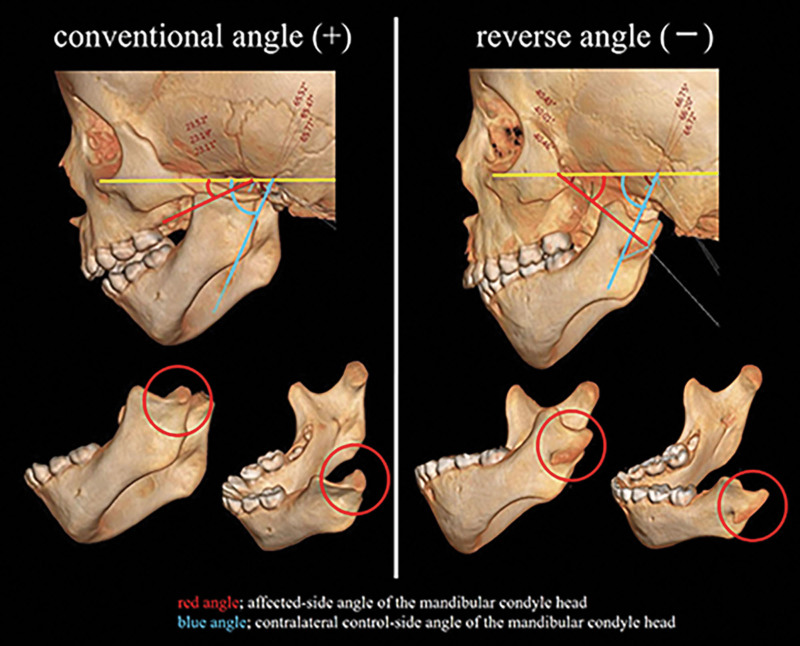
Fifty-three patients with HFM were classified according to the Pruzansky and Kaban classification (type I, IIA, IIB, and III) using computed tomographic scan images. 3D images of the mandible were isolated, and the 3D characteristics were observed; furthermore, the angle of inclination of the mandibular condyle was measured in 3D.

RESULTS

Subtypes of retroflexed mandibular condyle in 3D were observed in the Pruzansky and Kaban classification type IIA and IIB, termed as type IIAβ (33.3% in type IIA) and type IIBβ (100% in type IIB). Although some differences were observed in the inclination of the mandibular condyle between the control and the affected sides in type I and IIAα, multiple differences were observed in type IIAβ and IIBβ.

CONCLUSIONS

To the best of our knowledge, this is the first report that identified the retroflexed mandibular condyle as subtypes type IIAβ and IIBβ. Notably, this could not be identified in the two-dimensional images (lateral cephalogram) yet. We proposed to update the Pruzansky and Kaban classification as a 3D classification with a new 3D subtype. The angle of the retroflexed mandibular condyle may predict mandibular growth in HFM.

摘要

背景

先前已描述了半侧颜面短小畸形(HFM)的各种分类。尽管其中一些分类被广泛使用,但其他分类甚至在三维(3D)图像中也使用参考器官的外部轮廓。本研究的目的是研究HFM中下颌髁突的3D特征,并更新普鲁赞斯基和卡班分类作为一种3D分类。

方法

使用计算机断层扫描图像,根据普鲁赞斯基和卡班分类(I型、IIA、IIB和III型)对53例HFM患者进行分类。分离下颌骨的3D图像,观察其3D特征;此外,在3D中测量下颌髁突的倾斜角度。

结果

在普鲁赞斯基和卡班分类IIA和IIB型中观察到3D中后屈下颌髁突的亚型,分别称为IIAβ型(IIA型中占33.3%)和IIBβ型(IIB型中占100%)。虽然在I型和IIAα型中,患侧和对照侧下颌髁突的倾斜度存在一些差异,但在IIAβ型和IIBβ型中观察到多个差异。

结论

据我们所知,这是第一份将后屈下颌髁突确定为IIAβ型和IIBβ型亚型的报告。值得注意 的是,这在二维图像(侧位头影测量片)中尚未得到确认。我们建议将普鲁赞斯基和卡班分类更新为具有新3D亚型的3D分类。后屈下颌髁突的角度可能预测HFM中的下颌生长。

https://cdn.ncbi.nlm.nih.gov/pmc/blobs/74cd/11132385/36b945c7ef59/gox-12-e5810-g007.jpg
https://cdn.ncbi.nlm.nih.gov/pmc/blobs/74cd/11132385/f58d74a60192/gox-12-e5810-g001.jpg
https://cdn.ncbi.nlm.nih.gov/pmc/blobs/74cd/11132385/09dd369468b1/gox-12-e5810-g002.jpg
https://cdn.ncbi.nlm.nih.gov/pmc/blobs/74cd/11132385/4d69f4b473f0/gox-12-e5810-g003.jpg
https://cdn.ncbi.nlm.nih.gov/pmc/blobs/74cd/11132385/d99ce417a35c/gox-12-e5810-g004.jpg
https://cdn.ncbi.nlm.nih.gov/pmc/blobs/74cd/11132385/f2331a5b9ef3/gox-12-e5810-g005.jpg
https://cdn.ncbi.nlm.nih.gov/pmc/blobs/74cd/11132385/9e10d688321b/gox-12-e5810-g006.jpg
https://cdn.ncbi.nlm.nih.gov/pmc/blobs/74cd/11132385/36b945c7ef59/gox-12-e5810-g007.jpg

相似文献

1
Three-dimensional Hemifacial Microsomia Classification with New Subtypes Based on the Pruzansky and Kaban Classification.基于普鲁赞斯基和卡班分类法的具有新亚型的三维半侧颜面短小畸形分类
Plast Reconstr Surg Glob Open. 2024 May 28;12(5):e5810. doi: 10.1097/GOX.0000000000005810. eCollection 2024 May.
2
Effects of mandibular distraction osteogenesis on anesthetic implications in children with hemifacial microsomia.下颌骨牵引成骨术对单侧颜面短小症儿童麻醉影响。
Acta Anaesthesiol Scand. 2022 Aug;66(7):823-832. doi: 10.1111/aas.14073. Epub 2022 Apr 24.
3
The mandibular deformity in hemifacial microsomia: a reassessment of the Pruzansky and Kaban classification.半侧颜面短小畸形的下颌骨畸形:对 Pruzansky 和 Kaban 分类的再评估。
Plast Reconstr Surg. 2014 Feb;133(2):174e-181e. doi: 10.1097/01.prs.0000436858.63021.14.
4
Three-Dimensional Measurement of the Temporomandibular Joint in Pruzansky-Kaban Type IIa Hemifacial Microsomia.Pruzansky-Kaban Ⅱa 型半侧颜面短小患者颞下颌关节的三维测量
J Craniofac Surg. 2023;34(2):438-442. doi: 10.1097/SCS.0000000000008777. Epub 2022 Jul 22.
5
Three-dimensional longitudinal changes in craniofacial growth in untreated hemifacial microsomia patients with cone-beam computed tomography.锥形束计算机断层扫描评估未治疗单侧颜面短小患者颅颌面生长的三维纵向变化。
Am J Orthod Dentofacial Orthop. 2014 May;145(5):579-94. doi: 10.1016/j.ajodo.2013.09.015.
6
Morphological and quantitative study of the inferior alveolar nerve canal in hemifacial microsomia.颜面先天性畸形患者下颌神经管的形态和定量研究。
Sci Rep. 2024 Feb 27;14(1):4753. doi: 10.1038/s41598-024-54318-z.
7
Distribution and phenotypes of hemifacial microsomia and its association with other anomalies.半侧颜面短小畸形的分布、表型及其与其他异常的关联
Korean J Orthod. 2020 Jan;50(1):33-41. doi: 10.4041/kjod.2020.50.1.33. Epub 2020 Jan 22.
8
Morphologic Changes of the Temporomandibular Joint in Pruzansky-Kaban Type IIa Hemifacial Microsomia Postmandibular Distraction Osteogenesis.下颌骨牵引延长术后 Pruzansky-Kaban Ⅱa 型半侧颜面短小患者颞下颌关节的形态变化。
J Craniofac Surg. 2023;34(2):591-596. doi: 10.1097/SCS.0000000000008854. Epub 2022 Aug 1.
9
[Model test study on treatment of Pruzansky type B and hemifacial microsomia with artificial condyle-mandibular distractor complex].[人工髁突-下颌骨牵张复合体治疗普鲁赞斯基B型半侧颜面短小畸形的模型试验研究]
Zhongguo Xiu Fu Chong Jian Wai Ke Za Zhi. 2023 Oct 15;37(10):1270-1275. doi: 10.7507/1002-1892.202306022.
10
Treatment modalities for Korean patients with unilateral hemifacial microsomia according to Pruzansky-Kaban types and growth stages.根据普鲁赞斯基-卡班分型和生长阶段,韩国单侧半面短小畸形患者的治疗方式
Korean J Orthod. 2020 Sep 25;50(5):336-345. doi: 10.4041/kjod.2020.50.5.336.

本文引用的文献

1
Upper and Lower Limb Anomalies in Craniofacial Microsomia and Its Relation to the OMENS+ Classification: A Multicenter Study of 688 Patients.颅面小颌畸形中的上下肢异常及其与 OMENS+分类的关系:688 例患者的多中心研究。
Plast Reconstr Surg. 2023 May 1;151(5):1053-1061. doi: 10.1097/PRS.0000000000010090. Epub 2022 Dec 21.
2
Determination of Extra Craniofacial Abnormalities in Patients With Craniofacial Microsomia.颅面部微小畸形患者颅外异常的确定。
J Craniofac Surg. 2022;33(1):230-232. doi: 10.1097/SCS.0000000000007962.
3
Three-dimensional Coordinate System for Hemifacial Microsomia.
半侧颜面短小畸形的三维坐标系
Plast Reconstr Surg Glob Open. 2020 Apr 29;8(4):e2761. doi: 10.1097/GOX.0000000000002761. eCollection 2020 Apr.
4
Three-dimensional characterization of mandibular asymmetry in craniofacial microsomia.颅颌面小颌畸形中下颌骨不对称的三维特征。
Clin Oral Investig. 2020 Dec;24(12):4363-4372. doi: 10.1007/s00784-020-03302-8. Epub 2020 May 7.
5
Preferential Associated Malformation in Patients With Anotia and Microtia.无耳和小耳患者的优先相关畸形
J Craniofac Surg. 2019 Jan;30(1):66-70. doi: 10.1097/SCS.0000000000004915.
6
Craniofacial and extracraniofacial anomalies in craniofacial microsomia: a multicenter study of 755 patients’.颅面短小症的颅面和颅外异常:755 例多中心研究。
J Craniomaxillofac Surg. 2017 Aug;45(8):1302-1310. doi: 10.1016/j.jcms.2017.06.001. Epub 2017 Jun 8.
7
Three-dimensional longitudinal changes in craniofacial growth in untreated hemifacial microsomia patients with cone-beam computed tomography.锥形束计算机断层扫描评估未治疗单侧颜面短小患者颅颌面生长的三维纵向变化。
Am J Orthod Dentofacial Orthop. 2014 May;145(5):579-94. doi: 10.1016/j.ajodo.2013.09.015.
8
The mandibular deformity in hemifacial microsomia: a reassessment of the Pruzansky and Kaban classification.半侧颜面短小畸形的下颌骨畸形:对 Pruzansky 和 Kaban 分类的再评估。
Plast Reconstr Surg. 2014 Feb;133(2):174e-181e. doi: 10.1097/01.prs.0000436858.63021.14.
9
Evaluation of mandibular hypoplasia in patients with hemifacial microsomia: a comparison between panoramic radiography and three-dimensional computed tomography.半侧颜面短小畸形患者下颌骨发育不全的评估:全景X线摄影与三维计算机断层扫描的比较
Cleft Palate Craniofac J. 2013 Jul;50(4):381-7. doi: 10.1597/11-188. Epub 2012 Feb 5.
10
Variations in facial relationships; their significance in treatment and prognosis.面部关系的变异;它们在治疗和预后中的意义。
Am J Orthod. 1948 Oct;34(10):812-40. doi: 10.1016/0002-9416(48)90015-3.