Department of Neurosurgery, Nagoya City University Graduate School of Medical Science, Kawasumi 1, Mizuho-Cho, Mizuho-Ku, Nagoya, Aichi, 467-8601, Japan.
Interfaculty Initiative in Information Studies/Institute of Industrial Science, The University of Tokyo, Tokyo, Japan.
Fluids Barriers CNS. 2024 May 30;21(1):47. doi: 10.1186/s12987-024-00552-6.
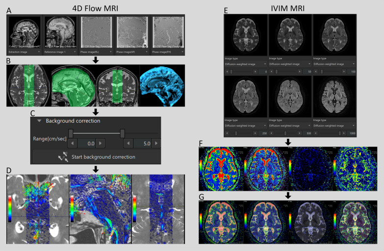
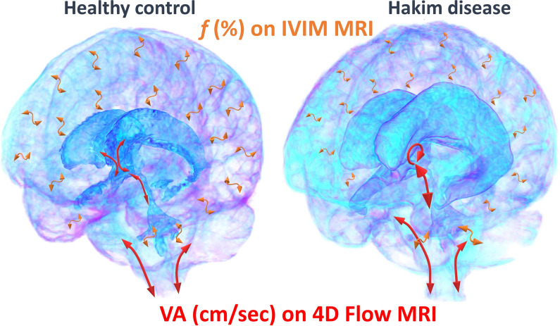
Bidirectional reciprocal motion of cerebrospinal fluid (CSF) was quantified using four-dimensional (4D) flow magnetic resonance imaging (MRI) and intravoxel incoherent motion (IVIM) MRI. To estimate various CSF motions in the entire intracranial region, we attempted to integrate the flow parameters calculated using the two MRI sequences. To elucidate how CSF dynamics deteriorate in Hakim's disease, an age-dependent chronic hydrocephalus, flow parameters were estimated from the two MRI sequences to assess CSF motion in the entire intracranial region.
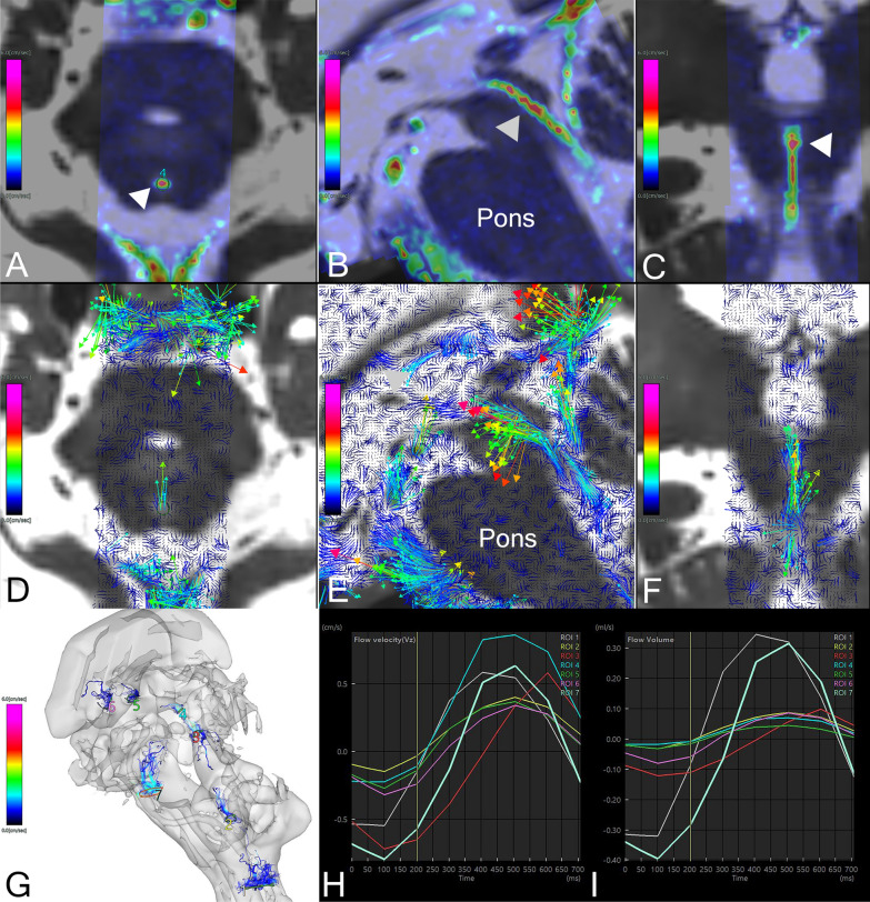
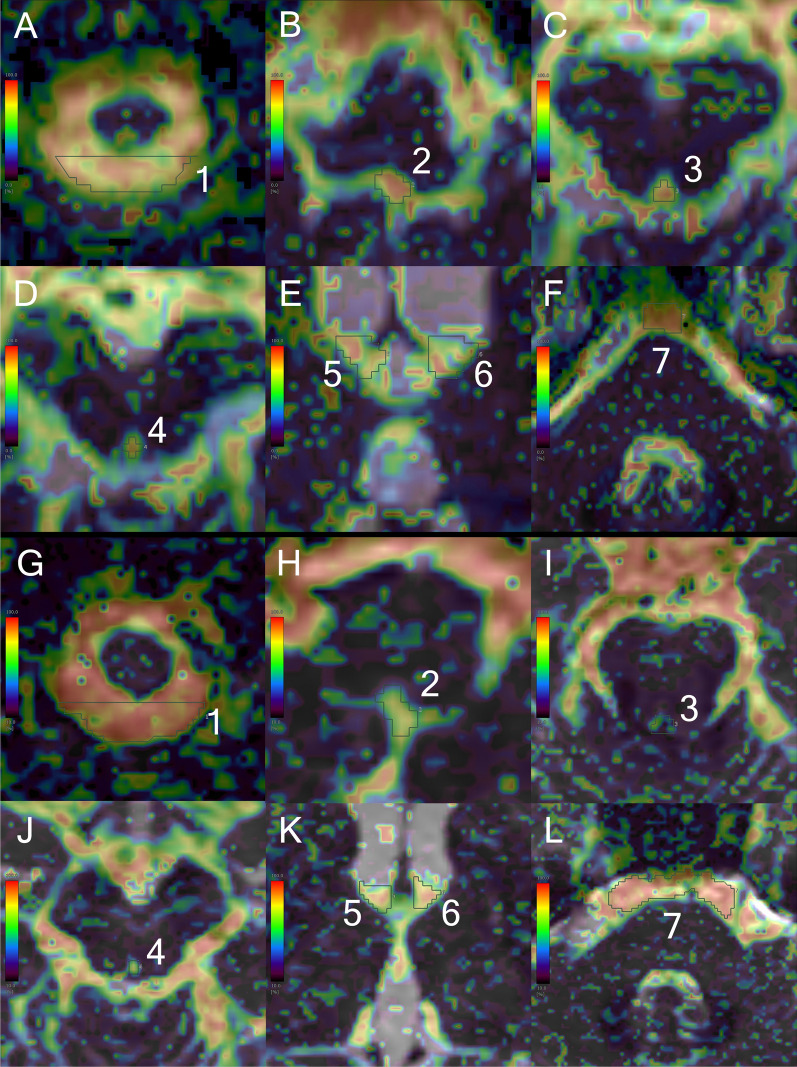
This study included 127 healthy volunteers aged ≥ 20 years and 44 patients with Hakim's disease. On 4D flow MRI for measuring CSF motion, velocity encoding was set at 5 cm/s. For the IVIM MRI analysis, the diffusion-weighted sequence was set at six b-values (i.e., 0, 50, 100, 250, 500, and 1000 s/mm), and the biexponential IVIM fitting method was adapted. The relationships between the fraction of incoherent perfusion (f) on IVIM MRI and 4D flow MRI parameters including velocity amplitude (VA), absolute maximum velocity, stroke volume, net flow volume, and reverse flow rate were comprehensively evaluated in seven locations in the ventricles and subarachnoid spaces. Furthermore, we developed a new parameter for fluid oscillation, the Fluid Oscillation Index (FOI), by integrating these two measurements. In addition, we investigated the relationship between the measurements and indices specific to Hakim's disease and the FOIs in the entire intracranial space.
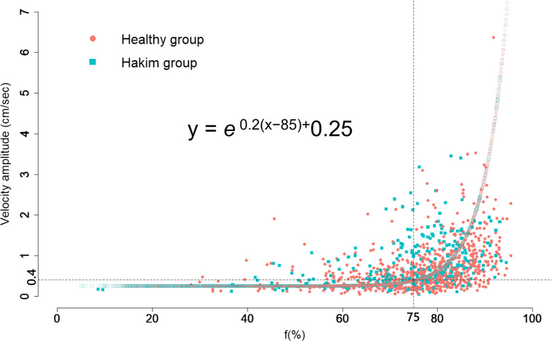
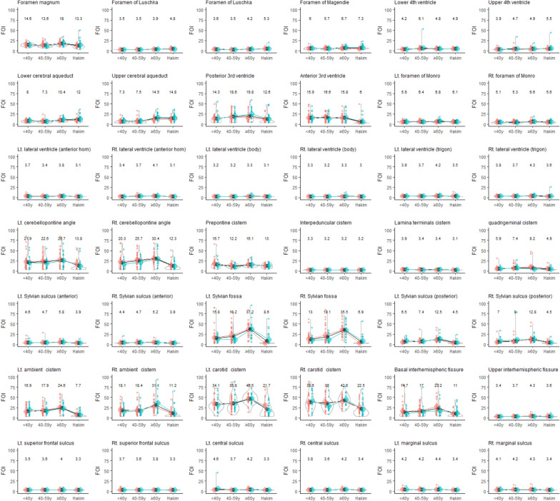
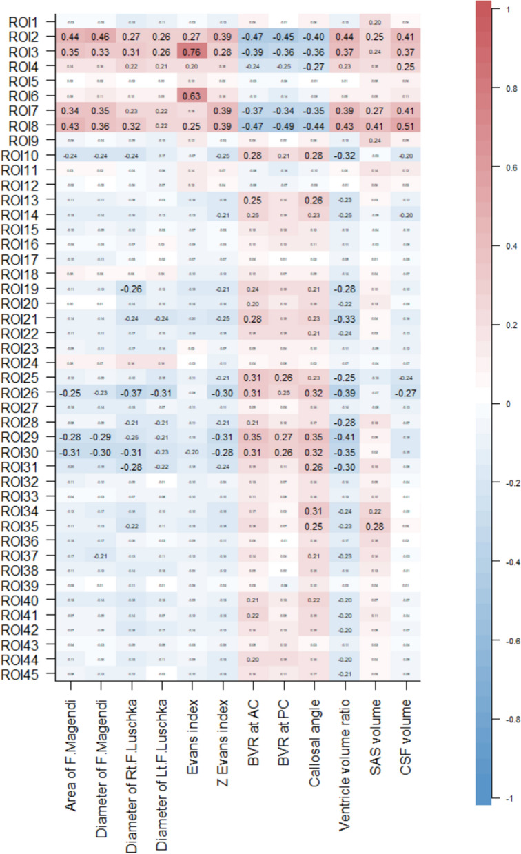
The VA on 4D flow MRI was significantly associated with the mean f-values on IVIM MRI. Therefore, we estimated VA that could not be directly measured on 4D flow MRI from the mean f-values on IVIM MRI in the intracranial CSF space, using the following formula; e + 0.25. To quantify fluid oscillation using one integrated parameter with weighting, FOI was calculated as VA × 10 + f × 0.02. In addition, the FOIs at the left foramen of Luschka had the strongest correlations with the Evans index (Pearson's correlation coefficient: 0.78). The other indices related with Hakim's disease were significantly associated with the FOIs at the cerebral aqueduct and bilateral foramina of Luschka. FOI at the cerebral aqueduct was also elevated in healthy controls aged ≥ 60 years.
We estimated pulsatile CSF movements in the entire intracranial CSF space in healthy individuals and patients with Hakim's disease using FOI integrating VA from 4D flow MRI and f-values from IVIM MRI. FOI is useful for quantifying the CSF oscillation.
使用四维(4D)流动磁共振成像(MRI)和体素内不相干运动(IVIM)MRI 来量化脑脊液(CSF)的双向往复运动。为了估计整个颅内区域的各种 CSF 运动,我们尝试整合使用两种 MRI 序列计算出的流量参数。为了阐明哈基姆病(一种与年龄相关的慢性脑积水)中 CSF 动力学如何恶化,我们使用两种 MRI 序列估计流量参数,以评估整个颅内区域的 CSF 运动。
本研究纳入了 127 名年龄≥20 岁的健康志愿者和 44 名哈基姆病患者。在用于测量 CSF 运动的 4D 流动 MRI 中,流速编码设置为 5cm/s。对于 IVIM MRI 分析,扩散加权序列设置为六个 b 值(即 0、50、100、250、500 和 1000s/mm),并采用双指数 IVIM 拟合方法。综合评估了七个脑室和蛛网膜下腔位置的 IVIM MRI 中不连贯灌注分数(f)与 4D 流动 MRI 参数(包括速度幅度(VA)、绝对最大速度、冲程体积、净流量体积和反向流速)之间的关系。此外,我们通过整合这两种测量方法,开发了一种新的流体振荡参数,即流体振荡指数(FOI)。此外,我们还研究了与哈基姆病相关的测量和指标以及整个颅内空间的 FOI 之间的关系。
4D 流动 MRI 的 VA 与 IVIM MRI 的平均 f 值显著相关。因此,我们使用以下公式从颅内 CSF 空间的 IVIM MRI 的平均 f 值估算 4D 流动 MRI 无法直接测量的 VA:e+0.25。为了使用加权的一个集成参数来量化流体振荡,将 FOI 计算为 VA×10+f×0.02。此外,左侧卢什卡孔的 FOI 与 Evans 指数(皮尔逊相关系数:0.78)相关性最强。与哈基姆病相关的其他指数与脑导水管和双侧卢什卡孔的 FOI 也有显著相关性。脑导水管的 FOI 在≥60 岁的健康对照组中也升高。
我们使用 FOI 整合了 4D 流动 MRI 的 VA 和 IVIM MRI 的 f 值,来估算健康个体和哈基姆病患者整个颅内 CSF 空间的搏动性 CSF 运动。FOI 可用于量化 CSF 振荡。