Faculty of Physical Therapy and Rehabilitation, Department of Musculoskeletal Physiotherapy and Rehabilitation, Hacettepe University, Ankara, Turkey.
Faculty of Health Science, Department of Physiotherapy and Rehabilitation, Ankara Medipol University, Ankara, Turkey.
J Foot Ankle Res. 2024 Jun;17(2):e12028. doi: 10.1002/jfa2.12028.
The aim of this study was to compare the plantar pressure distribution and knee and ankle muscle architecture in women with and without knee osteoarthritis (OA).
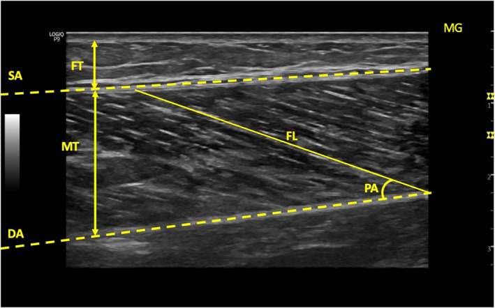
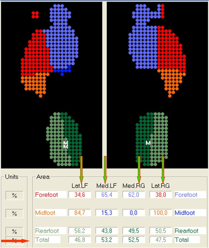
Fifty women with knee OA (mean age = 52.11 ± 4.96 years, mean Body mass index (BMI) = 30.94 ± 4.23 kg/m) and 50 healthy women as a control group (mean age = 50.93 ± 3.78 years, mean BMI = 29.06 ± 4.82 kg/m) were included in the study. Ultrasonography was used to evaluate knee and ankle muscles architecture and femoral cartilage thickness. The plantar pressure distribution was evaluated using the Digital Biometry Scanning System and Milleri software (DIASU, Italy). Static foot posture was evaluated using the Foot Posture Index (FPI), and pain severity was assessed using the Visual Analog Scale.
The OA group exhibited lower muscle thickness in Rectus Femoris (RF) (p = 0.003), Vastus Medialis (VM) (p = 0.004), Vastus Lateralis (p = 0.023), and Peroneus Longus (p = 0.002), as well as lower Medial Gastrocnemius pennation angle (p = 0.049) and higher Fat thickness (FT) in RF (p = 0.033) and VM (p = 0.037) compared to the control group. The OA group showed thinner femoral cartilage thickness (p = 0.001) and higher pain severity (p = 0.001) than the control groups. FPI scores were higher (p = 0.001) in OA group compared to the control group. The plantar pressure distribution results indicated an increase in total surface (p = 0.027), total load (p = 0.002), medial load (p = 0.005), and lateral load (p = 0.002) on dominant side in OA group compared to the control group.
Knee and ankle muscle architecture, knee extensor muscle FT, and plantar pressure distribution in the dominant foot differed in individuals with knee OA compared to the control group.
本研究旨在比较膝骨关节炎(OA)患者与无膝 OA 患者的足底压力分布、膝踝关节肌肉结构。
纳入 50 例膝 OA 患者(平均年龄 52.11±4.96 岁,平均体重指数(BMI)30.94±4.23kg/m²)和 50 例健康女性作为对照组(平均年龄 50.93±3.78 岁,平均 BMI 29.06±4.82kg/m²)。采用超声检查评估膝关节和踝关节肌肉结构及股骨软骨厚度。采用数字生物测量扫描系统和 Milleri 软件(DIASU,意大利)评估足底压力分布。采用足印指数(FPI)评估静态足姿,采用视觉模拟评分(VAS)评估疼痛严重程度。
OA 组股四头肌(RF)(p=0.003)、股内侧肌(VM)(p=0.004)、股外侧肌(p=0.023)和腓骨长肌(p=0.002)的肌肉厚度较低,以及内侧比目鱼肌的羽状角(p=0.049)和 RF(p=0.033)及 VM(p=0.037)的脂肪厚度较高。与对照组相比,OA 组的股骨软骨厚度较薄(p=0.001),疼痛程度较高(p=0.001)。与对照组相比,OA 组的 FPI 评分更高(p=0.001)。与对照组相比,OA 组的足底压力分布结果显示,在优势侧,总表面积(p=0.027)、总负荷(p=0.002)、内侧负荷(p=0.005)和外侧负荷(p=0.002)增加。
与对照组相比,膝骨关节炎患者的膝、踝关节肌肉结构、膝伸肌的 FT 和优势足的足底压力分布不同。