• 文献检索
  • 文档翻译
  • 深度研究
  • 学术资讯
  • Suppr Zotero 插件Zotero 插件
  • 邀请有礼
  • 套餐&价格
  • 历史记录
应用&插件
Suppr Zotero 插件Zotero 插件浏览器插件Mac 客户端Windows 客户端微信小程序
定价
高级版会员购买积分包购买API积分包
服务
文献检索文档翻译深度研究API 文档MCP 服务
关于我们
关于 Suppr公司介绍联系我们用户协议隐私条款
关注我们

Suppr 超能文献

核心技术专利:CN118964589B侵权必究
粤ICP备2023148730 号-1Suppr @ 2026

文献检索

告别复杂PubMed语法,用中文像聊天一样搜索,搜遍4000万医学文献。AI智能推荐,让科研检索更轻松。

立即免费搜索

文件翻译

保留排版,准确专业,支持PDF/Word/PPT等文件格式,支持 12+语言互译。

免费翻译文档

深度研究

AI帮你快速写综述,25分钟生成高质量综述,智能提取关键信息,辅助科研写作。

立即免费体验

脂毒性肝细胞来源的富含 LIMA1 的小细胞外囊泡通过抑制线粒体自噬促进肝星状细胞激活。

Lipotoxic hepatocyte derived LIMA1 enriched small extracellular vesicles promote hepatic stellate cells activation via inhibiting mitophagy.

机构信息

Department of Laboratory Medicine, Wujin Hospital Affiliated With Jiangsu University, Changzhou, 213017, China.

Department of Laboratory Medicine, School of Medicine, Jiangsu University, Zhenjiang, 212013, China.

出版信息

Cell Mol Biol Lett. 2024 May 31;29(1):82. doi: 10.1186/s11658-024-00596-4.

DOI:10.1186/s11658-024-00596-4
PMID:38822260
原文链接:https://pmc.ncbi.nlm.nih.gov/articles/PMC11140962/
Abstract

BACKGROUND

Hepatic stellate cells (HSCs) play a crucial role in the development of fibrosis in non-alcoholic fatty liver disease (NAFLD). Small extracellular vesicles (sEV) act as mediators for intercellular information transfer, delivering various fibrotic factors that impact the function of HSCs in liver fibrosis. In this study, we investigated the role of lipotoxic hepatocyte derived sEV (LTH-sEV) in HSCs activation and its intrinsic mechanisms.

METHODS

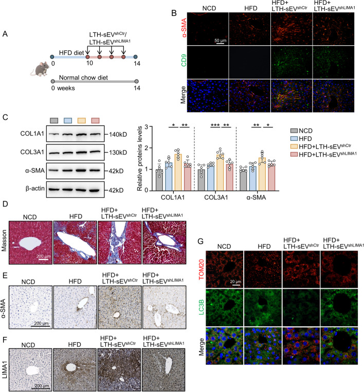
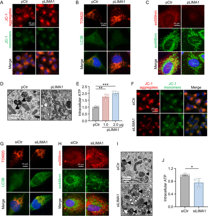
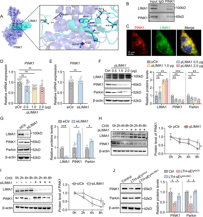
High-fat diet (HFD) mice model was constructed to confirm the expression of LIMA1. The relationship between LIMA1-enriched LTH-sEV and LX2 activation was evaluated by measurement of fibrotic markers and related genes. Levels of mitophagy were detected using mt-keima lentivirus. The interaction between LIMA1 and PINK1 was discovered through database prediction and molecular docking. Finally, sEV was injected to investigate whether LIMA1 can accelerate HFD induced liver fibrosis in mice.

RESULTS

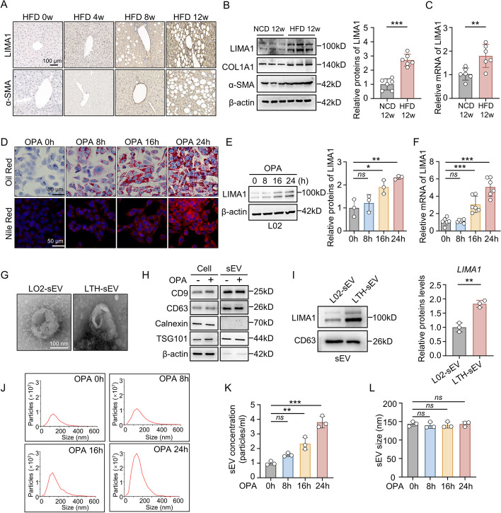
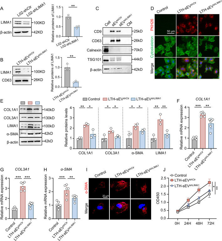
LIMA1 expression was upregulated in lipotoxic hepatocytes and was found to be positively associated with the expression of the HSCs activation marker α-SMA. Lipotoxicity induced by OPA led to an increase in both the level of LIMA1 protein in LTH-sEV and the release of LTH-sEV. When HSCs were treated with LTH-sEV, LIMA1 was observed to hinder LX2 mitophagy while facilitating LX2 activation. Further investigation revealed that LIMA1 derived from LTH-sEV may inhibit PINK1-Parkin-mediated mitophagy, consequently promoting HSCs activation. Knocking down LIMA1 significantly attenuates the inhibitory effects of LTH-sEV on mitophagy and the promotion of HSCs activation.

CONCLUSIONS

Lipotoxic hepatocyte-derived LIMA1-enriched sEVs play a crucial role in promoting HSCs activation in NAFLD-related liver fibrosis by negatively regulating PINK1 mediated mitophagy. These findings provide new insights into the pathological mechanisms involved in the development of fibrosis in NAFLD.

摘要

背景

肝星状细胞(HSCs)在非酒精性脂肪性肝病(NAFLD)纤维化的发展中起着至关重要的作用。小细胞外囊泡(sEV)作为细胞间信息传递的介质,传递各种纤维化因子,影响 HSCs 在肝纤维化中的功能。在这项研究中,我们研究了脂毒性肝细胞来源的 sEV(LTH-sEV)在 HSCs 激活中的作用及其内在机制。

方法

构建高脂肪饮食(HFD)小鼠模型,以证实 LIMA1 的表达。通过测量纤维化标志物和相关基因来评估富含 LIMA1 的 LTH-sEV 与 LX2 激活的关系。使用 mt-keima 慢病毒检测线粒体自噬水平。通过数据库预测和分子对接发现 LIMA1 与 PINK1 之间的相互作用。最后,通过注射 sEV 来研究 LIMA1 是否可以加速 HFD 诱导的小鼠肝纤维化。

结果

LIMA1 在脂毒性肝细胞中表达上调,并且与 HSCs 激活标志物 α-SMA 的表达呈正相关。OPA 诱导的脂毒性导致 LTH-sEV 中 LIMA1 蛋白水平升高和 LTH-sEV 释放增加。当 HSCs 用 LTH-sEV 处理时,观察到 LIMA1 阻碍 LX2 线粒体自噬,同时促进 LX2 激活。进一步研究表明,LTH-sEV 衍生的 LIMA1 可能抑制 PINK1-Parkin 介导的线粒体自噬,从而促进 HSCs 激活。敲低 LIMA1 可显著减轻 LTH-sEV 对线粒体自噬的抑制作用和对 HSCs 激活的促进作用。

结论

脂毒性肝细胞来源的富含 LIMA1 的 sEV 通过负向调节 PINK1 介导的线粒体自噬在促进 HSCs 激活中在 NAFLD 相关肝纤维化中起关键作用。这些发现为 NAFLD 纤维化发展中的病理机制提供了新的见解。

https://cdn.ncbi.nlm.nih.gov/pmc/blobs/0c0a/11140962/dae3646fe88d/11658_2024_596_Fig8_HTML.jpg
https://cdn.ncbi.nlm.nih.gov/pmc/blobs/0c0a/11140962/b1220866b672/11658_2024_596_Fig1_HTML.jpg
https://cdn.ncbi.nlm.nih.gov/pmc/blobs/0c0a/11140962/cc12c7019fe2/11658_2024_596_Fig2_HTML.jpg
https://cdn.ncbi.nlm.nih.gov/pmc/blobs/0c0a/11140962/6d1bc81f7a0d/11658_2024_596_Fig3_HTML.jpg
https://cdn.ncbi.nlm.nih.gov/pmc/blobs/0c0a/11140962/0bd363a590f3/11658_2024_596_Fig4_HTML.jpg
https://cdn.ncbi.nlm.nih.gov/pmc/blobs/0c0a/11140962/41631737aaed/11658_2024_596_Fig5_HTML.jpg
https://cdn.ncbi.nlm.nih.gov/pmc/blobs/0c0a/11140962/97bd380e38e8/11658_2024_596_Fig6_HTML.jpg
https://cdn.ncbi.nlm.nih.gov/pmc/blobs/0c0a/11140962/6c9b778726aa/11658_2024_596_Fig7_HTML.jpg
https://cdn.ncbi.nlm.nih.gov/pmc/blobs/0c0a/11140962/dae3646fe88d/11658_2024_596_Fig8_HTML.jpg

相似文献

1
Lipotoxic hepatocyte derived LIMA1 enriched small extracellular vesicles promote hepatic stellate cells activation via inhibiting mitophagy.脂毒性肝细胞来源的富含 LIMA1 的小细胞外囊泡通过抑制线粒体自噬促进肝星状细胞激活。
Cell Mol Biol Lett. 2024 May 31;29(1):82. doi: 10.1186/s11658-024-00596-4.
2
Aflatoxin B-exposed hepatocyte-derived extracellular vesicles: Initiating hepatic stellate cell-mediated liver fibrosis through a p53-Parkin-dependent mitophagy pathway.黄曲霉毒素 B 暴露的肝细胞衍生细胞外囊泡:通过 p53-Parkin 依赖的线粒体自噬途径引发肝星状细胞介导的肝纤维化。
Ecotoxicol Environ Saf. 2024 Jun 1;277:116363. doi: 10.1016/j.ecoenv.2024.116363. Epub 2024 Apr 25.
3
CircRNA608-microRNA222-PINK1 axis regulates the mitophagy of hepatic stellate cells in NASH related fibrosis.环状RNA608-微小RNA222-PINK1轴调控非酒精性脂肪性肝炎相关纤维化中肝星状细胞的线粒体自噬
Biochem Biophys Res Commun. 2022 Jun 25;610:35-42. doi: 10.1016/j.bbrc.2022.04.008. Epub 2022 Apr 5.
4
RNF31 alleviates liver steatosis by promoting p53/BNIP3-related mitophagy in hepatocytes.RNF31 通过促进肝细胞中 p53/BNIP3 相关的线粒体自噬来减轻肝脂肪变性。
Free Radic Biol Med. 2024 Jul;219:163-179. doi: 10.1016/j.freeradbiomed.2024.04.214. Epub 2024 Apr 13.
5
Hepatocyte-derived MASP1-enriched small extracellular vesicles activate HSCs to promote liver fibrosis.肝细胞衍生的富含甘露聚糖结合凝集素相关丝氨酸蛋白酶1的小细胞外囊泡激活肝星状细胞以促进肝纤维化。
Hepatology. 2023 Apr 1;77(4):1181-1197. doi: 10.1002/hep.32662. Epub 2022 Aug 8.
6
Acetyl-CoA carboxylase inhibition disrupts metabolic reprogramming during hepatic stellate cell activation.乙酰辅酶 A 羧化酶抑制破坏肝星状细胞激活过程中的代谢重编程。
J Hepatol. 2020 Oct;73(4):896-905. doi: 10.1016/j.jhep.2020.04.037. Epub 2020 May 4.
7
Extracellular vesicles derived from fat-laden hepatocytes undergoing chemical hypoxia promote a pro-fibrotic phenotype in hepatic stellate cells.脂变肝细胞来源的细胞外囊泡在化学缺氧下促进肝星状细胞的促纤维化表型。
Biochim Biophys Acta Mol Basis Dis. 2020 Oct 1;1866(10):165857. doi: 10.1016/j.bbadis.2020.165857. Epub 2020 Jun 5.
8
Hepatic stellate cell autophagy inhibits extracellular vesicle release to attenuate liver fibrosis.肝星状细胞自噬抑制细胞外囊泡释放以减轻肝纤维化。
J Hepatol. 2020 Nov;73(5):1144-1154. doi: 10.1016/j.jhep.2020.04.044. Epub 2020 May 8.
9
Extracellular Vesicles in the Development of the Non-Alcoholic Fatty Liver Disease: An Update.细胞外囊泡在非酒精性脂肪肝病发展中的作用:最新研究进展。
Biomolecules. 2020 Oct 30;10(11):1494. doi: 10.3390/biom10111494.
10
Hepatocyte Ninjurin2 promotes hepatic stellate cell activation and liver fibrosis through the IGF1R/EGR1/PDGF-BB signaling pathway.肝细胞 Ninjurin2 通过 IGF1R/EGR1/PDGF-BB 信号通路促进肝星状细胞活化和肝纤维化。
Metabolism. 2023 Mar;140:155380. doi: 10.1016/j.metabol.2022.155380. Epub 2022 Dec 20.

引用本文的文献

1
Pyrimethamine Inhibits Human Ovarian Cancer by Triggering Lethal Mitophagy via Activating the p38/JNK/ERK Pathway.乙胺嘧啶通过激活p38/JNK/ERK途径触发致死性线粒体自噬来抑制人卵巢癌。
Oncol Res. 2025 Aug 28;33(9):2421-2434. doi: 10.32604/or.2025.063724. eCollection 2025.
2
Communication initiated by hepatocytes: The driver of HSC activation and liver fibrosis.肝细胞引发的通讯:肝星状细胞激活和肝纤维化的驱动因素
Hepatol Commun. 2025 Jul 14;9(8). doi: 10.1097/HC9.0000000000000753. eCollection 2025 Aug 1.
3
Extracellular Vesicles as Biomarkers in Chronic Hepatobiliary Diseases: An Overview of Their Interplay.

本文引用的文献

1
The global epidemiology of nonalcoholic fatty liver disease (NAFLD) and nonalcoholic steatohepatitis (NASH): a systematic review.非酒精性脂肪性肝病(NAFLD)和非酒精性脂肪性肝炎(NASH)的全球流行病学:系统评价。
Hepatology. 2023 Apr 1;77(4):1335-1347. doi: 10.1097/HEP.0000000000000004. Epub 2023 Jan 3.
2
Hepatocyte-derived exosomes deliver H2AFJ to hepatic stellate cells and promote liver fibrosis via the MAPK/STMN1 axis activation.肝细胞来源的外泌体通过 MAPK/STMN1 轴激活将 H2AFJ 递送至肝星状细胞,促进肝纤维化。
Int Immunopharmacol. 2023 Feb;115:109605. doi: 10.1016/j.intimp.2022.109605. Epub 2023 Jan 4.
3
细胞外囊泡作为慢性肝胆疾病的生物标志物:它们相互作用的概述
Int J Mol Sci. 2025 Jun 30;26(13):6333. doi: 10.3390/ijms26136333.
4
From Genetic Findings to new Intestinal Molecular Targets in Lipid Metabolism.从基因发现到脂质代谢中的新型肠道分子靶点
Curr Atheroscler Rep. 2025 Jan 11;27(1):26. doi: 10.1007/s11883-024-01264-w.
Increased Tropism of Extracellular Vesicles Derived from Palmitic Acid-Treated Hepatocytes to Activated Hepatic Stellate Cells.
棕榈酸处理的肝细胞衍生的细胞外囊泡对活化肝星状细胞的嗜性增加。
Membranes (Basel). 2022 Oct 21;12(10):1023. doi: 10.3390/membranes12101023.
4
Hepatocyte-derived MASP1-enriched small extracellular vesicles activate HSCs to promote liver fibrosis.肝细胞衍生的富含甘露聚糖结合凝集素相关丝氨酸蛋白酶1的小细胞外囊泡激活肝星状细胞以促进肝纤维化。
Hepatology. 2023 Apr 1;77(4):1181-1197. doi: 10.1002/hep.32662. Epub 2022 Aug 8.
5
Current treatment of non-alcoholic fatty liver disease.非酒精性脂肪性肝病的治疗现状。
J Intern Med. 2022 Aug;292(2):190-204. doi: 10.1111/joim.13531. Epub 2022 Jul 7.
6
CircRNA608-microRNA222-PINK1 axis regulates the mitophagy of hepatic stellate cells in NASH related fibrosis.环状RNA608-微小RNA222-PINK1轴调控非酒精性脂肪性肝炎相关纤维化中肝星状细胞的线粒体自噬
Biochem Biophys Res Commun. 2022 Jun 25;610:35-42. doi: 10.1016/j.bbrc.2022.04.008. Epub 2022 Apr 5.
7
Hepatocyte-derived exosomal miR-27a activateshepatic stellate cells through the inhibitionof PINK1-mediated mitophagy in MAFLD.肝细胞来源的外泌体miR-27a通过抑制MAFLD中PINK1介导的线粒体自噬激活肝星状细胞。
Mol Ther Nucleic Acids. 2021 Nov 3;26:1241-1254. doi: 10.1016/j.omtn.2021.10.022. eCollection 2021 Dec 3.
8
MitoQ inhibits hepatic stellate cell activation and liver fibrosis by enhancing PINK1/parkin-mediated mitophagy.MitoQ通过增强PINK1/帕金介导的线粒体自噬来抑制肝星状细胞活化和肝纤维化。
Open Med (Wars). 2021 Nov 12;16(1):1718-1727. doi: 10.1515/med-2021-0394. eCollection 2021.
9
A Comparative Proteomic Analysis of Extracellular Vesicles Associated With Lipotoxicity.与脂毒性相关的细胞外囊泡的比较蛋白质组学分析
Front Cell Dev Biol. 2021 Nov 4;9:735001. doi: 10.3389/fcell.2021.735001. eCollection 2021.
10
The combined impact of decabromodiphenyl ether and high fat exposure on non-alcoholic fatty liver disease in vivo and in vitro.十溴二苯醚与高脂肪暴露对体内外非酒精性脂肪性肝病的联合影响
Toxicology. 2021 Dec;464:153015. doi: 10.1016/j.tox.2021.153015. Epub 2021 Oct 29.