Department of Ophthalmology, University Clinic Schleswig-Holstein (UKSH), University of Lübeck, Lübeck, Germany.
Department of Ophthalmology, The First Affiliated Hospital of Xiamen University, School of Medicine, Xiamen University, Xiamen, China.
Invest Ophthalmol Vis Sci. 2024 Jun 3;65(6):7. doi: 10.1167/iovs.65.6.7.
The purpose of this study was to analyze the extent of DNA breaks in primary uveal melanoma (UM) with regard to radiotherapy dose delivery (single-dose versus fractionated) and monosomy 3 status.
A total of 54 patients with UM were included. Stereotactic radiotherapy (SRT) was performed in 23 patients, with 8 undergoing single-dose SRT (sdSRT) treatment and 15 receiving fractionated SRT (fSRT). DNA breaks in the enucleated or endoresected tumors were visualized by a TUNEL assay and quantified by measuring the TUNEL-positive area. Protein expression was analyzed by immunohistochemistry. Co-detection of chromosome 3 with proteins was performed by immuno-fluorescent in situ hybridization.
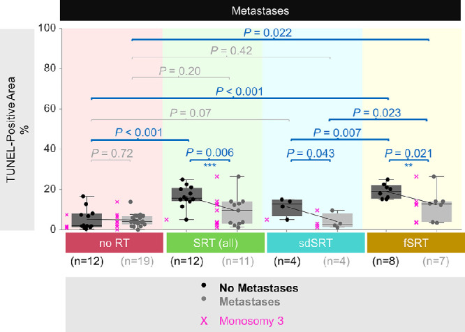
The amount of DNA breaks in the total irradiated group was increased by 2.7-fold (P < 0.001) compared to non-irradiated tissue. Tumors treated with fSRT were affected more severely, showing 2.1-fold more DNA damage (P = 0.007) compared to the cases after single (high) dose irradiation (sdSRT). Monosomy 3 tumors showed less DNA breaks compared to disomy 3 samples (P = 0.004). The presence of metastases after radiotherapy correlated with monosomy 3 and less DNA breaks compared to patients with non-metastatic cancer in the combined group with fSRT and sdSRT (P < 0.05).
Fractionated irradiation led to more DNA damage than single-dose treatment in primary UM. As tumors with monosomy 3 showed less DNA breaks than those with disomy 3, this may indicate that they are less radiosensitive, which may influence the efficacy of irradiation.
本研究旨在分析原发性葡萄膜黑色素瘤(UM)的 DNA 断裂程度与放疗剂量(单次剂量与分次剂量)和单体型 3 状态的关系。
共纳入 54 例 UM 患者。23 例患者接受立体定向放疗(SRT),其中 8 例接受单次剂量 SRT(sdSRT)治疗,15 例接受分次剂量 SRT(fSRT)治疗。通过 TUNEL 检测法观察离体或经内切除肿瘤中的 DNA 断裂,并通过测量 TUNEL 阳性面积进行定量分析。采用免疫组织化学法分析蛋白表达。通过免疫荧光原位杂交法检测染色体 3 与蛋白的共检测。
与未照射组织相比,总照射组的 DNA 断裂量增加了 2.7 倍(P < 0.001)。接受 fSRT 治疗的肿瘤受影响更为严重,与单次(高)剂量照射(sdSRT)后的病例相比,显示出 2.1 倍的更多 DNA 损伤(P = 0.007)。单体型 3 肿瘤的 DNA 断裂较二倍体 3 样本少(P = 0.004)。与 fSRT 和 sdSRT 联合组中无转移的癌症患者相比,放疗后发生转移的患者与单体型 3 存在相关性,且 DNA 断裂较少(P < 0.05)。
与单次剂量治疗相比,原发性 UM 中的分次照射导致更多的 DNA 损伤。由于单体型 3 的肿瘤比二倍体 3 的肿瘤显示出更少的 DNA 断裂,这可能表明它们的放射敏感性较低,这可能会影响照射的疗效。