Radiology Department, Hospital Universitari de Bellvitge, Barcelona, Spain.
Neuro-oncology Unit, Institut d'Investigació Biomèdica de Bellvitge- IDIBELL, Barcelona, Spain.
Neuroradiology. 2024 Aug;66(8):1267-1277. doi: 10.1007/s00234-024-03385-0. Epub 2024 Jun 5.
The presurgical discrimination of IDH-mutant astrocytoma grade 4 from IDH-wildtype glioblastoma is crucial for patient management, especially in younger adults, aiding in prognostic assessment, guiding molecular diagnostics and surgical planning, and identifying candidates for IDH-targeted trials. Despite its potential, the full capabilities of DSC-PWI remain underexplored. This research evaluates the differentiation ability of relative-cerebral-blood-volume (rCBV) percentile values for the enhancing and non-enhancing tumor regions compared to the more commonly used mean or maximum preselected rCBV values.
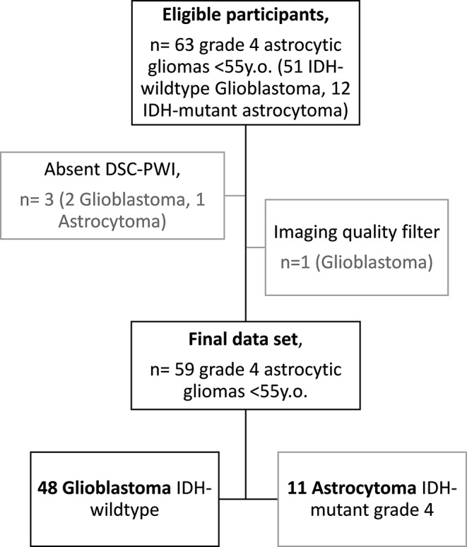
This retrospective study, spanning 2016-2023, included patients under 55 years (age threshold based on World Health Organization recommendations) with grade 4 astrocytic tumors and known IDH status, who underwent presurgical MR with DSC-PWI. Enhancing and non-enhancing regions were 3D-segmented to calculate voxel-level rCBV, deriving mean, maximum, and percentile values. Statistical analyses were conducted using the Mann-Whitney U test and AUC-ROC.
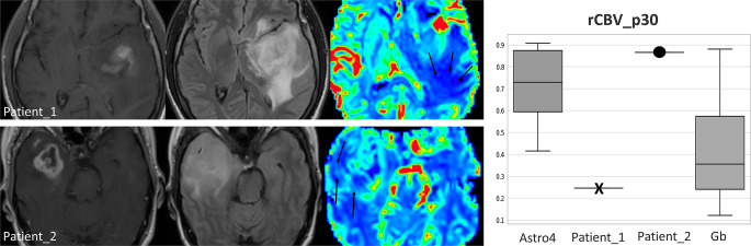
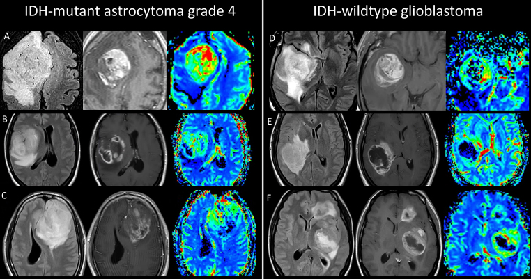
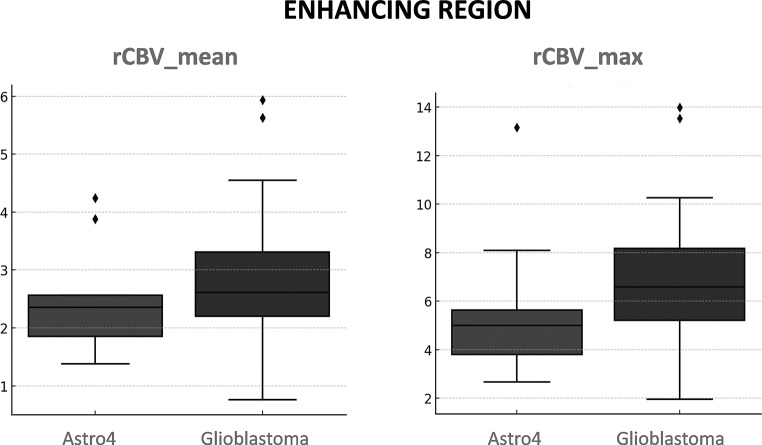
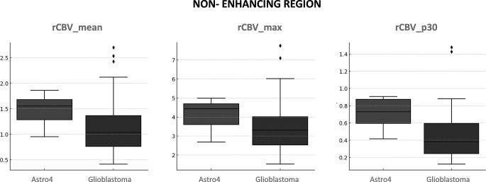
The cohort consisted of 59 patients (mean age 46; 34 male): 11 astrocytoma-4 and 48 glioblastoma. While glioblastoma showed higher rCBV in enhancing regions, the differences were not significant. However, non-enhancing astrocytoma-4 regions displayed notably higher rCBV, particularly in lower percentiles. The 30th rCBV percentile for non-enhancing regions was 0.705 in astrocytoma-4, compared to 0.458 in glioblastoma (p = 0.001, AUC-ROC = 0.811), outperforming standard mean and maximum values.
Employing an automated percentile-based approach for rCBV selection enhances differentiation capabilities, with non-enhancing regions providing more insightful data. Elevated rCBV in lower percentiles of non-enhancing astrocytoma-4 is the most distinguishable characteristic and may indicate lowly vascularized infiltrated edema, contrasting with glioblastoma's pure edema.
术前鉴别 IDH 突变型星形细胞瘤 4 级与 IDH 野生型胶质母细胞瘤对患者管理至关重要,尤其是在年轻患者中,有助于预后评估、指导分子诊断和手术计划,并识别 IDH 靶向试验的候选者。尽管其具有潜力,但 DSC-PWI 的全部功能仍未得到充分探索。本研究评估了与更常用的均值或最大预选 rCBV 值相比,增强和非增强肿瘤区域的相对脑血容量(rCBV)百分位数值的区分能力。
这项回顾性研究于 2016 年至 2023 年期间纳入了年龄在 55 岁以下(基于世界卫生组织建议的年龄阈值)、患有 4 级星形细胞瘤且 IDH 状态已知的患者,他们接受了 DSC-PWI 术前磁共振成像检查。通过 3D 分割增强和非增强区域以计算体素水平的 rCBV,得出均值、最大值和百分位数值。使用 Mann-Whitney U 检验和 AUC-ROC 进行统计分析。
该队列包括 59 名患者(平均年龄 46 岁;34 名男性):11 名星形细胞瘤-4 级和 48 名胶质母细胞瘤。虽然胶质母细胞瘤在增强区域显示出更高的 rCBV,但差异不显著。然而,非增强星形细胞瘤-4 区域显示出明显更高的 rCBV,特别是在较低的百分位数。非增强区域的第 30 个 rCBV 百分位数在星形细胞瘤-4 级中为 0.705,而在胶质母细胞瘤中为 0.458(p=0.001,AUC-ROC=0.811),优于标准均值和最大值。
采用基于自动百分位的 rCBV 选择方法可增强区分能力,非增强区域提供更具洞察力的数据。非增强星形细胞瘤-4 级中较低百分位的 rCBV 升高是最具区别特征,可能表明低度血管化浸润性水肿,与胶质母细胞瘤的纯水肿形成对比。