Zhou Jianan, Duan Shaofeng, Zhu Zhengyang, Wang Han, Tian Chuanshuai, Yang Huiquan, Chen Sixuan, Ye Meiping, Zhang Xin, Zhang Bing
Department of Radiology, Nanjing Drum Tower Hospital Clinical College of Nanjing Medical University, Nanjing, China.
Institute of Medical Imaging and Artificial Intelligence, Nanjing University, Nanjing, China.
Quant Imaging Med Surg. 2025 May 1;15(5):4734-4747. doi: 10.21037/qims-24-1459. Epub 2025 Apr 24.
There has been no research based on dynamic contrast-enhanced magnetic resonance imaging (DCE-MRI) radiomics for the stratification diagnosis and prognostic evaluation of gliomas. The study aimed to identify multiple glioma subtypes and decipher the gene expression profiles linked with different subtypes.
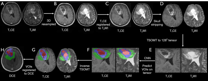
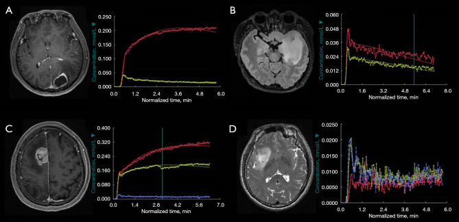
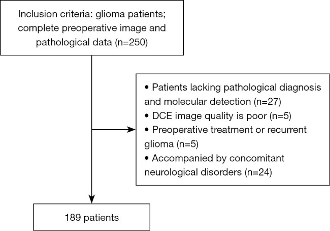
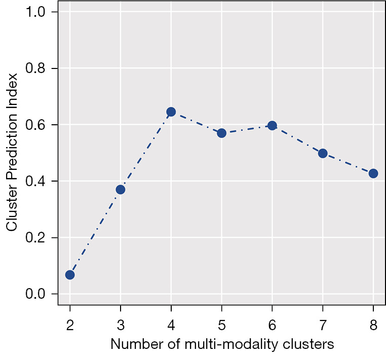
Cross-sectional and retrospective data of 189 patients were collected. The static radiomics features were obtained at three time points (0, 90, and 300 s) corresponding to pre-contrast, arterial, and delayed phases, respectively. The dynamic radiomics features were retrieved by determining the temporal anisotropy of these three phases. Multi-omics clustering was used to identify intrinsic radiomics subtypes within the cohort. The association between the radiomics clusters and gene expression profiles was evaluated through the analysis of variance.
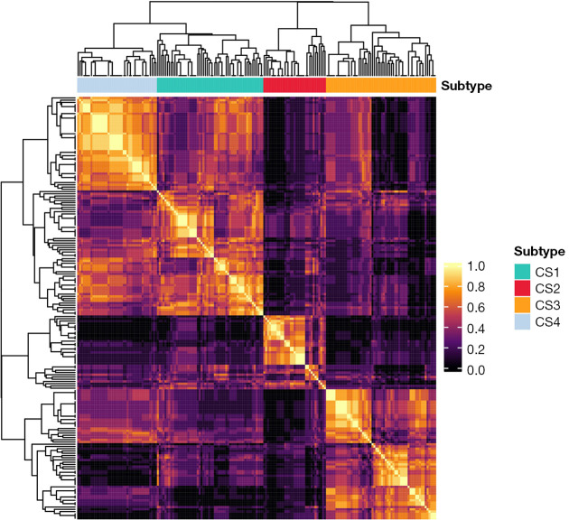
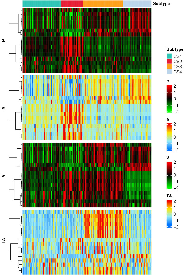
The patients in cluster 3 were oldest. Cluster 3 and cluster 1 had higher frequency of grade 4, high Ki-67 level, glioblastoma isocitrate dehydrogenase (IDH) wild-type, and unmethylated O6-methylguanine-DNA methyltransferase (MGMT) promoter. Cluster 3 had the highest frequency of epidermal growth factor receptor (EGFR) amplification and cyclin-dependent kinase inhibitor (CDKN) 2A/B homozygous deletion. Cluster 1 had the highest frequency of EGFR non-mutant. Cluster 4 and cluster 2 had a higher frequency of astrocytoma IDH-mutant. Cluster 4 had a higher frequency of grade 3, oligodendroglioma IDH-mutant and 1p/19q codeleted, MGMT promoter methylation, and EGFR non-amplification. Cluster 2 had a higher frequency of grade 2, low Ki-67 level, and patients without CDKN 2A/B homozygous deletion. There were no associations for other molecular markers between clusters.
The intrinsic imaging subtypes obtained from DCE-MRI radiomics features provide a new insight into glioma classification, potentially guiding the diagnosis.
目前尚无基于动态对比增强磁共振成像(DCE-MRI)影像组学对胶质瘤进行分层诊断和预后评估的研究。本研究旨在识别多种胶质瘤亚型,并解读与不同亚型相关的基因表达谱。
收集189例患者的横断面和回顾性数据。分别在对应于平扫、动脉期和延迟期的三个时间点(0、90和300秒)获取静态影像组学特征。通过确定这三个时期的时间各向异性来提取动态影像组学特征。采用多组学聚类方法识别队列中的内在影像组学亚型。通过方差分析评估影像组学聚类与基因表达谱之间的关联。
3组患者年龄最大。3组和1组的4级、高Ki-67水平、胶质母细胞瘤异柠檬酸脱氢酶(IDH)野生型和O6-甲基鸟嘌呤-DNA甲基转移酶(MGMT)启动子未甲基化的频率较高。3组表皮生长因子受体(EGFR)扩增和细胞周期蛋白依赖性激酶抑制剂(CDKN)2A/B纯合缺失的频率最高。1组EGFR非突变的频率最高。4组和2组星形细胞瘤IDH突变的频率较高。4组3级、少突胶质细胞瘤IDH突变和1p/19q共缺失、MGMT启动子甲基化以及EGFR非扩增的频率较高。2组2级、低Ki-67水平以及无CDKN 2A/B纯合缺失患者的频率较高。各聚类之间的其他分子标志物无关联。
从DCE-MRI影像组学特征获得的内在影像亚型为胶质瘤分类提供了新的见解,可能有助于指导诊断。