Shengli Clinical Medical College of Fujian Medical University, Fujian, Fuzhou, China.
Department of Radiology, Fujian Provincial Hospital, Fujian, Fuzhou, China.
Front Immunol. 2024 May 22;15:1391280. doi: 10.3389/fimmu.2024.1391280. eCollection 2024.
Currently, there is a lack of an objective quantitative measure to comprehensively evaluate the inflammatory activity of axSpA, which poses certain challenges in accurately assessing the disease activity.
To explore the value of combined-parameter models of sacroiliac joints (SIJs) MRI relaxometry and peripheral blood Mucosal-associated invariant T (MAIT) cells in evaluating the inflammatory activity of axial spondyloarthritis (axSpA).
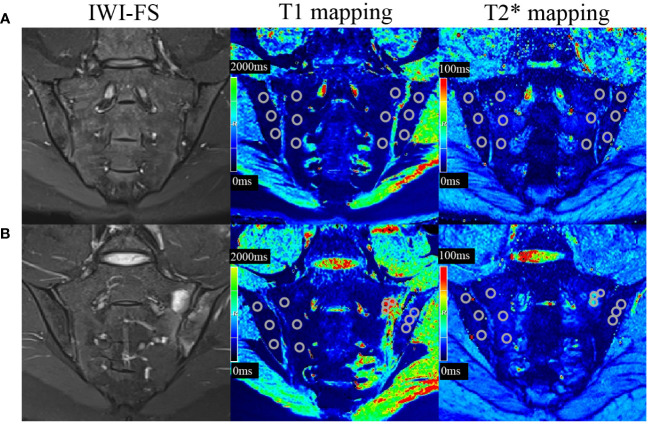
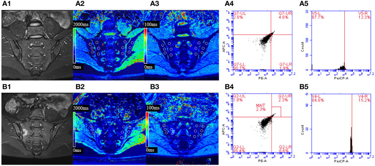
This retrospective clinical study included 88 axSpA patients (median age 31.0 (22.0, 41.8) years, 21.6% females) and 20 controls (median age 28.0 (20.5, 49.5) years, 40.0% females). The axSpA group was classified into active subgroup (n=50) and inactive subgroup (n=38) based on ASDAS-CRP. All participants underwent SIJs MRI examination including T1 and T2* mapping, and peripheral blood flow cytometry analysis of MAIT cells (defined as CD3Vα7.2CD161) and their activation markers (CD69). The T1 and T2* values, as were the percentages of MAIT cells and CD69MAIT cells were compared between different groups. Combined-parameter models were established using logistic regression, and ROC curves were employed to evaluate the diagnostic efficacy.
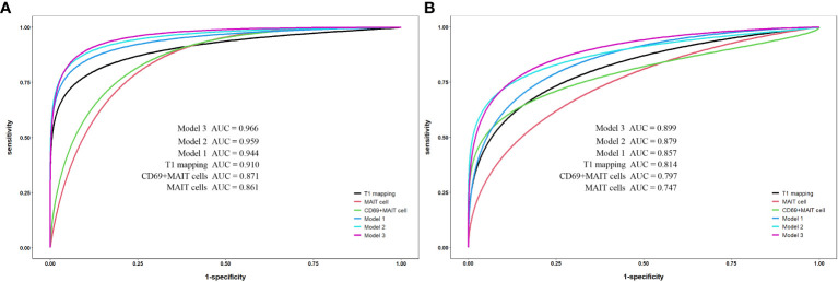
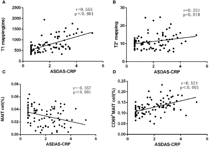
The T1 values of SIJs and %CD69MAIT cells in the axSpA group and its subgroup were higher than the control group (p<0.05), while %MAIT cells were lower than the control group (p<0.05). The T1 values and %CD69MAIT cells correlated positively, while %MAIT cells correlated negatively, with the ASDAS-CRP (r=0.555, 0.524, -0.357, p<0.001). Between the control and axSpA groups, and between the inactive and active subgroups, the combined-parameter model T1 mapping+%CD69MAIT cells has the best efficacy (AUC=0.959, 0.879, sensibility=88.6, 70%, specificity=95.0, 94.7%, respectively).
The combined-parameter model T1 mapping+%CD69MAIT cells allows a more accurate evaluation of the level of inflammatory activity.
目前,缺乏一种客观的定量方法来全面评估 axSpA 的炎症活动,这给准确评估疾病活动带来了一定的挑战。
探讨骶髂关节(SIJ)MRI 弛豫度和外周血黏膜相关不变 T(MAIT)细胞联合参数模型在评估轴性脊柱关节炎(axSpA)炎症活动中的价值。
本回顾性临床研究纳入了 88 例 axSpA 患者(中位年龄 31.0(22.0,41.8)岁,21.6%为女性)和 20 名对照者(中位年龄 28.0(20.5,49.5)岁,40.0%为女性)。根据 ASDAS-CRP,将 axSpA 组分为活动亚组(n=50)和非活动亚组(n=38)。所有参与者均接受 SIJ MRI 检查,包括 T1 和 T2* 成像,以及外周血 MAIT 细胞(定义为 CD3Vα7.2CD161)及其激活标志物(CD69)的流式细胞术分析。比较不同组间 SIJ 的 T1 和 T2* 值以及 MAIT 细胞和 CD69MAIT 细胞的百分比。使用逻辑回归建立联合参数模型,并采用 ROC 曲线评估诊断效能。
axSpA 组及其亚组的 SIJ T1 值和%CD69MAIT 细胞高于对照组(p<0.05),而 MAIT 细胞百分比低于对照组(p<0.05)。T1 值和%CD69MAIT 细胞与 ASDAS-CRP 呈正相关(r=0.555,0.524,p<0.001),而 MAIT 细胞与 ASDAS-CRP 呈负相关(r=-0.357,p<0.001)。在对照组与 axSpA 组之间,以及在非活动亚组与活动亚组之间,T1 映射+%CD69MAIT 细胞联合参数模型的效能最佳(AUC=0.959,0.879,敏感性=88.6%,特异性=95.0%;AUC=0.879,0.700,敏感性=70%,特异性=94.7%)。
T1 映射+%CD69MAIT 细胞联合参数模型可更准确地评估炎症活动水平。