• 文献检索
  • 文档翻译
  • 深度研究
  • 学术资讯
  • Suppr Zotero 插件Zotero 插件
  • 邀请有礼
  • 套餐&价格
  • 历史记录
应用&插件
Suppr Zotero 插件Zotero 插件浏览器插件Mac 客户端Windows 客户端微信小程序
定价
高级版会员购买积分包购买API积分包
服务
文献检索文档翻译深度研究API 文档MCP 服务
关于我们
关于 Suppr公司介绍联系我们用户协议隐私条款
关注我们

Suppr 超能文献

核心技术专利:CN118964589B侵权必究
粤ICP备2023148730 号-1Suppr @ 2026

文献检索

告别复杂PubMed语法,用中文像聊天一样搜索,搜遍4000万医学文献。AI智能推荐,让科研检索更轻松。

立即免费搜索

文件翻译

保留排版,准确专业,支持PDF/Word/PPT等文件格式,支持 12+语言互译。

免费翻译文档

深度研究

AI帮你快速写综述,25分钟生成高质量综述,智能提取关键信息,辅助科研写作。

立即免费体验

MRI T1、T2 和 T2* 图谱与组织学比较评估羊模型椎间盘退变。

Comparison of MRI T1, T2, and T2* mapping with histology for assessment of intervertebral disc degeneration in an ovine model.

机构信息

INSERM, UMRS 1229, Regenerative Medicine and Skeleton (RMeS), Université de Nantes, ONIRIS, 44042, Nantes, France.

Université de Nantes, UFR Odontologie, 44042, Nantes, France.

出版信息

Sci Rep. 2022 Mar 30;12(1):5398. doi: 10.1038/s41598-022-09348-w.

DOI:10.1038/s41598-022-09348-w
PMID:35354902
原文链接:https://pmc.ncbi.nlm.nih.gov/articles/PMC8967912/
Abstract

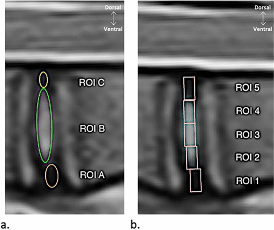
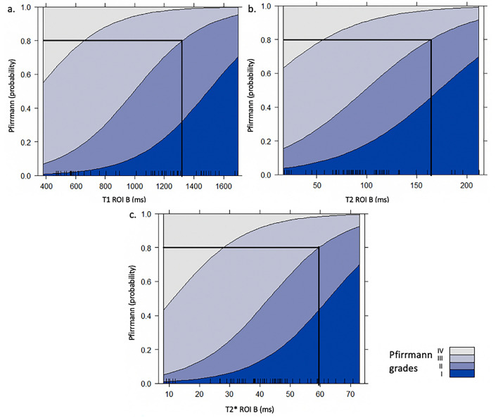
An easy, reliable, and time-efficient standardized approach for assessing lumbar intervertebral disc (IVD) degeneration with relaxation times measurements in pre-clinical and clinical studies is lacking. This prospective study aims to determine the most appropriate method for lumbar IVD degeneration (IDD) assessment in sheep by comparing three quantitative MRI sequences (variable-flip-angle T1 mapping, and multi-echo T2 and T2* mapping), correlating them with Pfirrmann grading and histology. Strong intra- and interrater agreements were found for Nucleus pulposus (NP) regions-of-interest (ROI). T1, T2, and T2* mapping correlated with Pfirrmann grading and histological scoring (p < 0.05) except for the most ventral rectangular ROI on T2 maps. Correlations were excellent for all of the T1 ROIs and the T2* NP ROIs. Highly significant differences in T1 values were found between all Pfirrmann grades except between grades I/II and between grades III/IV. Significant differences were identified in the T2 and the T2* values between all grades except between grades I/III. T1, T2, and T2* relaxation times measurements of the NP are an accurate and time-efficient tool to assess lumbar IDD in sheep. Variable-flip-angle T1 mapping may be further considered as a valuable method to investigate IDD and to assess the efficacy of regenerative treatments in longitudinal studies.

摘要

在临床前和临床研究中,缺乏一种简单、可靠且高效的评估腰椎间盘(IVD)退变的标准化方法,该方法可通过弛豫时间测量来实现。本前瞻性研究旨在通过比较三种定量 MRI 序列(可变翻转角 T1 映射、多回波 T2 和 T2映射),评估它们与 Pfirrmann 分级和组织学的相关性,从而确定评估绵羊腰椎 IVD 退变(IDD)的最合适方法。NP 感兴趣区(ROI)的核(NP)区域具有很强的内部和组内一致性。T1、T2 和 T2 映射与 Pfirrmann 分级和组织学评分相关(p<0.05),除了 T2 图上最腹侧的矩形 ROI 外。所有 T1 ROI 和 T2NP ROI 的相关性均较好。除 I/II 级和 III/IV 级之间外,所有 Pfirrmann 分级之间的 T1 值均存在显著差异。除 I/III 级之间外,所有分级之间的 T2 和 T2 值均存在显著差异。NP 的 T1、T2 和 T2*弛豫时间测量是评估绵羊腰椎 IDD 的一种准确且高效的工具。可变翻转角 T1 映射可进一步考虑作为一种有价值的方法来研究 IDD,并在纵向研究中评估再生治疗的疗效。

https://cdn.ncbi.nlm.nih.gov/pmc/blobs/044f/8967912/62eb403994d1/41598_2022_9348_Fig6_HTML.jpg
https://cdn.ncbi.nlm.nih.gov/pmc/blobs/044f/8967912/10836f1a479a/41598_2022_9348_Fig1_HTML.jpg
https://cdn.ncbi.nlm.nih.gov/pmc/blobs/044f/8967912/18e5ecef9ad3/41598_2022_9348_Fig2_HTML.jpg
https://cdn.ncbi.nlm.nih.gov/pmc/blobs/044f/8967912/19e3d1c4bee7/41598_2022_9348_Fig3_HTML.jpg
https://cdn.ncbi.nlm.nih.gov/pmc/blobs/044f/8967912/9efc3a66a7d1/41598_2022_9348_Fig4_HTML.jpg
https://cdn.ncbi.nlm.nih.gov/pmc/blobs/044f/8967912/20ce117942c0/41598_2022_9348_Fig5_HTML.jpg
https://cdn.ncbi.nlm.nih.gov/pmc/blobs/044f/8967912/62eb403994d1/41598_2022_9348_Fig6_HTML.jpg

相似文献

1
Comparison of MRI T1, T2, and T2* mapping with histology for assessment of intervertebral disc degeneration in an ovine model.MRI T1、T2 和 T2* 图谱与组织学比较评估羊模型椎间盘退变。
Sci Rep. 2022 Mar 30;12(1):5398. doi: 10.1038/s41598-022-09348-w.
2
Variable flip angle T1 mapping and multi-echo T2 and T2* mapping magnetic resonance imaging sequences allow quantitative assessment of canine lumbar disc degeneration.可变翻转角 T1 映射和多回波 T2 和 T2* 映射磁共振成像序列可定量评估犬腰椎间盘退变。
Vet Radiol Ultrasound. 2023 Sep;64(5):864-872. doi: 10.1111/vru.13288. Epub 2023 Aug 7.
3
Quantitative T2 evaluation at 3.0T compared to morphological grading of the lumbar intervertebral disc: a standardized evaluation approach in patients with low back pain.3.0T 下定量 T2 评估与腰椎间盘形态学分级的比较:腰痛患者的标准化评估方法。
Eur J Radiol. 2012 Feb;81(2):324-30. doi: 10.1016/j.ejrad.2010.12.093. Epub 2011 Feb 18.
4
Quantitative synthetic MRI for evaluation of the lumbar intervertebral disk degeneration in patients with chronic low back pain.定量磁共振成像在慢性下腰痛患者腰椎间盘退变评估中的应用。
Eur J Radiol. 2020 Mar;124:108858. doi: 10.1016/j.ejrad.2020.108858. Epub 2020 Jan 29.
5
Ultrashort time-to-echo T2* and T2* relaxometry for evaluation of lumbar disc degeneration: a comparative study.用于评估腰椎间盘退变的超短回波时间T2*及T2*弛豫测量法:一项对比研究
BMC Musculoskelet Disord. 2022 Jun 1;23(1):524. doi: 10.1186/s12891-022-05481-9.
6
T1ρ, T2 and T2* mapping of lumbar intervertebral disc degeneration: a comparison study.腰椎间盘退变的 T1ρ、T2 和 T2* 图谱:一项对比研究。
BMC Musculoskelet Disord. 2022 Dec 27;23(1):1135. doi: 10.1186/s12891-022-06040-y.
7
T2 relaxation time for intervertebral disc degeneration in patients with upper back pain: initial results on the clinical use of 3.0 Tesla MRI.上背部疼痛患者椎间盘退变的T2弛豫时间:3.0特斯拉磁共振成像临床应用的初步结果
BMC Med Imaging. 2017 Jan 31;17(1):9. doi: 10.1186/s12880-017-0182-z.
8
Quantitative T2 magnetic resonance imaging compared to morphological grading of the early cervical intervertebral disc degeneration: an evaluation approach in asymptomatic young adults.定量 T2 磁共振成像与早期颈椎间盘退变的形态学分级比较:无症状年轻成年人的评估方法。
PLoS One. 2014 Feb 3;9(2):e87856. doi: 10.1371/journal.pone.0087856. eCollection 2014.
9
T2 mapping of lumbar intervertebral disc: quantitative evaluation of degeneration in relation to Pfirrmann grading system and a template for intervertebral disc segmentation.腰椎间盘的T2映射:与Pfirrmann分级系统相关的退变定量评估及椎间盘分割模板
Hippokratia. 2023 Jul-Sep;27(3):75-81.
10
Identification of intervertebral disc regeneration with magnetic resonance imaging after a long-term follow-up in patients treated with percutaneous diode laser nucleoplasty: a retrospective clinical and radiological analysis of 14 patients.经皮二极管激光髓核成形术治疗后的长期随访中,通过磁共振成像识别椎间盘再生:14 例患者的回顾性临床和放射学分析。
Eur Spine J. 2014 May;23(5):1044-51. doi: 10.1007/s00586-014-3194-1. Epub 2014 Jan 31.

引用本文的文献

1
Physiological Ageing of the Lumbar Intervertebral Disc Based on Magnetic Resonance Imaging, a Systematic Literature Review.基于磁共振成像的腰椎间盘生理性老化:一项系统文献综述
Medicina (Kaunas). 2025 Aug 8;61(8):1430. doi: 10.3390/medicina61081430.
2
T2 mapping and q-Dixon for assessment of intervertebral disc degeneration in lower back pain.用于评估下腰痛患者椎间盘退变的T2映射和定量迪克森技术
BMC Musculoskelet Disord. 2025 Mar 11;26(1):234. doi: 10.1186/s12891-025-08453-x.
3
Quantitative observation based on magnetic resonance imaging: the effects of marathon running on lumbar vertebrae, paraspinal muscles, and lumbar disc components.

本文引用的文献

1
T2* Mapping of the Adult Intervertebral Lumbar Disc: Normative Data and Analysis of Diurnal Effects.成人腰椎间盘 T2* 映射:正常数据和昼夜效应分析。
J Orthop Res. 2019 Sep;37(9):1956-1962. doi: 10.1002/jor.24327. Epub 2019 May 20.
2
Evaluation of Lumbar Intervertebral Disc Degeneration Using T1ρ and T2 Magnetic Resonance Imaging in a Rabbit Disc Injury Model.在兔椎间盘损伤模型中使用T1ρ和T2磁共振成像评估腰椎间盘退变
Asian Spine J. 2018 Apr;12(2):317-324. doi: 10.4184/asj.2018.12.2.317. Epub 2018 Apr 16.
3
T1 and T2 Mapping in Cardiology: "Mapping the Obscure Object of Desire".
基于磁共振成像的定量观察:马拉松跑步对腰椎、椎旁肌肉和腰椎间盘各部分的影响。
Quant Imaging Med Surg. 2024 Dec 5;14(12):8811-8823. doi: 10.21037/qims-24-1053. Epub 2024 Nov 13.
4
Magnetic resonance imaging classification in a percutaneous needle injury rat model of intervertebral disc degeneration.磁共振成像分类在经皮穿刺针损伤椎间盘退变大鼠模型中的应用。
J Orthop Surg Res. 2024 Oct 7;19(1):632. doi: 10.1186/s13018-024-05110-2.
5
Qualitative and Quantitative MR Imaging of the Cartilaginous Endplate: A Review.软骨终板的定性和定量磁共振成像:综述
J Magn Reson Imaging. 2025 Apr;61(4):1552-1571. doi: 10.1002/jmri.29562. Epub 2024 Aug 20.
6
Mitochondria-engine with self-regulation to restore degenerated intervertebral disc cells via bioenergetic robust hydrogel design.具有自我调节功能的线粒体引擎,通过生物能量强大的水凝胶设计来恢复退变的椎间盘细胞。
Bioact Mater. 2024 May 31;40:1-18. doi: 10.1016/j.bioactmat.2024.05.044. eCollection 2024 Oct.
7
Inflammatory activity evaluation in patients with axial spondyloarthritis using MRI relaxometry and mucosal-associated invariant T cells.采用 MRI 弛豫率和黏膜相关不变 T 细胞评估中轴型脊柱关节炎患者的炎症活动度。
Front Immunol. 2024 May 22;15:1391280. doi: 10.3389/fimmu.2024.1391280. eCollection 2024.
8
Plant-Derived Senotherapeutics for the Prevention and Treatment of Intervertebral Disc Degeneration and Aging.用于预防和治疗椎间盘退变与衰老的植物源衰老治疗药物
Metabolites. 2024 Feb 28;14(3):146. doi: 10.3390/metabo14030146.
9
Characterization of Age-Related and Sex-Related Differences of Relaxation Parameters in the Intervertebral Disc Using MR-Fingerprinting.利用磁共振指纹技术对椎间盘弛豫参数的年龄相关性和性别相关性差异进行特征描述。
J Magn Reson Imaging. 2024 Apr;59(4):1312-1324. doi: 10.1002/jmri.28925. Epub 2023 Aug 23.
10
T1ρ, T2 and T2* mapping of lumbar intervertebral disc degeneration: a comparison study.腰椎间盘退变的 T1ρ、T2 和 T2* 图谱:一项对比研究。
BMC Musculoskelet Disord. 2022 Dec 27;23(1):1135. doi: 10.1186/s12891-022-06040-y.
心脏病学中的T1和T2映射:“描绘难以捉摸的心仪对象”
Cardiology. 2017;138(4):207-217. doi: 10.1159/000478901. Epub 2017 Aug 17.
4
Real T1 relaxation time measurement and diurnal variation analysis of intervertebral discs in a healthy population of 50 volunteers.50名健康志愿者椎间盘的真实T1弛豫时间测量及日变化分析
Eur J Radiol. 2017 Feb;87:13-19. doi: 10.1016/j.ejrad.2016.12.001. Epub 2016 Dec 6.
5
Functional MR Imaging Mapping of Human Articular Cartilage Response to Loading.功能性磁共振成像对人体关节软骨负重反应的映射。
Radiology. 2017 Feb;282(2):464-474. doi: 10.1148/radiol.2016160053. Epub 2016 Aug 26.
6
Prevalence of Age-Related Changes in Ovine Lumbar Intervertebral Discs during Computed Tomography and Magnetic Resonance Imaging.绵羊腰椎间盘在计算机断层扫描和磁共振成像过程中与年龄相关变化的患病率
Comp Med. 2016;66(4):300-7.
7
T1 Mapping: Basic Techniques and Clinical Applications.T1 映射:基本技术与临床应用。
JACC Cardiovasc Imaging. 2016 Jan;9(1):67-81. doi: 10.1016/j.jcmg.2015.11.005.
8
T2* mapping of ovine intervertebral discs: Normative data for cervical and lumbar spine.绵羊椎间盘的T2* 成像:颈椎和腰椎的正常数据
J Orthop Res. 2016 Apr;34(4):717-24. doi: 10.1002/jor.23071. Epub 2015 Oct 26.
9
Comparison of T1ρ and T2* Relaxation Mapping in Patients with Different Grades of Disc Degeneration at 3T MR.3T磁共振成像下不同椎间盘退变程度患者的T1ρ与T2*弛豫成像比较
Med Sci Monit. 2015 Jul 4;21:1934-41. doi: 10.12659/MSM.894406.
10
MRI T2* mapping correlates with biochemistry and histology in intervertebral disc degeneration in a large animal model.在大型动物模型中,MRI T2* 映射与椎间盘退变中的生物化学和组织学相关。
Eur Spine J. 2015 Sep;24(9):1935-43. doi: 10.1007/s00586-014-3498-1. Epub 2014 Aug 5.