Mallio Carlo A, Volterrani Claudia, Bernetti Caterina, Stiffi Massimo, Greco Federico, Beomonte Zobel Bruno
Fondazione Policlinico Universitario Campus Bio-Medico, Rome, Italy.
Research Unit of Radiology, Department of Medicine and Surgery, Università Campus Bio-Medico di Roma, Rome, Italy.
Quant Imaging Med Surg. 2024 Jun 1;14(6):4189-4201. doi: 10.21037/qims-23-1770. Epub 2024 Apr 15.
Computed tomography (CT) and magnetic resonance imaging (MRI) of the spine are fundamental non-invasive tools to investigate the status of the bone and soft tissue in vivo. A novel and promising approach is to investigate the quality and quantity of paraspinal muscles even beyond the clinical question. The aim of the present review is to summarize current evidence on CT and MRI about the relationship between paraspinal muscular status and bone health in osteoporosis (OP) and fracture risk.
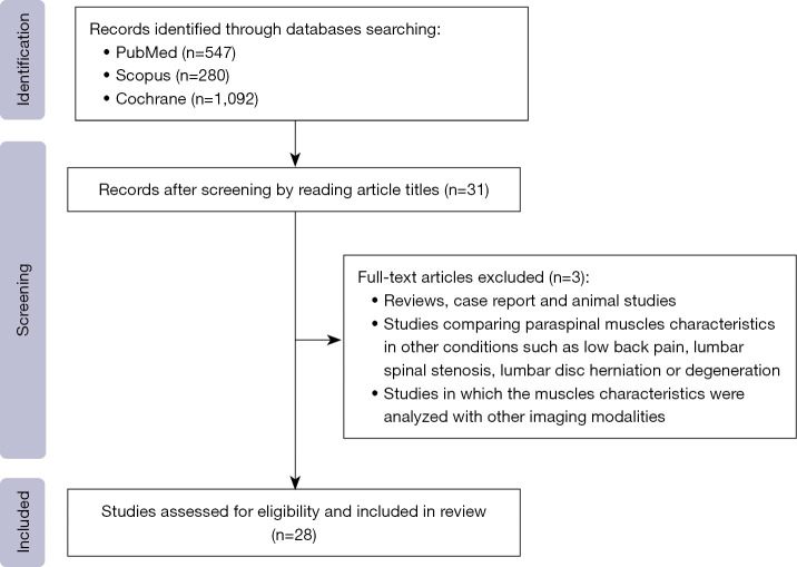
Literature research was carried out on September 2023 using PubMed, Scopus, and Cochrane databases.
Research investigating the intricate interplay between musculature and bone health reveals that degenerating paraspinal muscles, characterized by shrinking and fatty infiltration, are associated with lower bone mineral density (BMD) and the development of OP. Additionally, research indicates that weaker paraspinal muscles are linked to a higher risk of fractures, including those at the spine.
The findings suggest that paraspinal muscle health may be a significant factor in identifying individuals at risk for OP and fractures. Further investigation is needed to explore the potential of paraspinal muscles in preventing these conditions.
脊柱计算机断层扫描(CT)和磁共振成像(MRI)是研究体内骨骼和软组织状况的基本无创工具。一种新颖且有前景的方法是,甚至超出临床问题范畴来研究椎旁肌肉的质量和数量。本综述的目的是总结当前关于CT和MRI的证据,以探讨骨质疏松症(OP)中椎旁肌肉状态与骨骼健康之间的关系以及骨折风险。
2023年9月利用PubMed、Scopus和Cochrane数据库进行文献研究。
对肌肉组织与骨骼健康之间复杂相互作用的研究表明,以萎缩和脂肪浸润为特征的退化性椎旁肌肉与较低的骨矿物质密度(BMD)以及OP的发生有关。此外,研究表明较弱的椎旁肌肉与更高的骨折风险相关,包括脊柱骨折。
研究结果表明,椎旁肌肉健康可能是识别OP和骨折风险个体的一个重要因素。需要进一步研究以探索椎旁肌肉在预防这些病症方面的潜力。