Subramonian Sakthi Ganesh, R K Karpagam, Raja Sam, Lucas Ajay, Natarajan Paarthipan
Department of Radio-Diagnosis, Saveetha Medical College and Hospital, Saveetha Institute of Medical and Technical Sciences (SIMATS) Saveetha University, Chennai, IND.
Cureus. 2024 May 11;16(5):e60097. doi: 10.7759/cureus.60097. eCollection 2024 May.
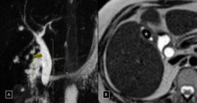
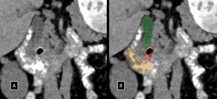
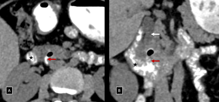
Lemmel syndrome, characterized by biliary and pancreatic duct obstruction secondary to a periampullary duodenal diverticulum (PAD), remains a rare and often overlooked diagnosis. Although duodenal diverticula are found in approximately 23% of the population, only about 5% of these cases lead to complications such as obstructions. A new case of Lemmel syndrome is demonstrated in the article about a middle-aged woman with chronic epigastric pain and right upper abdominal quadrant initially misdiagnosed as cholelithiasis. The accuracy of diagnosis was made possible by multimodal imaging methods, such as ultrasound, magnetic resonance cholangiopancreatography (MRCP), and computed tomography (CT) with oral contrast when a diffuse common bile duct was compressed by a PAD. Additionally, it highlights the necessity of including Lemmel syndrome in cases where patients have dilated bile ducts without calculi or mass lesions while emphasizing advanced imaging techniques for the revelation of structural malformations that underlay these conditions. The endoscopic intervention was minimally invasive but effective in relieving symptoms through sphincterotomy followed by laparoscopic cholecystectomy and biliary stent placement, thus making a point of the need for multiple disciplinary approaches toward treatment rare phenomenon like this one. This case report not only sheds light on the diagnostic and therapeutic avenues for Lemmel syndrome but also serves as a valuable educational resource for healthcare professionals. It emphasizes the need for heightened clinical vigilance and the adept use of imaging modalities in cases of biliary obstruction with obscure etiology. By contributing to the growing knowledge of this rare condition, we aim to facilitate timely diagnosis and optimize patient outcomes.
莱梅尔综合征,其特征为继发于壶腹周围十二指肠憩室(PAD)的胆管和胰管梗阻,仍然是一种罕见且常被忽视的诊断。尽管十二指肠憩室在约23%的人群中被发现,但这些病例中只有约5%会导致诸如梗阻等并发症。本文展示了一例新的莱梅尔综合征病例,患者为一名中年女性,最初因慢性上腹部疼痛和右上腹误诊为胆石症。当PAD压迫胆总管导致弥漫性改变时,通过多模态成像方法,如超声、磁共振胰胆管造影(MRCP)以及口服造影剂的计算机断层扫描(CT),得以准确诊断。此外,它强调了在患者胆管扩张但无结石或肿块病变的情况下考虑莱梅尔综合征的必要性,同时强调先进成像技术对于揭示这些情况背后结构畸形的作用。内镜干预微创且有效,通过括约肌切开术,随后进行腹腔镜胆囊切除术和胆管支架置入来缓解症状,从而凸显了针对此类罕见现象采取多学科治疗方法的必要性。本病例报告不仅阐明了莱梅尔综合征的诊断和治疗途径,也为医护人员提供了宝贵的教育资源。它强调在病因不明的胆管梗阻病例中提高临床警惕性和熟练使用成像方式的必要性。通过增加对这种罕见病症的认识,我们旨在促进及时诊断并优化患者预后。