Frauenfelder Giulia, Maraziti Annamaria, Ciccone Vincenzo, Maraziti Giuliano, Caleo Oliviero, Giurazza Francesco, Zobel Bruno Beomonte, Carbone Mattia
Departments of Radiology, Università Campus-Bio Medico di Roma, Via A. del Portillo, Rome.
Departments of Radiology, San Giovanni e Ruggi D'Aragona Hospital, Ospedale, Via San Leonardo, Salerno.
J Clin Imaging Sci. 2019 May 24;9:23. doi: 10.25259/JCIS-17-2019. eCollection 2019.
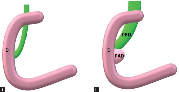
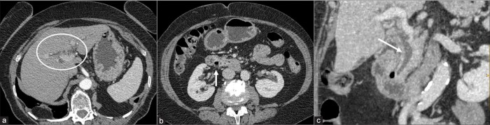
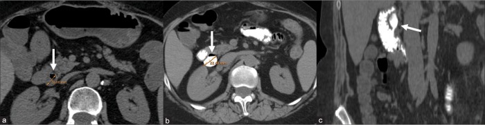
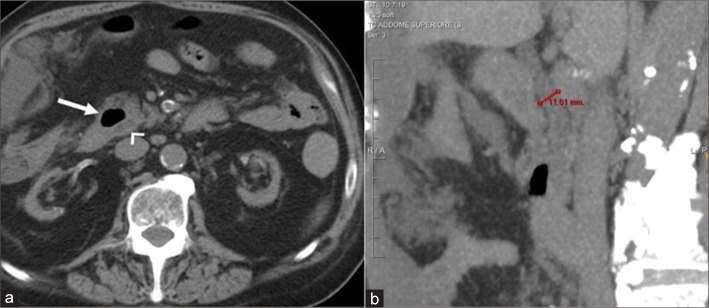
Lemmel syndrome is a rare and misdiagnosed cause of acute abdominal pain due to a juxtapapillary duodenal diverticulum causing mechanical obstruction of the common bile duct. Frequently, patients suffering from Lemmel syndrome have a history of recurrent access to the emergency room for acute abdominal pain referable to a biliopancreatic obstruction, in the absence of lithiasis nuclei or solid lesions at radiological examinations. Ultrasonography (US) may be helpful in evaluation of upstream dilatation of extra-/intra-hepatic biliary duct, but computed tomography (CT) is the reference imaging modality for the diagnosis of periampullary duodenal diverticula compressing the intrapancreatic portion of the common bile duct. Recognition of this entity is crucial for targeted, timely therapy avoiding mismanagement and therapeutic delay. The aim of this paper is to report CT imaging findings and our experience in two patients affected by Lemmel syndrome.
莱梅尔综合征是一种罕见且易被误诊的急性腹痛病因,由十二指肠乳头旁憩室导致胆总管机械性梗阻引起。通常,患有莱梅尔综合征的患者有因胆胰管梗阻导致的急性腹痛而反复前往急诊室就诊的病史,而影像学检查未发现结石核心或实性病变。超声检查(US)可能有助于评估肝内外胆管的上游扩张情况,但计算机断层扫描(CT)是诊断压迫胆总管胰内段的壶腹周围十二指肠憩室的参考成像方式。认识到这一实体对于进行有针对性的及时治疗至关重要,可避免管理不当和治疗延误。本文旨在报告两名莱梅尔综合征患者的CT影像学表现及我们的经验。