Instituto de Neurociencias, Centro Interdisciplinario de Neurociencias, Universidad de Valparaíso, Valparaíso 2381850, Chile.
Center for Bioinformatics and Integrative Biology, Facultad de Ciencias de la Vida, Universidad Andrés Bello, Santiago 8370146, Chile.
Proc Natl Acad Sci U S A. 2024 Jun 18;121(25):e2405468121. doi: 10.1073/pnas.2405468121. Epub 2024 Jun 11.
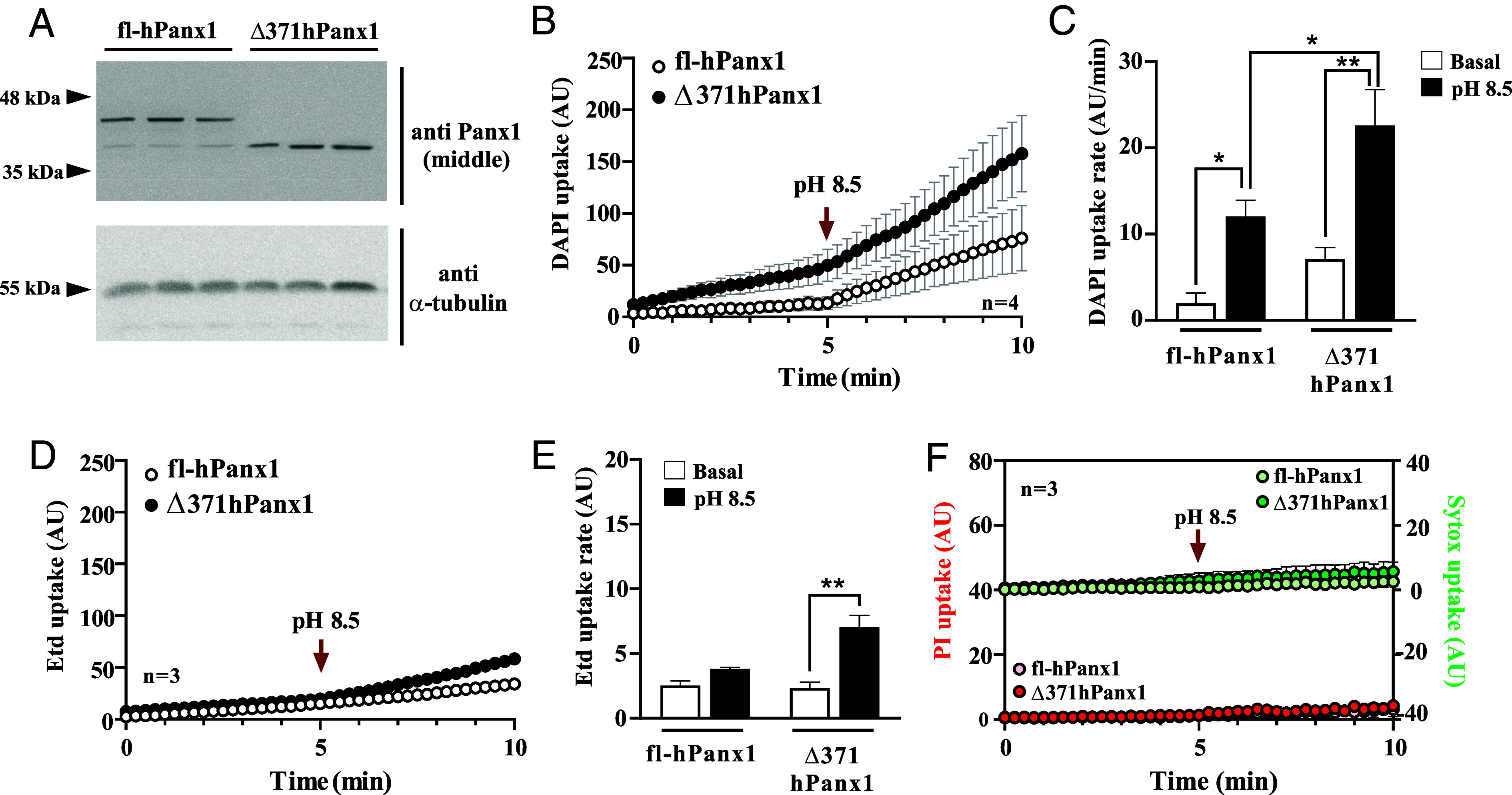
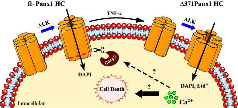
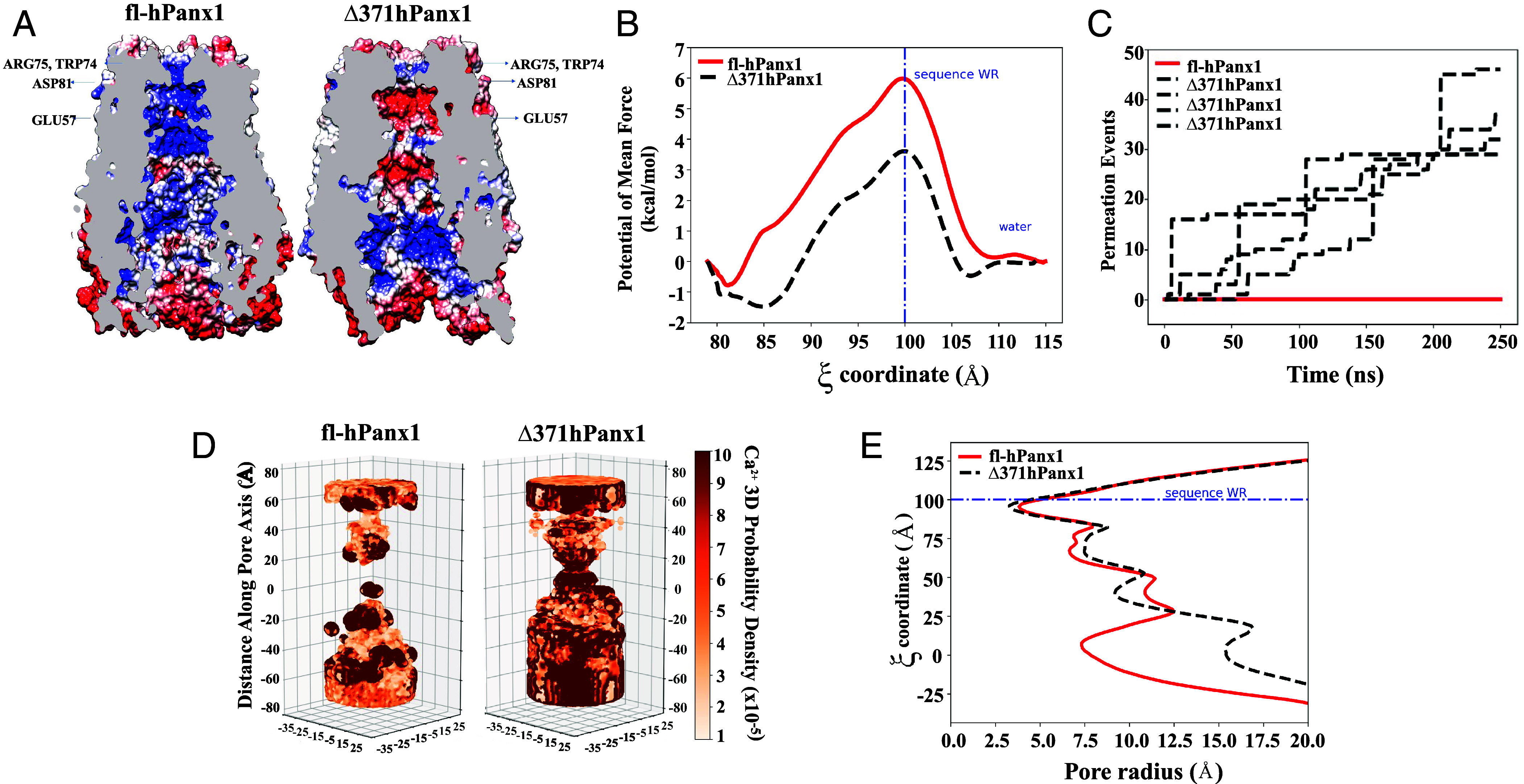
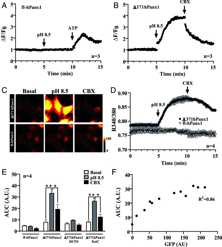
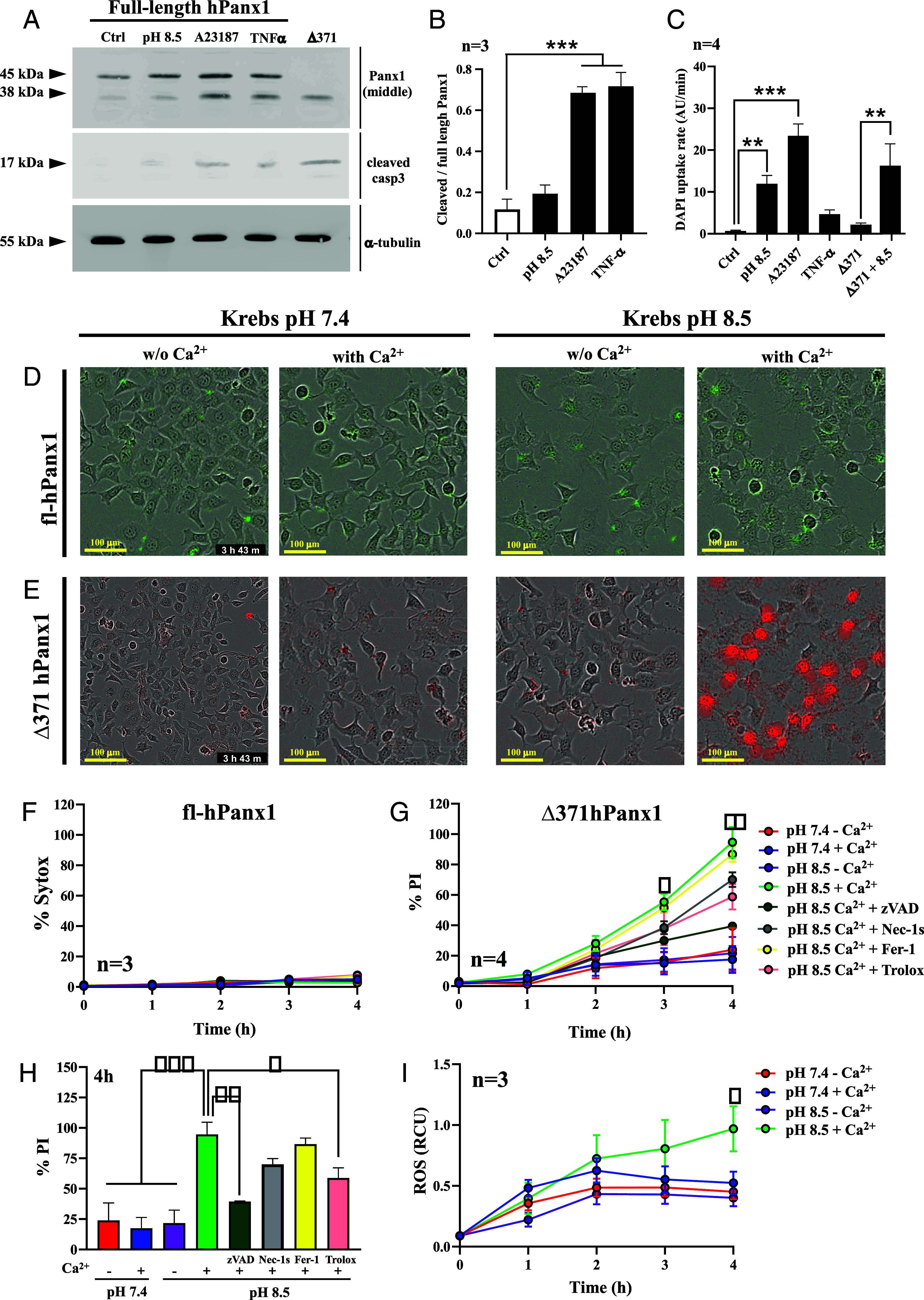
Pannexin1 hemichannels (Panx1 HCs) are found in the membrane of most mammalian cells and communicate the intracellular and extracellular spaces, enabling the passive transfer of ions and small molecules. They are involved in physiological and pathophysiological conditions. During apoptosis, the C-terminal tail of Panx1 is proteolytically cleaved, but the permeability features of hemichannels and their role in cell death remain elusive. To address these topics, HeLa cells transfected with full-length human Panx1 (fl-hPanx1) or C-terminal truncated hPanx1 (Δ371hPanx1) were exposed to alkaline extracellular saline solution, increasing the activity of Panx1 HCs. The Δ371hPanx1 HC was permeable to DAPI and Etd, but not to propidium iodide, whereas fl-hPanx1 HC was only permeable to DAPI. Furthermore, the cytoplasmic Ca signal increased only in Δ371hPanx1 cells, which was supported by bioinformatics approaches. The influx of Ca through Δ371hPanx1 HCs was necessary to promote cell death up to about 95% of cells, whereas the exposure to alkaline saline solution without Ca failed to induce cell death, and the Ca ionophore A23187 promoted more than 80% cell death even in fl-hPanx1 transfectants. Moreover, cell death was prevented with carbenoxolone or Panx1 in Δ371hPanx1 cells, whereas it was undetectable in HeLa Panx1 cells. Pretreatment with Ferrostatin-1 and necrostatin-1 did not prevent cell death, suggesting that ferroptosis or necroptosis was not involved. In comparison, zVAD-FMK, a pancaspase inhibitor, reduced death by ~60%, suggesting the involvement of apoptosis. Therefore, alkaline pH increases the activity of Δ371hPanx1HCs, leading to a critical intracellular free-Ca overload that promotes cell death.
缝隙连接蛋白 1 半通道(Panx1 HCs)存在于大多数哺乳动物细胞的膜中,沟通细胞内外空间,使离子和小分子被动转移。它们参与生理和病理生理条件。在细胞凋亡过程中,Panx1 的 C 端尾部被蛋白水解切割,但半通道的通透性特征及其在细胞死亡中的作用仍不清楚。为了解决这些问题,用全长人 Panx1(fl-hPanx1)或 C 端截断的 hPanx1(Δ371hPanx1)转染的 HeLa 细胞暴露于碱性细胞外盐溶液中,增加 Panx1 HCs 的活性。Δ371hPanx1 HC 对 DAPI 和 Etd 是可渗透的,但对碘化丙啶不可渗透,而 fl-hPanx1 HC 仅对 DAPI 是可渗透的。此外,只有在Δ371hPanx1 细胞中细胞质 Ca 信号增加,这得到了生物信息学方法的支持。通过Δ371hPanx1 HCs 流入的 Ca 对于促进细胞死亡是必要的,直到约 95%的细胞死亡,而没有 Ca 的碱性盐溶液暴露不能诱导细胞死亡,并且 Ca 离子载体 A23187 甚至在 fl-hPanx1 转染物中促进超过 80%的细胞死亡。此外,用 carbenoxolone 或 Panx1 在Δ371hPanx1 细胞中阻止细胞死亡,而在 HeLa Panx1 细胞中则无法检测到。用 Ferrostatin-1 和 necrostatin-1 预处理不能防止细胞死亡,表明铁死亡或坏死性凋亡不参与。相比之下,pan-caspase 抑制剂 zVAD-FMK 将死亡减少了约 60%,表明涉及细胞凋亡。因此,碱性 pH 值增加了Δ371hPanx1HCs 的活性,导致关键的细胞内游离 Ca 超载,从而促进细胞死亡。