Rigato Annafrancesca, Meng Huicheng, Chardes Claire, Runions Adam, Abouakil Faris, Smith Richard S, LeGoff Loïc
Aix Marseille Univ, CNRS, Centrale Marseille, Institut Fresnel UMR7249, Turing Center for Living Systems, Marseille, France.
Aix Marseille Univ, CNRS, IBDM UMR7288, Turing Center for Living Systems, Marseille, France.
PLoS Biol. 2024 Jun 13;22(6):e3002662. doi: 10.1371/journal.pbio.3002662. eCollection 2024 Jun.
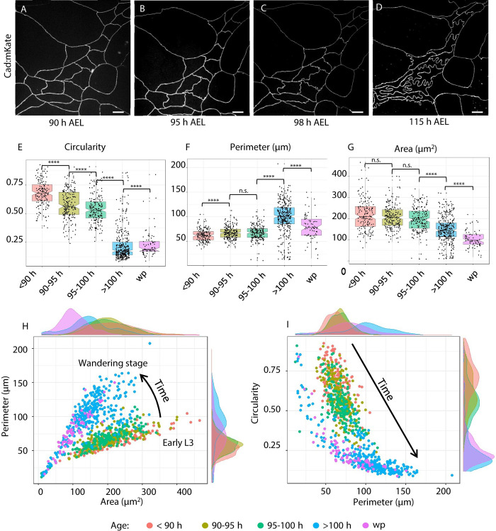
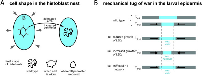
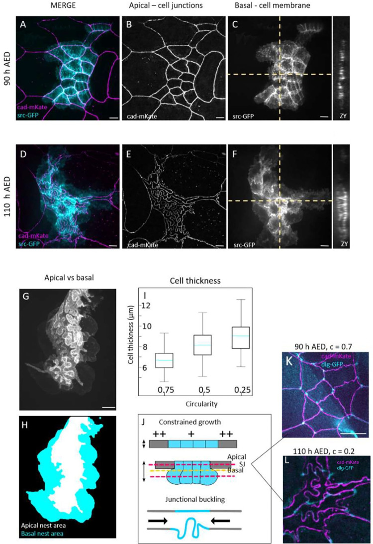
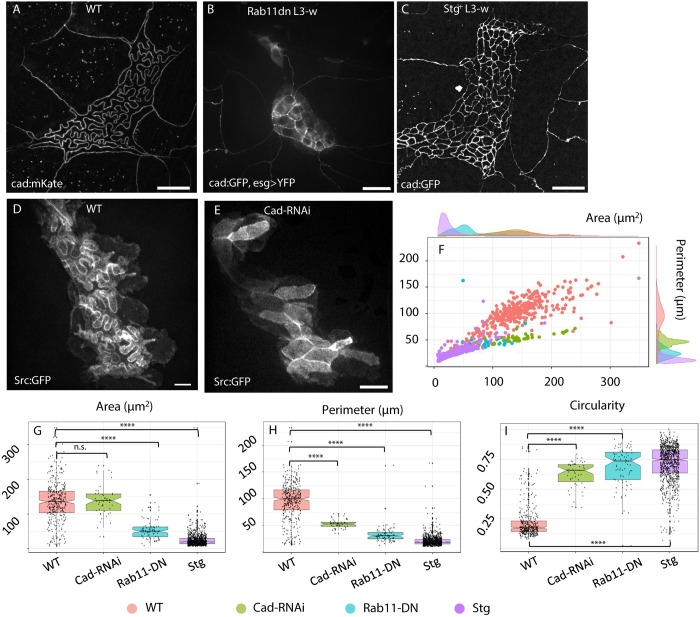
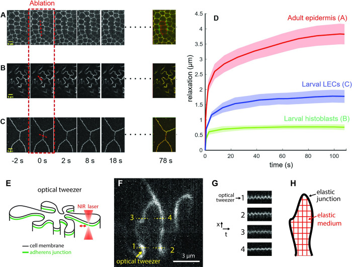
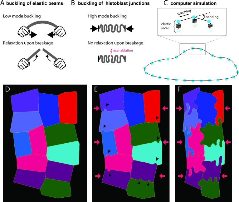
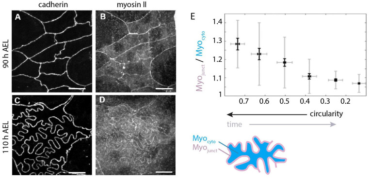
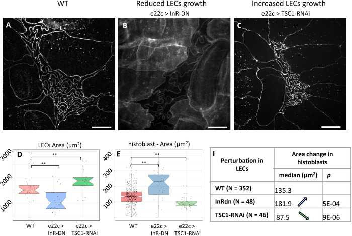
The polygonal shape of cells in proliferating epithelia is a result of the tensile forces of the cytoskeletal cortex and packing geometry set by the cell cycle. In the larval Drosophila epidermis, two cell populations, histoblasts and larval epithelial cells, compete for space as they grow on a limited body surface. They do so in the absence of cell divisions. We report a striking morphological transition of histoblasts during larval development, where they change from a tensed network configuration with straight cell outlines at the level of adherens junctions to a highly folded morphology. The apical surface of histoblasts shrinks while their growing adherens junctions fold, forming deep lobules. Volume increase of growing histoblasts is accommodated basally, compensating for the shrinking apical area. The folded geometry of apical junctions resembles elastic buckling, and we show that the imbalance between the shrinkage of the apical domain of histoblasts and the continuous growth of junctions triggers buckling. Our model is supported by laser dissections and optical tweezer experiments together with computer simulations. Our analysis pinpoints the ability of histoblasts to store mechanical energy to a much greater extent than most other epithelial cell types investigated so far, while retaining the ability to dissipate stress on the hours time scale. Finally, we propose a possible mechanism for size regulation of histoblast apical size through the lateral pressure of the epidermis, driven by the growth of cells on a limited surface. Buckling effectively compacts histoblasts at their apical plane and may serve to avoid physical harm to these adult epidermis precursors during larval life. Our work indicates that in growing nondividing cells, compressive forces, instead of tension, may drive cell morphology.
增殖上皮细胞的多边形形状是细胞骨架皮层的张力和细胞周期设定的堆积几何结构的结果。在果蝇幼虫表皮中,两种细胞群体,即组织母细胞和幼虫上皮细胞,在有限的体表上生长时会争夺空间。它们在没有细胞分裂的情况下这样做。我们报告了组织母细胞在幼虫发育过程中一个显著的形态转变,它们从粘着连接水平上具有笔直细胞轮廓的紧张网络构型转变为高度折叠的形态。组织母细胞的顶端表面收缩,而其不断生长的粘着连接折叠,形成深小叶。生长中的组织母细胞的体积增加在基部得到补偿,以弥补顶端面积的缩小。顶端连接的折叠几何形状类似于弹性屈曲,我们表明组织母细胞顶端区域的收缩与连接的持续生长之间的不平衡触发了屈曲。我们的模型得到了激光切割和光镊实验以及计算机模拟的支持。我们的分析指出,与迄今为止研究的大多数其他上皮细胞类型相比,组织母细胞储存机械能的能力要强得多,同时还保留了在数小时时间尺度上消散应力的能力。最后,我们提出了一种可能的机制,通过表皮的侧向压力来调节组织母细胞顶端大小,这种压力是由细胞在有限表面上的生长驱动的。屈曲有效地在顶端平面压缩组织母细胞,并可能有助于避免在幼虫期对这些成体表皮前体造成物理伤害。我们的工作表明,在生长的非分裂细胞中,压缩力而非张力可能驱动细胞形态。