Cooper Medical School of Rowan University, 3 Cooper plaza, suite 312, Camden, NJ 08103, USA.
Cooper Medical School of Rowan University, Camden, NJ, USA.
Ther Adv Respir Dis. 2024 Jan-Dec;18:17534666241259369. doi: 10.1177/17534666241259369.
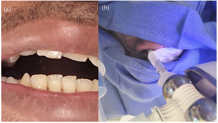
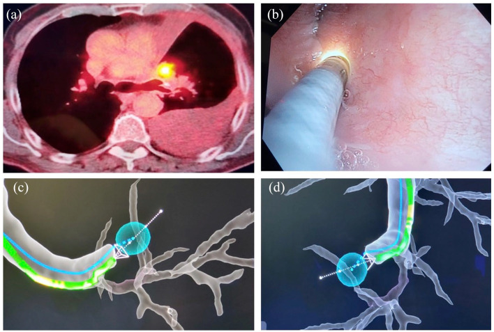
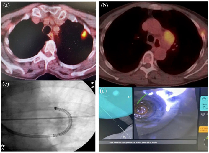
Robotic-assisted bronchoscopy (RAB) was recently added to the armamentarium of tools used in sampling peripheral lung nodules. Protocols and guidelines have since been published advocating use of large oral artificial airways, use of confirmatory technologies such as radial endobronchial ultrasound (R-EBUS), and preferably limiting sampling to pulmonary parenchymal lesions. We present three clinical cases where RAB was used unconventionally to sample pulmonary nodules in unusual locations and in patients with challenging airway anatomy. In case 1, we introduced the ion catheter through a nasal airway in a patient with trismus. In case 2, we established a diagnosis by sampling a station 5 lymph node, and in case 3, we sampled a lesion located behind an airway stump from previous thoracic surgery. All three patients would have presented significant challenges for alternative biopsy modalities such as CT-guided needle biopsy or video-assisted thoracic surgery.
机器人辅助支气管镜检查 (RAB) 最近被添加到用于采样外周肺结节的工具中。此后,已经发布了协议和指南,提倡使用大的口腔人工气道,使用径向支气管内超声 (R-EBUS) 等确认技术,并最好将采样限于肺实质病变。我们提出了三个临床病例,其中 RAB 非常规地用于采样位于不常见位置和具有挑战性气道解剖结构的患者的肺结节。在病例 1 中,我们通过患有牙关紧闭的患者的鼻气道引入了离子导管。在病例 2 中,我们通过采样第 5 站淋巴结来建立诊断,在病例 3 中,我们从以前的胸部手术后的气道残端后面采样了一个病变。对于其他活检方式,如 CT 引导下的针吸活检或电视辅助胸腔镜手术,所有这三个患者都将带来重大挑战。