Department of Biomedical Engineering, Istituto di Ricerche Farmacologiche Mario Negri IRCCS, Ranica, Italy.
Department of Biomedical Engineering, Istituto di Ricerche Farmacologiche Mario Negri IRCCS, Ranica, Italy.
Neuroimage Clin. 2024;43:103631. doi: 10.1016/j.nicl.2024.103631. Epub 2024 Jun 12.
The COVID-19 pandemic has affected millions worldwide, causing mortality and multi-organ morbidity. Neurological complications have been recognized. This study aimed to assess brain structural, microstructural, and connectivity alterations in patients with COVID-19-related olfactory or cognitive impairment using post-acute (time from onset: 264[208-313] days) multi-directional diffusion-weighted MRI (DW-MRI).
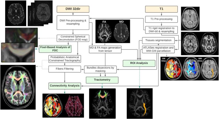
The study included 16 COVID-19 patients with cognitive impairment (COVID-CM), 35 COVID-19 patients with olfactory disorder (COVID-OD), and 14 controls. A state-of-the-art processing pipeline was developed for DW-MRI pre-processing, mean diffusivity and fractional anisotropy computation, fiber density and cross-section analysis, and tractography of white-matter bundles. Brain parcellation required for probing network connectivity, region-specific microstructure and volume, and cortical thickness was based on T1-weighted scans and anatomical atlases.
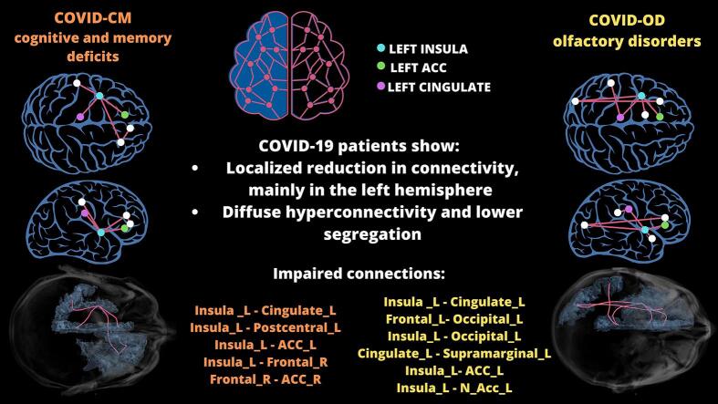
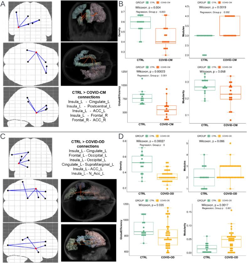
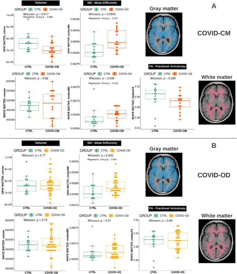
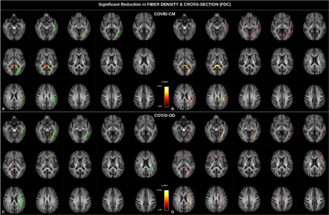
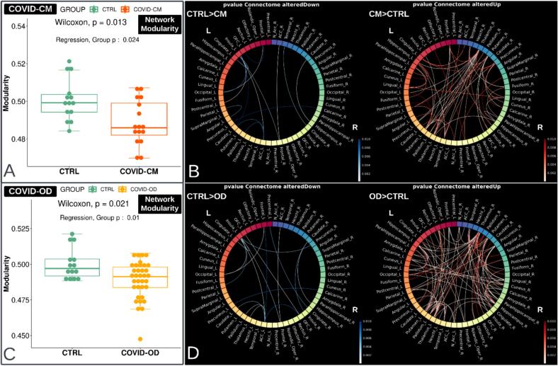
Compared to controls, COVID-CM patients showed overall gray matter atrophy (age and sex corrected p = 0.004), and both COVID-19 patient groups showed regional atrophy and cortical thinning. Both groups presented an increase in gray matter mean diffusivity (corrected p = 0.001), decrease in white matter fiber density and cross-section (corrected p < 0.05), , and COVID-CM patients also displayed an overall increased diffusivity (p = 0.022) and decreased anisotropy (corrected p = 0.038) in white matter. Graph-based analysis revealed reduced network modularity, with an extensive pattern of connectivity increase, in conjunction with a localized reduction in a few connections, mainly located in the left hemisphere. The left cingulate, anterior cingulate, and insula were primarily involved.
Expanding upon previous findings, this study further investigated significant alterations in brain morphology, microstructure, and connectivity in COVID-19 patients with olfactory or cognitive disfunction. These findings suggest underlying neurodegeneration, neuroinflammation, and concomitant compensatory mechanisms. Future longitudinal studies are required to monitor the alterations over time and assess their transient or permanent nature.
COVID-19 大流行已在全球范围内影响数百万人,导致死亡率和多器官发病。已认识到神经系统并发症。本研究旨在使用急性后(发病后时间:264[208-313]天)多方向扩散加权 MRI(DW-MRI)评估与 COVID-19 相关的嗅觉或认知障碍患者的脑结构、微观结构和连接变化。
该研究纳入了 16 名认知障碍(COVID-CM)COVID-19 患者、35 名嗅觉障碍(COVID-OD)COVID-19 患者和 14 名对照组。为 DW-MRI 预处理、平均弥散度和各向异性分数计算、纤维密度和横截面分析以及白质束追踪开发了一种最先进的处理流水线。用于探测网络连接、区域特定微观结构和体积以及皮质厚度的脑分割基于 T1 加权扫描和解剖图谱。
与对照组相比,COVID-CM 患者表现出总体灰质萎缩(年龄和性别校正后 p=0.004),两组 COVID-19 患者均表现出区域性萎缩和皮质变薄。两组患者均表现出灰质平均弥散度增加(校正后 p=0.001),白质纤维密度和横截面降低(校正后 p<0.05),COVID-CM 患者还表现出白质整体弥散度增加(p=0.022)和各向异性降低(校正后 p=0.038)。基于图的分析显示网络模块性降低,连接增加呈广泛模式,同时少数连接减少,主要位于左半球。左扣带回、前扣带回和岛叶主要受累。
在以前研究的基础上,本研究进一步研究了 COVID-19 嗅觉或认知功能障碍患者脑形态、微观结构和连接的显著变化。这些发现表明潜在的神经退行性变、神经炎症和伴随的代偿机制。需要进行未来的纵向研究来监测随时间的变化,并评估其暂时或永久性性质。