Chiesa Scott T, Rader Lydia, Garfield Victoria, Foote Isabelle, Suri Sana, Davey Smith George, Hughes Alun D, Richardson Tom G
Medical Research Council Unit for Lifelong Health and Ageing at UCL, Institute of Cardiovascular Science, UCL, London WC1E 7HB, UK.
Institute for Behavioral Genetics, University of Colorado Boulder, Boulder, CO 80309, USA.
Brain. 2025 Jan 7;148(1):133-142. doi: 10.1093/brain/awae198.
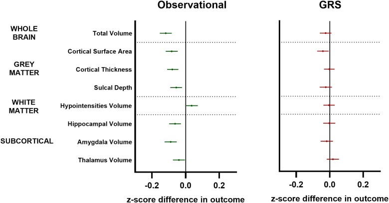
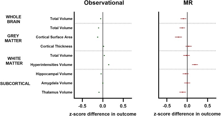
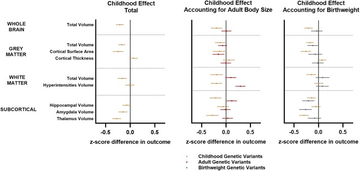
Obese adults are often reported to have smaller brain volumes than their non-obese peers. Whether this represents evidence of accelerations in obesity-driven atrophy or is instead a legacy of developmental differences established earlier in the lifespan remains unclear. This study investigated whether early-life differences in adiposity explain differences in numerous adult brain traits commonly attributed to mid-life obesity. We used a two-sample life course Mendelian randomization study in 37 501 adults recruited to UK Biobank (UKB) imaging centres from 2014, with secondary analyses in 6996 children assessed in the Adolescent Brain Cognitive Development Study (ABCD) recruited from 2018. Exposures were genetic variants for childhood (266 variants) and adult (470 variants) adiposity derived from a genome-wide association study (GWAS) of 407 741 UKB participants. Primary outcomes were: adult total brain volume; grey matter volume, thickness and surface area; white matter volume and hyperintensities; and hippocampus, amygdala and thalamus volumes at mean age 55 in the UKB. Secondary outcomes were equivalent childhood measures collected at mean age 10 in ABCD. In the UKB, individuals who were genetically predicted to have had higher levels of adiposity in childhood were found to have multiple smaller adult brain volumes relative to intracranial volume [e.g. z-score difference in normalized brain volume per category increase in adiposity-95% confidence interval (CI) = -0.20 (-0.28, -0.12); P = 4 × 10⁻⁶]. These effect sizes remained essentially unchanged after accounting for birthweight or current adult obesity in multivariable models, whereas most observed adult effects attenuated towards null [e.g. adult z-score (95% CI) for total volume = 0.06 (-0.05, 0.17); P = 0.3]. Observational analyses in ABCD showed a similar pattern of changes already present in those with a high body mass index by age 10 [z-score (95% CI) = -0.10 (-0.13, -0.07); P = 8 × 10-13], with follow-up genetic risk score analyses providing some evidence for a causal effect already at this early age. Sensitivity analyses revealed that many of these effects were likely due to the persistence of larger head sizes established in those who gained excess weight in childhood [childhood z-score (95% CI) for intracranial volume = 0.14 (0.05, 0.23); P = 0.002], rather than smaller brain sizes per se. Our data suggest that the persistence of early-life developmental differences across the life course may underlie numerous neuroimaging traits commonly attributed to obesity-related atrophy in later life.
据报道,肥胖成年人的脑容量通常比非肥胖同龄人小。这是肥胖驱动的脑萎缩加速的证据,还是生命早期建立的发育差异的遗留影响,目前尚不清楚。本研究调查了生命早期肥胖差异是否能解释许多通常归因于中年肥胖的成人大脑特征差异。我们进行了一项两样本生命历程孟德尔随机化研究,研究对象为2014年招募到英国生物银行(UKB)成像中心的37501名成年人,并对2018年招募的青少年大脑认知发展研究(ABCD)中评估的6996名儿童进行了二次分析。暴露因素是从407741名UKB参与者的全基因组关联研究(GWAS)中得出的儿童期(266个变异)和成年期(470个变异)肥胖的基因变异。主要结局包括:成人大脑总体积;灰质体积、厚度和表面积;白质体积和高强度信号;以及UKB中平均年龄55岁时海马体、杏仁核和丘脑的体积。次要结局是在ABCD中平均年龄10岁时收集的等效儿童测量指标。在UKB中,相对于颅内体积,基因预测儿童期肥胖水平较高的个体被发现有多个较小的成人大脑体积[例如,肥胖程度每增加一个类别,标准化脑体积的z评分差异-95%置信区间(CI)=-0.20(-0.28,-0.12);P = 4×10⁻⁶]。在多变量模型中考虑出生体重或当前成人肥胖后,这些效应大小基本保持不变,而大多数观察到的成人效应减弱至无效[例如,总体积的成人z评分(95%CI)= 0.06(-0.05,0.17);P = 0.3]。ABCD中的观察性分析显示,到10岁时体重指数较高的个体中已经存在类似的变化模式[z评分(95%CI)=-0.10(-0.13,-0.07);P = 8×10-13],后续的遗传风险评分分析提供了一些证据表明在这个早期年龄就已经存在因果效应。敏感性分析表明,这些效应中的许多可能是由于童年期体重增加者中较大头围的持续存在[颅内体积的儿童期z评分(95%CI)= 0.14(0.05,0.23);P = 0.002],而不是脑体积本身较小。我们的数据表明,生命历程中早期发育差异的持续存在可能是许多通常归因于晚年肥胖相关萎缩的神经影像学特征的基础。