• 文献检索
  • 文档翻译
  • 深度研究
  • 学术资讯
  • Suppr Zotero 插件Zotero 插件
  • 邀请有礼
  • 套餐&价格
  • 历史记录
应用&插件
Suppr Zotero 插件Zotero 插件浏览器插件Mac 客户端Windows 客户端微信小程序
定价
高级版会员购买积分包购买API积分包
服务
文献检索文档翻译深度研究API 文档MCP 服务
关于我们
关于 Suppr公司介绍联系我们用户协议隐私条款
关注我们

Suppr 超能文献

核心技术专利:CN118964589B侵权必究
粤ICP备2023148730 号-1Suppr @ 2026

文献检索

告别复杂PubMed语法,用中文像聊天一样搜索,搜遍4000万医学文献。AI智能推荐,让科研检索更轻松。

立即免费搜索

文件翻译

保留排版,准确专业,支持PDF/Word/PPT等文件格式,支持 12+语言互译。

免费翻译文档

深度研究

AI帮你快速写综述,25分钟生成高质量综述,智能提取关键信息,辅助科研写作。

立即免费体验

对 3 例具有小脑皮质变性的猫的临床、影像和组织病理学特征进行描述。

Clinical, imaging and histopathological characterization of a series of three cats with cerebellar cortical degeneration.

机构信息

Department of clinical sciences, Faculty of veterinary medicine, Université de Montréal, Saint-Hyacinthe, QC, J2S 2M2, Canada.

Department of pathology and microbiology, Faculty of veterinary medicine, Université de Montréal, Saint-Hyacinthe, QC, J2S 2M2, Canada.

出版信息

BMC Vet Res. 2024 Jun 19;20(1):263. doi: 10.1186/s12917-024-04127-3.

DOI:10.1186/s12917-024-04127-3
PMID:38890680
原文链接:https://pmc.ncbi.nlm.nih.gov/articles/PMC11186075/
Abstract

BACKGROUND

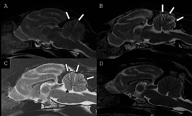
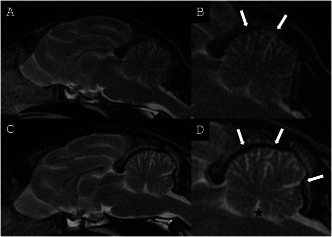
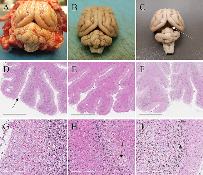
Neurological inherited disorders are rare in domestic animals. Cerebellar cortical degeneration remains amongst the most common of these disorders. The condition is defined as the premature loss of fully differentiated cerebellar components due to genetic or metabolic defects. It has been studied in dogs and cats, and various genetic defects and diagnostic tests (including magnetic resonance imaging (MRI)) have been refined in these species. Cases in cats remain rare and mostly individual, and few diagnostic criteria, other than post-mortem exam, have been evaluated in reports with multiple cases. Here, we report three feline cases of cerebellar cortical degeneration with detailed clinical, diagnostic imaging and post-mortem findings.

CASE PRESENTATION

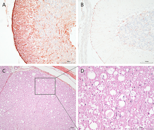
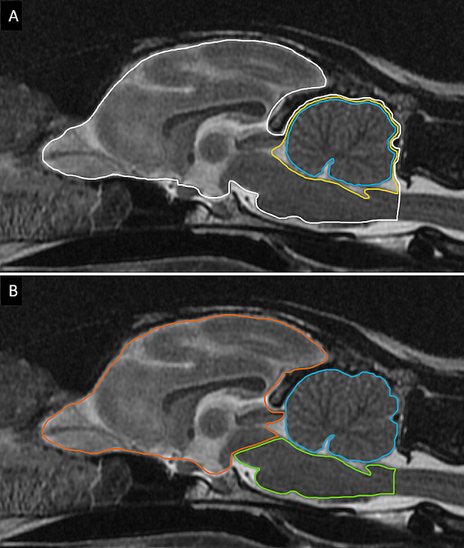
The three cases were directly (siblings, case #1 and #2) or indirectly related (same farm, case #3) and showed early-onset of the disease, with clinical signs including cerebellar ataxia and tremors. Brain MRI was highly suggestive of cerebellar cortical degeneration on all three cases. The relative cerebrospinal fluid (CSF) space, relative cerebellum size, brainstem: cerebellum area ratio, and cerebellum: total brain area ratio, were measured and compared to a control group of cats and reference cut-offs for dogs in the literature. For the relative cerebellum size and cerebellum: total brain area ratio, all affected cases had a lower value than the control group. For the relative CSF space and brainstem: cerebellum area ratio, the more affected cases (#2 and #3) had higher values than the control group, while the least affected case (#3) had values within the ranges of the control group, but a progression was visible over time. Post-mortem examination confirmed the diagnosis of cerebellar cortical degeneration, with marked to complete loss of Purkinje cells and associated granular layer depletion and proliferation of Bergmann glia. One case also had Wallerian-like degeneration in the spinal cord, suggestive of spinocerebellar degeneration.

CONCLUSION

Our report further supports a potential genetic component for the disease in cats. For the MRI examination, the relative cerebellum size and cerebellum: total brain area ratio seem promising, but further studies are needed to establish specific feline cut-offs. Post-mortem evaluation of the cerebellum remains the gold standard for the final diagnosis.

摘要

背景

神经遗传性疾病在宠物中较为罕见。小脑皮质变性是此类疾病中最常见的一种。该疾病是指由于遗传或代谢缺陷,完全分化的小脑成分过早丧失。在犬和猫中对其进行了研究,并且在这些物种中已经对各种遗传缺陷和诊断测试(包括磁共振成像(MRI))进行了改进。猫科动物的病例仍然很少且多为个体病例,并且在具有多个病例的报告中,除了尸检之外,很少有诊断标准得到评估。在这里,我们报告了三例具有详细临床,诊断成像和死后发现的猫小脑皮质变性病例。

病例介绍

这三个病例直接(兄弟姐妹,病例#1和#2)或间接(同一农场,病例#3)相关,并且具有疾病的早期发作,临床症状包括小脑共济失调和震颤。脑部 MRI 在所有三个病例中均高度提示小脑皮质变性。相对脑脊髓液(CSF)空间,相对小脑大小,脑干:小脑区域比以及小脑:大脑总区域比,均进行了测量,并与猫的对照组和文献中犬的参考截止值进行了比较。对于相对小脑大小和小脑:大脑总区域比,所有受影响的病例的值均低于对照组。对于相对 CSF 空间和脑干:小脑区域比,受影响更严重的病例(#2 和#3)的值高于对照组,而受影响最小的病例(#3)的值在对照组范围内,但随着时间的推移,可见到进展。尸检证实了小脑皮质变性的诊断,出现明显至完全丧失浦肯野细胞以及相关的颗粒层耗竭和伯格曼神经胶质增生。一例脊髓中还出现了华勒氏样变性,提示脊髓小脑变性。

结论

我们的报告进一步支持了猫科动物小脑皮质变性的潜在遗传因素。对于 MRI 检查,相对小脑大小和小脑:大脑总区域比似乎很有前途,但是需要进一步的研究来建立特定的猫科动物截止值。对小脑进行死后评估仍然是最终诊断的金标准。

https://cdn.ncbi.nlm.nih.gov/pmc/blobs/559c/11186075/2da2deef5b28/12917_2024_4127_Fig5_HTML.jpg
https://cdn.ncbi.nlm.nih.gov/pmc/blobs/559c/11186075/5ad1ed4a5785/12917_2024_4127_Fig1_HTML.jpg
https://cdn.ncbi.nlm.nih.gov/pmc/blobs/559c/11186075/9a8bb38908f4/12917_2024_4127_Fig2_HTML.jpg
https://cdn.ncbi.nlm.nih.gov/pmc/blobs/559c/11186075/f451a8c18e9e/12917_2024_4127_Fig3_HTML.jpg
https://cdn.ncbi.nlm.nih.gov/pmc/blobs/559c/11186075/89dde51dab8e/12917_2024_4127_Fig4_HTML.jpg
https://cdn.ncbi.nlm.nih.gov/pmc/blobs/559c/11186075/2da2deef5b28/12917_2024_4127_Fig5_HTML.jpg

相似文献

1
Clinical, imaging and histopathological characterization of a series of three cats with cerebellar cortical degeneration.对 3 例具有小脑皮质变性的猫的临床、影像和组织病理学特征进行描述。
BMC Vet Res. 2024 Jun 19;20(1):263. doi: 10.1186/s12917-024-04127-3.
2
Computer-assisted magnetic resonance imaging brain morphometry in American Staffordshire Terriers with cerebellar cortical degeneration.患有小脑皮质变性的美国斯塔福郡梗犬的计算机辅助磁共振成像脑形态测量学
J Vet Intern Med. 2008 Jul-Aug;22(4):969-75. doi: 10.1111/j.1939-1676.2008.0138.x.
3
Histopathological and ultrastructural features of feline hereditary cerebellar cortical atrophy: a novel animal model of human spinocerebellar degeneration.猫遗传性小脑皮质萎缩的组织病理学和超微结构特征:一种人类脊髓小脑变性的新型动物模型。
Acta Neuropathol. 1998 Oct;96(4):379-87. doi: 10.1007/s004010050908.
4
Late onset cerebellar degeneration in a middle-aged cat.一只中年猫的迟发性小脑变性
J Feline Med Surg. 2006 Dec;8(6):424-9. doi: 10.1016/j.jfms.2006.04.007. Epub 2006 Jun 15.
5
Diagnosis of cerebellar cortical degeneration in a Scottish terrier using magnetic resonance imaging.使用磁共振成像诊断一只苏格兰梗犬的小脑皮质变性。
J Small Anim Pract. 2001 Aug;42(8):409-12. doi: 10.1111/j.1748-5827.2001.tb02491.x.
6
Cerebellar cortical degeneration associated with feline leukemia virus infection and cerebellar lymphoma in a young cat.一只幼猫中与猫白血病病毒感染及小脑淋巴瘤相关的小脑皮质变性。
Open Vet J. 2019 Oct;9(3):246-252. doi: 10.4314/ovj.v9i3.9. Epub 2019 Aug 29.
7
Cerebellar cortical degeneration with selective granule cell loss in Bavarian mountain dogs.巴伐利亚山地犬小脑皮质变性伴选择性颗粒细胞丢失
J Small Anim Pract. 2007 Aug;48(8):462-5. doi: 10.1111/j.1748-5827.2006.00257.x.
8
Granuloprival cerebellar cortical degeneration in a Yorkshire Terrier and Lagotto Romagnolo dog.在一只约克夏梗犬和拉戈托罗马阁诺犬中发现了颗粒缺失性小脑皮质变性。
J Vet Intern Med. 2024 Jul-Aug;38(4):2368-2372. doi: 10.1111/jvim.17091. Epub 2024 Apr 25.
9
Study of hereditary cerebellar degeneration in cats.猫遗传性小脑变性的研究。
Am J Vet Res. 1996 Mar;57(3):296-301.
10
Rostral cerebellar arterial infarct in two cats.两只猫的延髓小脑动脉梗死
J Feline Med Surg. 2007 Jun;9(3):246-53. doi: 10.1016/j.jfms.2006.12.003. Epub 2007 Feb 20.

引用本文的文献

1
Exosomes in Regulating miRNAs for Biomarkers of Neurodegenerative Disorders.外泌体在调节用于神经退行性疾病生物标志物的微小RNA中的作用
Mol Neurobiol. 2025 Jun;62(6):7576-7596. doi: 10.1007/s12035-025-04733-8. Epub 2025 Feb 7.

本文引用的文献

1
Phenotypic and genetic aspects of hereditary ataxia in dogs.遗传性犬共济失调的表型和遗传学研究
J Vet Intern Med. 2023 Jul-Aug;37(4):1306-1322. doi: 10.1111/jvim.16742. Epub 2023 Jun 21.
2
Cerebellar Abiotrophy in Australian Working Kelpies Is Associated with Two Major Risk Loci.澳大利亚工作牧羊犬小脑萎缩症与两个主要风险基因座相关。
Genes (Basel). 2022 Sep 23;13(10):1709. doi: 10.3390/genes13101709.
3
The inherited cerebellar ataxias: an update.遗传性小脑共济失调:更新。
J Neurol. 2023 Jan;270(1):208-222. doi: 10.1007/s00415-022-11383-6. Epub 2022 Sep 24.
4
Toxoplasma gondii multifocal central nervous system infiltration in an apparently immunocompetent cat in Italy.意大利一例貌似免疫健全的猫体内弓形体多灶性中枢神经系统浸润
Vet Parasitol Reg Stud Reports. 2021 Jul;25:100605. doi: 10.1016/j.vprsr.2021.100605. Epub 2021 Jun 24.
5
Conventional MRI findings in hereditary degenerative ataxias: a pictorial review.遗传性退行性共济失调的传统MRI表现:图谱综述
Neuroradiology. 2021 Jul;63(7):983-999. doi: 10.1007/s00234-021-02682-2. Epub 2021 Mar 17.
6
A Missense Variant in in Alpine Dachsbracke Dogs Affected by Spinocerebellar Ataxia.在患有脊髓小脑共济失调的阿尔卑斯山达克斯布拉克犬中, 中的错义变异。
Genes (Basel). 2019 May 10;10(5):362. doi: 10.3390/genes10050362.
7
Immunohistochemical Evaluation of Canine Pituitary Adenomas Obtained by Transsphenoidal Hypophysectomy.经蝶窦垂体切除术获取的犬垂体腺瘤的免疫组织化学评估
Vet Pathol. 2018 Nov;55(6):889-895. doi: 10.1177/0300985818784160. Epub 2018 Jun 20.
8
MRI findings of neuronal ceroid lipofuscinosis in a cat.一只猫的神经元蜡样脂褐质沉积症的磁共振成像结果
JFMS Open Rep. 2018 Mar 5;4(1):2055116918757330. doi: 10.1177/2055116918757330. eCollection 2018 Jan-Jun.
9
A retrospective study of the neuropathology and diagnosis of naturally occurring feline infectious peritonitis.自然发生的猫传染性腹膜炎的神经病理学与诊断的回顾性研究
J Vet Diagn Invest. 2018 May;30(3):392-399. doi: 10.1177/1040638718755833. Epub 2018 Feb 7.
10
Variation in MUTYH expression in Arabian horses with Cerebellar Abiotrophy.患有小脑性共济失调的阿拉伯马中MUTYH表达的变化。
Brain Res. 2018 Jan 1;1678:330-336. doi: 10.1016/j.brainres.2017.10.034. Epub 2017 Nov 2.