National Laboratory of Solid State Microstructure, Department of Physics, Nanjing University, Nanjing 210093, China.
School of Physics, Zhejiang University, Hangzhou 310027, China.
Int J Mol Sci. 2024 Jun 4;25(11):6198. doi: 10.3390/ijms25116198.
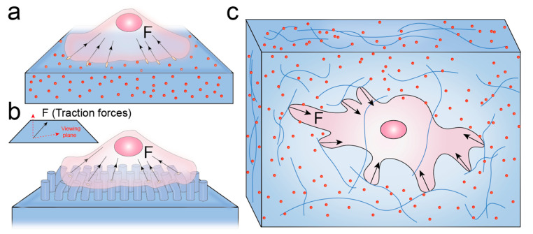
The mechanical forces exerted by cells on their surrounding microenvironment are known as cellular traction forces. These forces play crucial roles in various biological processes, such as tissue development, wound healing and cell functions. However, it is hard for traditional techniques to measure cellular traction forces accurately because their magnitude (from pN to nN) and the length scales over which they occur (from nm to μm) are extremely small. In order to fully understand mechanotransduction, highly sensitive tools for measuring cellular forces are needed. Current powerful techniques for measuring traction forces include traction force microscopy (TFM) and fluorescent molecular force sensors (FMFS). In this review, we elucidate the force imaging principles of TFM and FMFS. Then we highlight the application of FMFS in a variety of biological processes and offer our perspectives and insights into the potential applications of FMFS.
细胞对其周围微环境施加的机械力被称为细胞牵引力。这些力在各种生物过程中起着至关重要的作用,例如组织发育、伤口愈合和细胞功能。然而,由于其大小(从皮牛顿到毫牛顿)和发生的长度尺度(从纳米到微米)非常小,传统技术很难准确测量细胞牵引力。为了充分理解力转导,需要用于测量细胞力的高灵敏度工具。目前用于测量牵引力的强大技术包括牵引力显微镜(TFM)和荧光分子力传感器(FMFS)。在这篇综述中,我们阐明了 TFM 和 FMFS 的力成像原理。然后,我们重点介绍了 FMFS 在各种生物过程中的应用,并对 FMFS 的潜在应用提出了我们的观点和见解。