• 文献检索
  • 文档翻译
  • 深度研究
  • 学术资讯
  • Suppr Zotero 插件Zotero 插件
  • 邀请有礼
  • 套餐&价格
  • 历史记录
应用&插件
Suppr Zotero 插件Zotero 插件浏览器插件Mac 客户端Windows 客户端微信小程序
定价
高级版会员购买积分包购买API积分包
服务
文献检索文档翻译深度研究API 文档MCP 服务
关于我们
关于 Suppr公司介绍联系我们用户协议隐私条款
关注我们

Suppr 超能文献

核心技术专利:CN118964589B侵权必究
粤ICP备2023148730 号-1Suppr @ 2026

文献检索

告别复杂PubMed语法,用中文像聊天一样搜索,搜遍4000万医学文献。AI智能推荐,让科研检索更轻松。

立即免费搜索

文件翻译

保留排版,准确专业,支持PDF/Word/PPT等文件格式,支持 12+语言互译。

免费翻译文档

深度研究

AI帮你快速写综述,25分钟生成高质量综述,智能提取关键信息,辅助科研写作。

立即免费体验

中央软骨性骨肿瘤的定量骨SPECT/CT:SUVmax与亨氏单位中骨密度的关系

Quantitative Bone SPECT/CT of Central Cartilaginous Bone Tumors: Relationship between SUVmax and Radiodensity in Hounsfield Unit.

作者信息

Yoon Hyukjin, Lee Seul Ki, Kim Jee-Young, Joo Min Wook

机构信息

Division of Nuclear Medicine, Department of Radiology, St. Vincent's Hospital, College of Medicine, The Catholic University of Korea, Seoul 06591, Republic of Korea.

Department of Radiology, St. Vincent's Hospital, College of Medicine, The Catholic University of Korea, Seoul 06591, Republic of Korea.

出版信息

Cancers (Basel). 2024 May 22;16(11):1968. doi: 10.3390/cancers16111968.

DOI:10.3390/cancers16111968
PMID:38893090
原文链接:https://pmc.ncbi.nlm.nih.gov/articles/PMC11171356/
Abstract

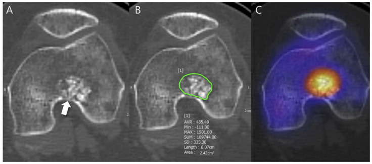
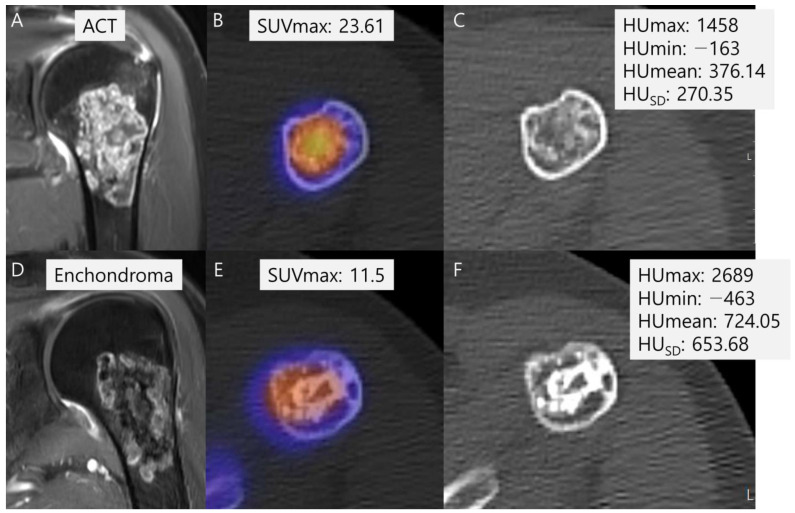
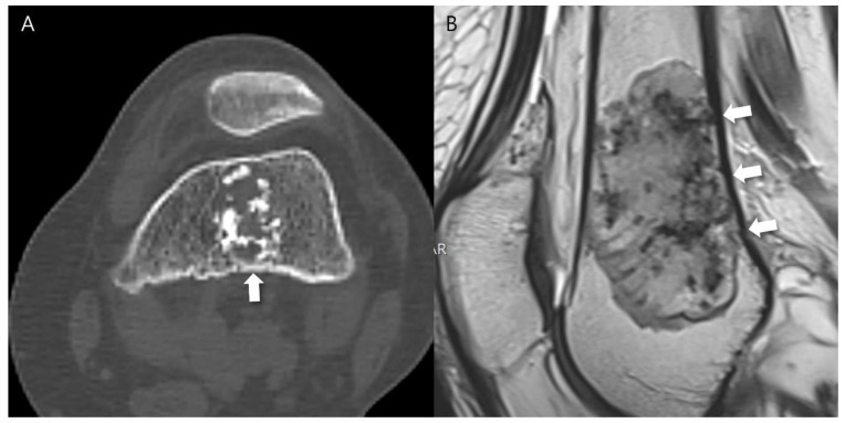
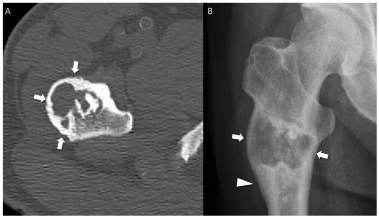
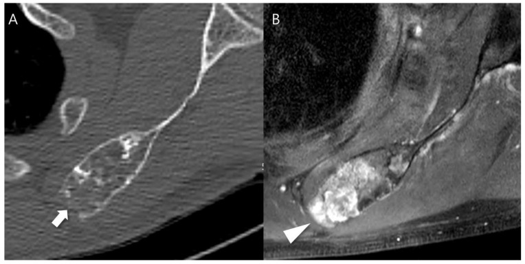
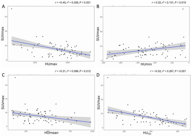
(1) Background: it is challenging to determine the accurate grades of cartilaginous bone tumors. Using bone single photon emission computed tomography (SPECT)/computed tomography (CT), maximum standardized uptake value (SUVmax) was found to be significantly associated with different grades of cartilaginous bone tumor. The inquiry focused on the effect of the tumor matrix on SUVmax. (2) Methods: a total of 65 patients from 2017 to 2022 with central cartilaginous bone tumors, including enchondromas and low-to-intermediate grade chondrosarcomas, who had undergone bone SPECT/CT were retrospectively enrolled. The SUVmax was recorded and any aggressive CT findings of cartilaginous bone tumor and Hounsfield units (HU) of the chondroid matrix as mean, minimum, maximum, and standard deviation (SD) were reviewed on CT scans. Pearson's correlation analysis was performed to determine the relationship between CT features and SUVmax. Subgroup analysis was also performed between the benign group (enchondroma) and the malignant group (grade 1 and 2 chondrosarcoma) for comparison of HU values and SUVmax. (3) Results: a significant negative correlation between SUVmax and HU measurements, including HUmax, HUmean, and HU, was found. The subgroup analysis showed significantly higher SUVmax in the malignant group, with more frequent CT aggressive features, and significantly lower HU in the malignant group than in the benign group. (4) Conclusions: it was observed that higher SUVmax and lower HU were associated with a higher probability of having a low-to-intermediate chondrosarcoma with aggressive features and a less calcified tumor matrix.

摘要

(1) 背景:确定软骨性骨肿瘤的准确分级具有挑战性。使用骨单光子发射计算机断层扫描(SPECT)/计算机断层扫描(CT)发现,最大标准化摄取值(SUVmax)与不同分级的软骨性骨肿瘤显著相关。该研究聚焦于肿瘤基质对SUVmax的影响。(2) 方法:回顾性纳入了2017年至2022年期间共65例患有中央型软骨性骨肿瘤(包括内生软骨瘤和低至中级软骨肉瘤)且接受过骨SPECT/CT检查的患者。记录SUVmax,并在CT扫描上复查软骨性骨肿瘤的任何侵袭性CT表现以及类软骨基质的亨氏单位(HU),包括平均值、最小值、最大值和标准差(SD)。进行Pearson相关性分析以确定CT特征与SUVmax之间的关系。还在良性组(内生软骨瘤)和恶性组(1级和2级软骨肉瘤)之间进行亚组分析,以比较HU值和SUVmax。(3) 结果:发现SUVmax与HU测量值(包括HUmax、HUmean和HU)之间存在显著负相关。亚组分析显示,恶性组的SUVmax显著更高,具有更频繁的CT侵袭性特征,且恶性组的HU显著低于良性组。(4) 结论:观察到较高的SUVmax和较低的HU与具有侵袭性特征且肿瘤基质钙化较少的低至中级软骨肉瘤的可能性较高相关。

https://cdn.ncbi.nlm.nih.gov/pmc/blobs/3f12/11171356/a76f6ffbab59/cancers-16-01968-g006.jpg
https://cdn.ncbi.nlm.nih.gov/pmc/blobs/3f12/11171356/2e6a9a2031b2/cancers-16-01968-g001.jpg
https://cdn.ncbi.nlm.nih.gov/pmc/blobs/3f12/11171356/98b093d03f18/cancers-16-01968-g002.jpg
https://cdn.ncbi.nlm.nih.gov/pmc/blobs/3f12/11171356/001463f8077f/cancers-16-01968-g003.jpg
https://cdn.ncbi.nlm.nih.gov/pmc/blobs/3f12/11171356/71b328be8553/cancers-16-01968-g004.jpg
https://cdn.ncbi.nlm.nih.gov/pmc/blobs/3f12/11171356/9e0b2651f420/cancers-16-01968-g005.jpg
https://cdn.ncbi.nlm.nih.gov/pmc/blobs/3f12/11171356/a76f6ffbab59/cancers-16-01968-g006.jpg

相似文献

1
Quantitative Bone SPECT/CT of Central Cartilaginous Bone Tumors: Relationship between SUVmax and Radiodensity in Hounsfield Unit.中央软骨性骨肿瘤的定量骨SPECT/CT:SUVmax与亨氏单位中骨密度的关系
Cancers (Basel). 2024 May 22;16(11):1968. doi: 10.3390/cancers16111968.
2
Correlation between the maximum standard uptake value and mean Hounsfield unit on single-photon emission computed tomography-computed tomography to discriminate benign and metastatic lesions among patients with breast cancer.单光子发射计算机断层扫描-计算机断层扫描中最大标准摄取值与平均亨氏单位之间的相关性,以鉴别乳腺癌患者的良性和转移性病变。
Asian Spine J. 2024 Jun;18(3):398-406. doi: 10.31616/asj.2022.0451. Epub 2024 Jun 25.
3
Quantitative SPECT/CT for differentiating between enchondroma and grade I chondrosarcoma.定量 SPECT/CT 鉴别内生软骨瘤和 I 级软骨肉瘤。
Sci Rep. 2020 Jun 29;10(1):10587. doi: 10.1038/s41598-020-67506-4.
4
Standardized uptake values of Tc-MDP in normal vertebrae assessed using quantitative SPECT/CT for differentiation diagnosis of benign and malignant bone lesions.采用定量 SPECT/CT 评估 Tc-MDP 标准化摄取值在正常椎体中对良恶性骨病变进行鉴别诊断的应用。
BMC Med Imaging. 2021 Feb 27;21(1):39. doi: 10.1186/s12880-021-00569-5.
5
F-18 FDG PET differentiation of benign from malignant chondroid neoplasms: a systematic review of the literature.F-18 FDG PET对软骨样肿瘤良恶性的鉴别:文献系统综述
Skeletal Radiol. 2017 Sep;46(9):1233-1239. doi: 10.1007/s00256-017-2685-7. Epub 2017 Jun 12.
6
Skeletal standardized uptake values obtained using quantitative SPECT/CT for the detection of bone metastases in patients with lung adenocarcinoma.使用定量SPECT/CT获取的骨骼标准化摄取值,用于检测肺腺癌患者的骨转移。
Front Med (Lausanne). 2023 Feb 2;10:1119214. doi: 10.3389/fmed.2023.1119214. eCollection 2023.
7
Quantitative bone SPECT/CT applications for cartilaginous bone neoplasms.定量骨SPECT/CT在软骨性骨肿瘤中的应用
Hell J Nucl Med. 2020 May-Aug;23(2):133-137. doi: 10.1967/s002449912102. Epub 2020 Jul 27.
8
F-FDG PET/CT in the evaluation of cartilaginous bone neoplasms: the added value of tumor grading.F-FDG PET/CT 评估软骨性骨肿瘤:肿瘤分级的附加价值。
Ann Nucl Med. 2019 Nov;33(11):813-821. doi: 10.1007/s12149-019-01392-3. Epub 2019 Aug 8.
9
The diagnostic value of quantitative bone SPECT/CT in solitary undetermined bone lesions.定量骨SPECT/CT在孤立性未确诊骨病变中的诊断价值
Front Oncol. 2023 Nov 9;13:1205379. doi: 10.3389/fonc.2023.1205379. eCollection 2023.
10
Comparison of bone single-photon emission computed tomography (SPECT)/CT and bone scintigraphy in assessing knee joints.骨单光子发射计算机断层扫描(SPECT)/CT 与骨闪烁显像术评估膝关节的比较。
BMC Med Imaging. 2021 Mar 26;21(1):60. doi: 10.1186/s12880-021-00590-8.

引用本文的文献

1
Whole-body imaging for distant staging of bone chondrosarcoma: a systematic review.骨软骨肉瘤远处分期的全身成像:一项系统评价
Radiol Med. 2025 Jul 23. doi: 10.1007/s11547-025-02054-3.

本文引用的文献

1
SPECT/CT Radiomics for Differentiating between Enchondroma and Grade I Chondrosarcoma.SPECT/CT 影像组学在软骨瘤与 I 级软骨肉瘤鉴别诊断中的应用
Tomography. 2023 Oct 16;9(5):1868-1875. doi: 10.3390/tomography9050148.
2
Classification of Chondrosarcoma: From Characteristic to Challenging Imaging Findings.软骨肉瘤的分类:从特征性影像表现到具有挑战性的影像发现
Cancers (Basel). 2023 Mar 10;15(6):1703. doi: 10.3390/cancers15061703.
3
Differentiating Enchondromas and Atypical Cartilaginous Tumors in Long Bones with Computed Tomography and Magnetic Resonance Imaging.
利用计算机断层扫描和磁共振成像鉴别长骨内生软骨瘤和非典型软骨肿瘤
Diagnostics (Basel). 2022 Sep 9;12(9):2186. doi: 10.3390/diagnostics12092186.
4
Diagnostic Value of CT- and MRI-Based Texture Analysis and Imaging Findings for Grading Cartilaginous Tumors in Long Bones.基于CT和MRI的纹理分析及影像学表现对长骨软骨肿瘤分级的诊断价值
Front Oncol. 2021 Oct 14;11:700204. doi: 10.3389/fonc.2021.700204. eCollection 2021.
5
Effects of Interobserver Variability on 2D and 3D CT- and MRI-Based Texture Feature Reproducibility of Cartilaginous Bone Tumors.观察者间变异性对基于二维和三维 CT 和 MRI 的软骨性骨肿瘤纹理特征可重复性的影响。
J Digit Imaging. 2021 Aug;34(4):820-832. doi: 10.1007/s10278-021-00498-3. Epub 2021 Aug 17.
6
A Radiological Scoring System for Differentiation between Enchondroma and Chondrosarcoma.一种用于鉴别内生软骨瘤和软骨肉瘤的放射学评分系统。
Cancers (Basel). 2021 Jul 16;13(14):3558. doi: 10.3390/cancers13143558.
7
CT radiomics-based machine learning classification of atypical cartilaginous tumours and appendicular chondrosarcomas.基于 CT 影像组学的机器学习分类方法,用于鉴别非典型性软骨肿瘤和四肢部软骨肉瘤。
EBioMedicine. 2021 Jun;68:103407. doi: 10.1016/j.ebiom.2021.103407. Epub 2021 May 26.
8
Development and Validation of a Radiomics Model for Differentiating Bone Islands and Osteoblastic Bone Metastases at Abdominal CT.基于腹部 CT 影像的骨岛与成骨性骨转移瘤的影像组学模型的建立与验证
Radiology. 2021 Jun;299(3):626-632. doi: 10.1148/radiol.2021203783. Epub 2021 Mar 30.
9
Assessment of central cartilaginous tumor of the appendicular bone: inter-observer and intermodality agreement and comparison of diagnostic performance of CT and MRI.四肢骨中央软骨肿瘤的评估:观察者间和模态间的一致性以及CT与MRI诊断性能的比较
Acta Radiol. 2022 Mar;63(3):376-386. doi: 10.1177/0284185121996268. Epub 2021 Feb 27.
10
Chondrogenic Bone Tumors: The Importance of Imaging Characteristics.软骨源性骨肿瘤:影像学特征的重要性。
Rofo. 2021 Mar;193(3):262-275. doi: 10.1055/a-1288-1209. Epub 2020 Nov 5.