• 文献检索
  • 文档翻译
  • 深度研究
  • 学术资讯
  • Suppr Zotero 插件Zotero 插件
  • 邀请有礼
  • 套餐&价格
  • 历史记录
应用&插件
Suppr Zotero 插件Zotero 插件浏览器插件Mac 客户端Windows 客户端微信小程序
定价
高级版会员购买积分包购买API积分包
服务
文献检索文档翻译深度研究API 文档MCP 服务
关于我们
关于 Suppr公司介绍联系我们用户协议隐私条款
关注我们

Suppr 超能文献

核心技术专利:CN118964589B侵权必究
粤ICP备2023148730 号-1Suppr @ 2026

文献检索

告别复杂PubMed语法,用中文像聊天一样搜索,搜遍4000万医学文献。AI智能推荐,让科研检索更轻松。

立即免费搜索

文件翻译

保留排版,准确专业,支持PDF/Word/PPT等文件格式,支持 12+语言互译。

免费翻译文档

深度研究

AI帮你快速写综述,25分钟生成高质量综述,智能提取关键信息,辅助科研写作。

立即免费体验

定量 SPECT/CT 鉴别内生软骨瘤和 I 级软骨肉瘤。

Quantitative SPECT/CT for differentiating between enchondroma and grade I chondrosarcoma.

机构信息

Division of Nuclear Medicine, Department of Radiology, St. Vincent's Hospital, College of Medicine, The Catholic University of Korea, Seoul, Republic of Korea.

Division of Nuclear Medicine, Department of Radiology, Yeouido St. Mary's Hospital, College of Medicine, The Catholic University of Korea, Seoul, Republic of Korea.

出版信息

Sci Rep. 2020 Jun 29;10(1):10587. doi: 10.1038/s41598-020-67506-4.

DOI:10.1038/s41598-020-67506-4
PMID:32601314
原文链接:https://pmc.ncbi.nlm.nih.gov/articles/PMC7324365/
Abstract

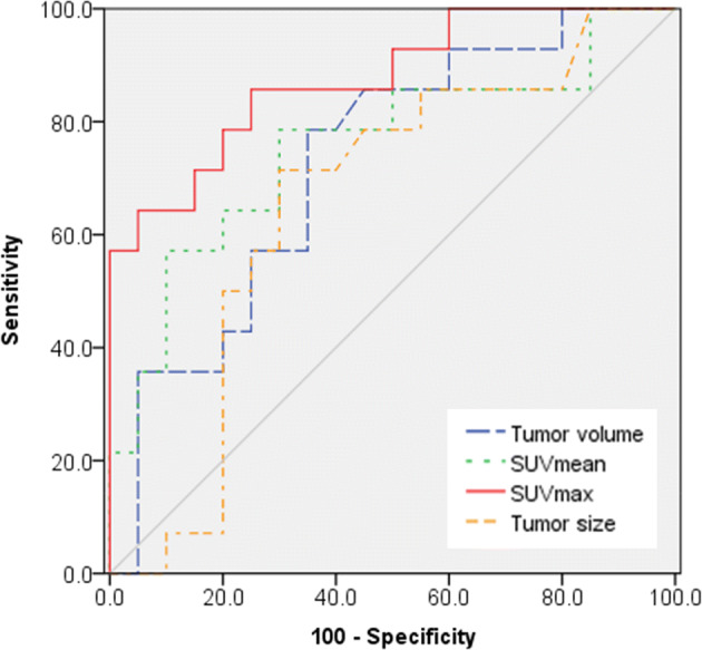
Although differentiation between central chondroid tumors is important, their parallelism makes it a diagnostic conundrum for clinicians and radiologists. The objective of this study was to evaluate the efficiency of quantitative single photon emission computed tomography (SPECT)/computed tomography (CT) in differentiating grade I chondrosarcomas from enchondromas. We reviewed SPECT/CT images of patients with enchondromas and grade I chondrosarcomas arising in the long bones. Volume, mean standardized uptake value (SUVmean), and maximum standardized uptake value (SUVmax) of tumors were calculated from SPECT/CT images. In addition, clinical characteristics and radiological information were assessed. Of a total of 34 patients, 14 had chondrosarcomas. Chondrosarcoma group had significantly larger volume, and higher SUVmean and SUVmax of tumors than enchondroma group. There was no significant difference in age and tumor size between two groups. Areas under the receiver-operating characteristic curve (AUCs) for tumor volume, SUVmean, and SUVmax were 0.727, 0.757, and 0.875. In pairwise analyses, SUVmax had larger AUC than SUVmean (p = 0.0216). With a cut-off value of 15.6 for SUVmax, its sensitivity and specificity were 86% and 75% for differentiating between enchondroma and grade I chondrosarcoma. Quantitative SPECT/CT is a potential method to differentiate grade I chondroarcomas from enchondromas in patients with central chondroid tumors.

摘要

虽然中央性软骨肿瘤的鉴别很重要,但它们的相似性使得临床医生和放射科医生在诊断上感到困惑。本研究的目的是评估定量单光子发射计算机断层扫描(SPECT)/计算机断层扫描(CT)在区分 I 级软骨肉瘤和内生软骨瘤中的效率。我们回顾了长骨内生软骨瘤和 I 级软骨肉瘤患者的 SPECT/CT 图像。从 SPECT/CT 图像中计算肿瘤的体积、平均标准化摄取值(SUVmean)和最大标准化摄取值(SUVmax)。此外,还评估了临床特征和影像学信息。在总共 34 名患者中,有 14 名患有软骨肉瘤。与内生软骨瘤组相比,软骨肉瘤组肿瘤的体积、SUVmean 和 SUVmax 均显著增大。两组间的年龄和肿瘤大小无显著差异。肿瘤体积、SUVmean 和 SUVmax 的受试者工作特征曲线(AUC)下面积分别为 0.727、0.757 和 0.875。两两分析中,SUVmax 的 AUC 大于 SUVmean(p=0.0216)。SUVmax 的截断值为 15.6 时,其鉴别内生软骨瘤和 I 级软骨肉瘤的灵敏度和特异性分别为 86%和 75%。定量 SPECT/CT 是区分中央性软骨肿瘤患者 I 级软骨肉瘤和内生软骨瘤的一种有潜力的方法。

https://cdn.ncbi.nlm.nih.gov/pmc/blobs/05a6/7324365/c7661defea81/41598_2020_67506_Fig3_HTML.jpg
https://cdn.ncbi.nlm.nih.gov/pmc/blobs/05a6/7324365/dd617ce39fb0/41598_2020_67506_Fig1_HTML.jpg
https://cdn.ncbi.nlm.nih.gov/pmc/blobs/05a6/7324365/1c11d4ef1337/41598_2020_67506_Fig2_HTML.jpg
https://cdn.ncbi.nlm.nih.gov/pmc/blobs/05a6/7324365/c7661defea81/41598_2020_67506_Fig3_HTML.jpg
https://cdn.ncbi.nlm.nih.gov/pmc/blobs/05a6/7324365/dd617ce39fb0/41598_2020_67506_Fig1_HTML.jpg
https://cdn.ncbi.nlm.nih.gov/pmc/blobs/05a6/7324365/1c11d4ef1337/41598_2020_67506_Fig2_HTML.jpg
https://cdn.ncbi.nlm.nih.gov/pmc/blobs/05a6/7324365/c7661defea81/41598_2020_67506_Fig3_HTML.jpg

相似文献

1
Quantitative SPECT/CT for differentiating between enchondroma and grade I chondrosarcoma.定量 SPECT/CT 鉴别内生软骨瘤和 I 级软骨肉瘤。
Sci Rep. 2020 Jun 29;10(1):10587. doi: 10.1038/s41598-020-67506-4.
2
F-18 FDG PET differentiation of benign from malignant chondroid neoplasms: a systematic review of the literature.F-18 FDG PET对软骨样肿瘤良恶性的鉴别:文献系统综述
Skeletal Radiol. 2017 Sep;46(9):1233-1239. doi: 10.1007/s00256-017-2685-7. Epub 2017 Jun 12.
3
SPECT/CT Radiomics for Differentiating between Enchondroma and Grade I Chondrosarcoma.SPECT/CT 影像组学在软骨瘤与 I 级软骨肉瘤鉴别诊断中的应用
Tomography. 2023 Oct 16;9(5):1868-1875. doi: 10.3390/tomography9050148.
4
Usefulness of radiography in differentiating enchondroma from central grade 1 chondrosarcoma.X线摄影在鉴别内生软骨瘤与中央型1级软骨肉瘤中的作用。
AJR Am J Roentgenol. 1997 Oct;169(4):1097-104. doi: 10.2214/ajr.169.4.9308471.
5
What are the differentiating clinical and MRI-features of enchondromas from low-grade chondrosarcomas?软骨瘤与低级别软骨肉瘤的临床和 MRI 特征有何区别?
Eur Radiol. 2018 Jan;28(1):398-409. doi: 10.1007/s00330-017-4947-0. Epub 2017 Jul 10.
6
Chondroid Tumors as Incidental Findings and Differential Diagnosis between Enchondromas and Low-grade Chondrosarcomas.软骨样肿瘤作为偶然发现以及内生软骨瘤与低级别软骨肉瘤之间的鉴别诊断
Semin Musculoskelet Radiol. 2019 Feb;23(1):3-18. doi: 10.1055/s-0038-1675550. Epub 2019 Jan 30.
7
Differentiating clinical and radiographic features of enchondroma and secondary chondrosarcoma in the foot.足部内生软骨瘤与继发性软骨肉瘤的临床及影像学特征鉴别
Foot Ankle Int. 2006 Apr;27(4):240-4. doi: 10.1177/107110070602700403.
8
CT radiomics-based machine learning model for differentiating between enchondroma and low-grade chondrosarcoma.基于 CT 放射组学的机器学习模型鉴别内生软骨瘤和低级别软骨肉瘤。
Medicine (Baltimore). 2024 Aug 16;103(33):e39311. doi: 10.1097/MD.0000000000039311.
9
A retrospective external validation study of the Birmingham Atypical Cartilage Tumour Imaging Protocol (BACTIP) for the management of solitary central cartilage tumours of the proximal humerus and around the knee.一项回顾性外部验证研究,评估伯明翰非典型软骨肿瘤成像方案(BACTIP)在肱骨头和膝关节周围单发中心性软骨肿瘤管理中的应用。
Eur Radiol. 2024 Aug;34(8):4988-5006. doi: 10.1007/s00330-024-10604-y. Epub 2024 Feb 6.
10
Radiographic differentiation of enchondroma from low-grade chondrosarcoma in the fibula.腓骨内生软骨瘤与低级别软骨肉瘤的影像学鉴别
Skeletal Radiol. 2004 Aug;33(8):458-66. doi: 10.1007/s00256-004-0791-9. Epub 2004 Jun 23.

引用本文的文献

1
Whole-body imaging for distant staging of bone chondrosarcoma: a systematic review.骨软骨肉瘤远处分期的全身成像:一项系统评价
Radiol Med. 2025 Jul 23. doi: 10.1007/s11547-025-02054-3.
2
Imaging bone turnover assessment through volumetric density-adjusted standardized uptake value using quantitative bone SPECT/CT in osteoporosis.在骨质疏松症中,通过定量骨SPECT/CT使用体积密度调整后的标准化摄取值进行骨转换评估成像。
EJNMMI Res. 2024 Aug 25;14(1):75. doi: 10.1186/s13550-024-01137-y.
3
Quantitative Bone SPECT/CT of Central Cartilaginous Bone Tumors: Relationship between SUVmax and Radiodensity in Hounsfield Unit.

本文引用的文献

1
Quantification evaluation of Tc-MDP concentration in the lumbar spine with SPECT/CT: compare with bone mineral density.SPECT/CT 定量评估腰椎 Tc-MDP 浓度:与骨密度比较。
Ann Nucl Med. 2020 Feb;34(2):136-143. doi: 10.1007/s12149-019-01425-x. Epub 2019 Nov 25.
2
Intralesional treatment versus wide resection for central low-grade chondrosarcoma of the long bones.长骨中央型低级别软骨肉瘤的瘤内治疗与广泛切除
Cochrane Database Syst Rev. 2019 Mar 7;3(3):CD010778. doi: 10.1002/14651858.CD010778.pub2.
3
What are the differentiating clinical and MRI-features of enchondromas from low-grade chondrosarcomas?
中央软骨性骨肿瘤的定量骨SPECT/CT:SUVmax与亨氏单位中骨密度的关系
Cancers (Basel). 2024 May 22;16(11):1968. doi: 10.3390/cancers16111968.
4
SPECT/CT Radiomics for Differentiating between Enchondroma and Grade I Chondrosarcoma.SPECT/CT 影像组学在软骨瘤与 I 级软骨肉瘤鉴别诊断中的应用
Tomography. 2023 Oct 16;9(5):1868-1875. doi: 10.3390/tomography9050148.
5
Tc-methylene diphosphonate SPECT/CT imaging of gout spondylitis: a case report.Tc-亚甲基二膦酸盐 SPECT/CT 成像在痛风性脊柱炎中的应用:一例报告。
J Int Med Res. 2022 Oct;50(10):3000605221129557. doi: 10.1177/03000605221129557.
6
Clinical significance of quantitative bone SPECT/CT in the evaluation of hand and wrist pain in patients with rheumatic disease.定量骨 SPECT/CT 在评估风湿性疾病患者手和腕部疼痛中的临床意义。
Sci Rep. 2022 Jan 10;12(1):327. doi: 10.1038/s41598-021-03874-9.
7
A Radiological Scoring System for Differentiation between Enchondroma and Chondrosarcoma.一种用于鉴别内生软骨瘤和软骨肉瘤的放射学评分系统。
Cancers (Basel). 2021 Jul 16;13(14):3558. doi: 10.3390/cancers13143558.
8
Chondrosarcoma-from Molecular Pathology to Novel Therapies.软骨肉瘤——从分子病理学到新型疗法
Cancers (Basel). 2021 May 14;13(10):2390. doi: 10.3390/cancers13102390.
9
Comparison of bone single-photon emission computed tomography (SPECT)/CT and bone scintigraphy in assessing knee joints.骨单光子发射计算机断层扫描(SPECT)/CT 与骨闪烁显像术评估膝关节的比较。
BMC Med Imaging. 2021 Mar 26;21(1):60. doi: 10.1186/s12880-021-00590-8.
软骨瘤与低级别软骨肉瘤的临床和 MRI 特征有何区别?
Eur Radiol. 2018 Jan;28(1):398-409. doi: 10.1007/s00330-017-4947-0. Epub 2017 Jul 10.
4
SUV measurement of normal vertebrae using SPECT/CT with Tc-99m methylene diphosphonate.使用锝-99m亚甲基二膦酸盐的SPECT/CT对正常椎体进行SUV测量。
Am J Nucl Med Mol Imaging. 2016 Sep 22;6(5):262-268. eCollection 2016.
5
Can imaging criteria distinguish enchondroma from grade 1 chondrosarcoma?影像学标准能否区分内生软骨瘤和1级软骨肉瘤?
Eur J Radiol. 2015 Nov;84(11):2222-30. doi: 10.1016/j.ejrad.2015.06.033. Epub 2015 Jul 17.
6
Quantitative SPECT/CT: SPECT joins PET as a quantitative imaging modality.定量 SPECT/CT:SPECT 与 PET 一起成为定量成像方式。
Eur J Nucl Med Mol Imaging. 2014 May;41 Suppl 1:S17-25. doi: 10.1007/s00259-013-2542-4. Epub 2013 Sep 14.
7
Dynamic contrast-enhanced MR imaging for differentiation between enchondroma and chondrosarcoma.动态对比增强磁共振成像用于鉴别内生软骨瘤和软骨肉瘤
Eur Radiol. 2013 Nov;23(11):3140-52. doi: 10.1007/s00330-013-2913-z. Epub 2013 Jun 17.
8
Quantification of 99mTc-DPD concentration in the lumbar spine with SPECT/CT.SPECT/CT 定量腰椎骨 99mTc-DPD 浓度。
EJNMMI Res. 2013 Jun 5;3(1):45. doi: 10.1186/2191-219X-3-45.
9
Chondrosarcoma: A Diagnostic Imager's Guide to Decision Making and Patient Management.软骨肉瘤:诊断成像医师的决策与患者管理指南
Semin Musculoskelet Radiol. 2013 Apr;17(2):101-15. doi: 10.1055/s-0033-1342967. Epub 2013 May 14.
10
An evidence-based review of quantitative SPECT imaging and potential clinical applications.基于证据的定量 SPECT 成像及其潜在临床应用的综述。
J Nucl Med. 2013 Jan;54(1):83-9. doi: 10.2967/jnumed.112.111476.