Angstwurm Pia, Hense Katharina, Rosengarth Katharina, Strotzer Quirin, Schmidt Nils Ole, Bumes Elisabeth, Hau Peter, Pukrop Tobias, Wendl Christina
Faculty of Medicine, University of Regensburg, 93053 Regensburg, Germany.
Center for Neuroradiology, Institute for Diagnostic Radiology, University Hospital Regensburg, 93053 Regensburg, Germany.
Cancers (Basel). 2024 May 25;16(11):2010. doi: 10.3390/cancers16112010.
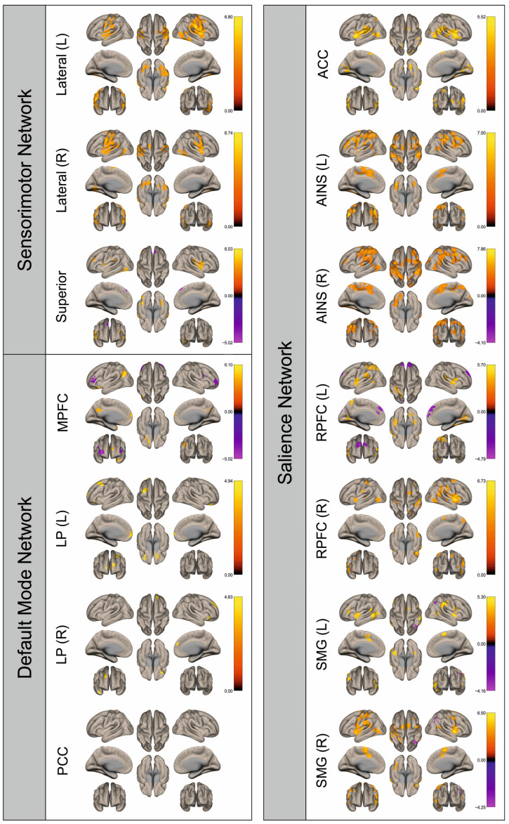
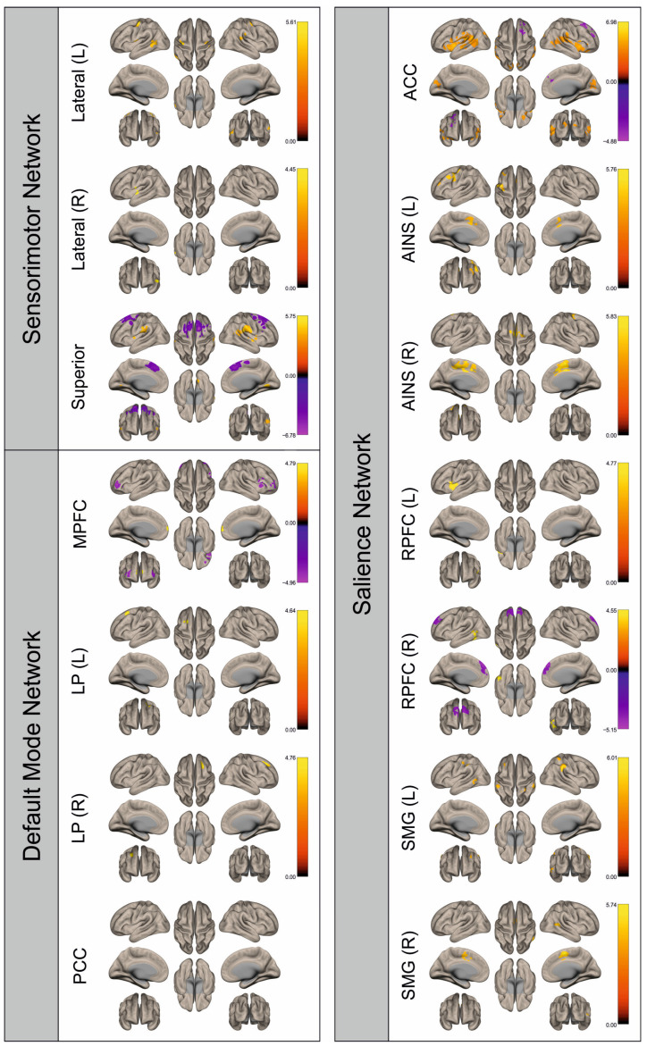
To date, there are almost no investigations addressing functional connectivity (FC) in patients with brain metastases (BM). In this retrospective study, we investigate the influence of BM on hemodynamic brain signals derived from functional magnetic resonance imaging (fMRI) and FC. Motor-fMRI data of 29 patients with BM and 29 matched healthy controls were analyzed to assess percent signal changes (PSC) in the ROIs motor cortex, premotor cortex, and supplementary motor cortex and FC in the sensorimotor, default mode, and salience networks using Statistical Parametric Mapping (SPM12) and marsbar and CONN toolboxes. In the PSC analysis, an attenuation of the BOLD signal in the metastases-affected hemisphere compared to the contralateral hemisphere was significant only in the supplementary motor cortex during hand movement. In the FC analysis, we found alterations in patients' FC compared to controls in all examined networks, also in the hemisphere contralateral to the metastasis. This indicates a qualitative attenuation of the BOLD signal in the affected hemisphere and also that FC is altered by the presence of BM, similarly to what is known for primary brain tumors. This transformation is not only visible in the infiltrated hemisphere, but also in the contralateral one, suggesting an influence of BM beyond local damage.
迄今为止,几乎没有针对脑转移瘤(BM)患者功能连接性(FC)的研究。在这项回顾性研究中,我们调查了BM对源自功能磁共振成像(fMRI)的脑血流动力学信号和FC的影响。分析了29例BM患者和29例匹配的健康对照的运动fMRI数据,以使用统计参数映射(SPM12)以及marsbar和CONN工具箱评估运动皮层、运动前区皮层和辅助运动皮层感兴趣区域(ROI)的信号变化百分比(PSC),以及感觉运动、默认模式和突显网络中的FC。在PSC分析中,与对侧半球相比,在手部运动期间,仅在辅助运动皮层中,转移瘤影响半球的血氧水平依赖(BOLD)信号衰减显著。在FC分析中,我们发现与对照组相比,在所有检查的网络中,患者的FC均有改变,在转移瘤对侧半球也是如此。这表明受影响半球中BOLD信号存在质性衰减,并且BM的存在也会改变FC,这与原发性脑肿瘤的情况类似。这种改变不仅在浸润半球中可见,在对侧半球中也可见,表明BM的影响超出了局部损伤。